. 2023 Feb;24(2):267-279.
doi: 10.1038/s41590-022-01379-9. Epub 2022 Dec 21.

Hypoxia drives CD39-dependent suppressor function in exhausted T cells to limit antitumor immunity

Affiliations

Hypoxia drives CD39-dependent suppressor function in exhausted T cells to limit antitumor immunity

Paolo D A Vignali et al. Nat Immunol. 2023 Feb.

Abstract

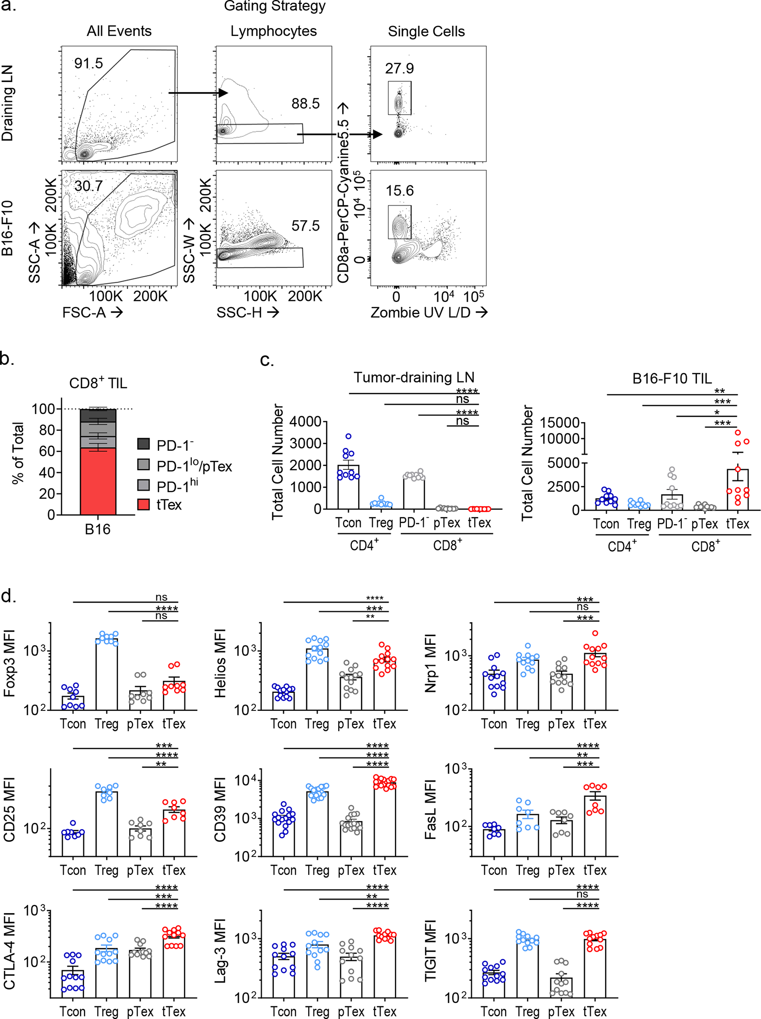
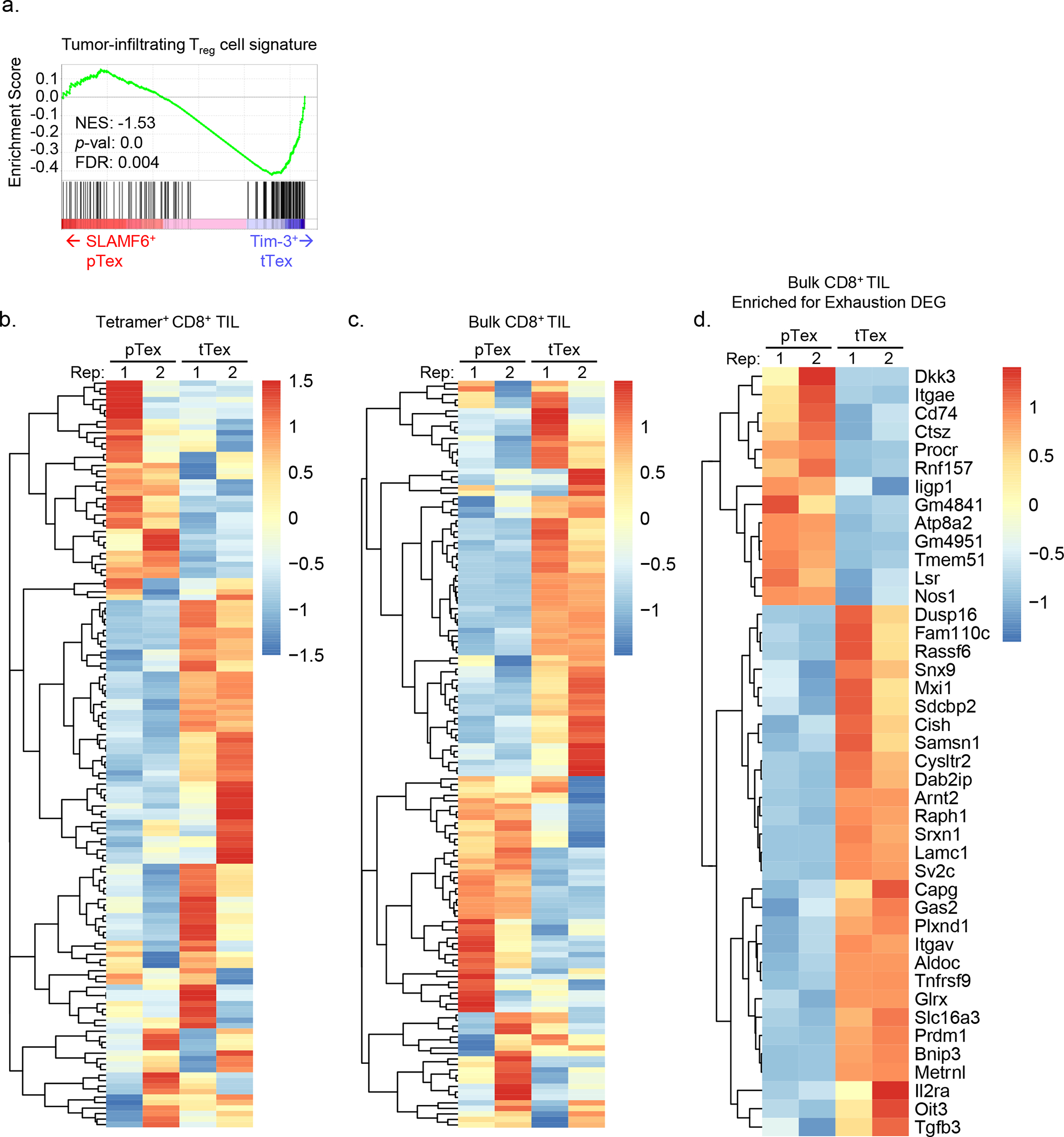
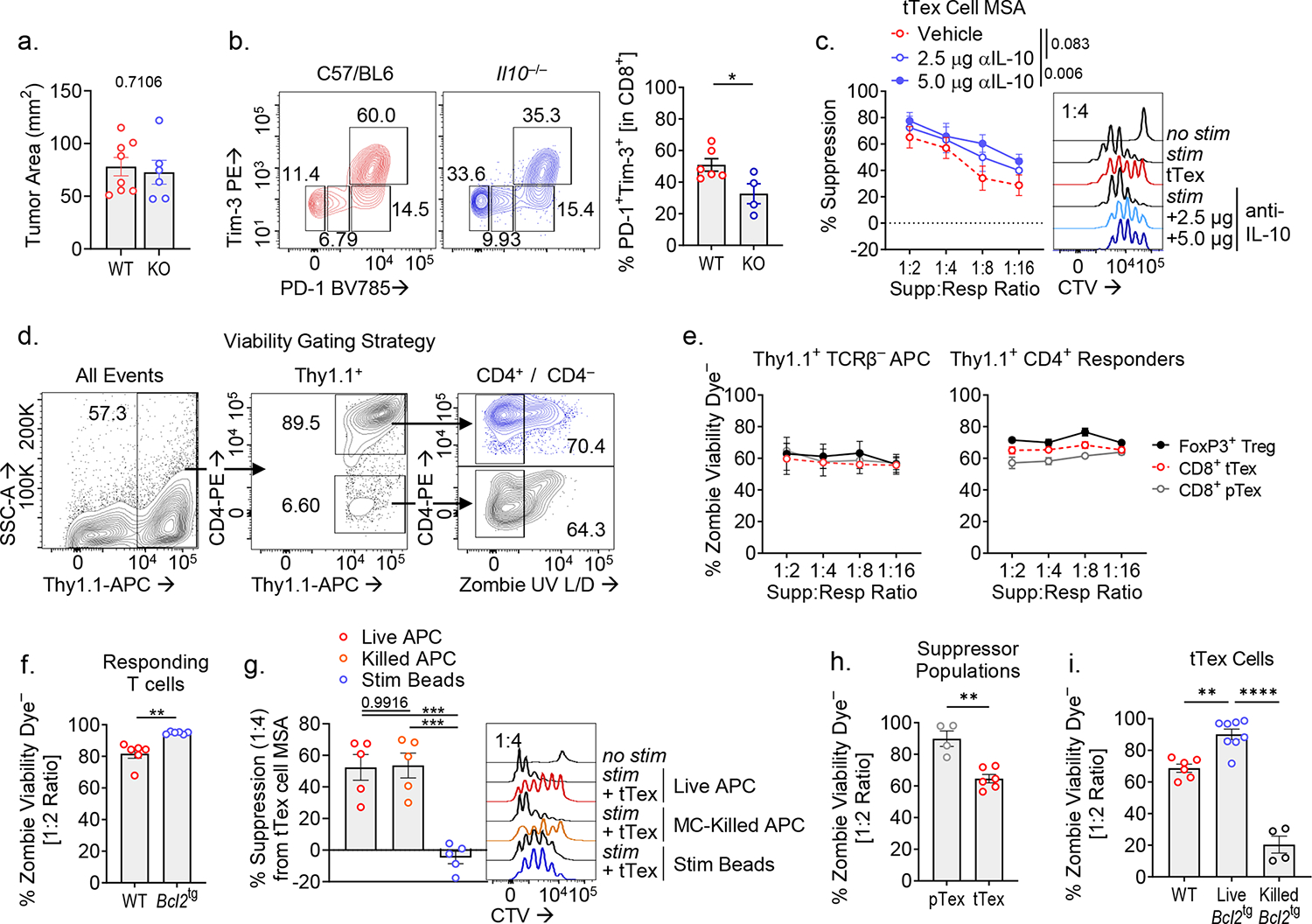
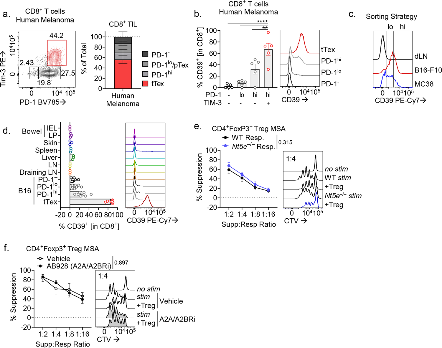
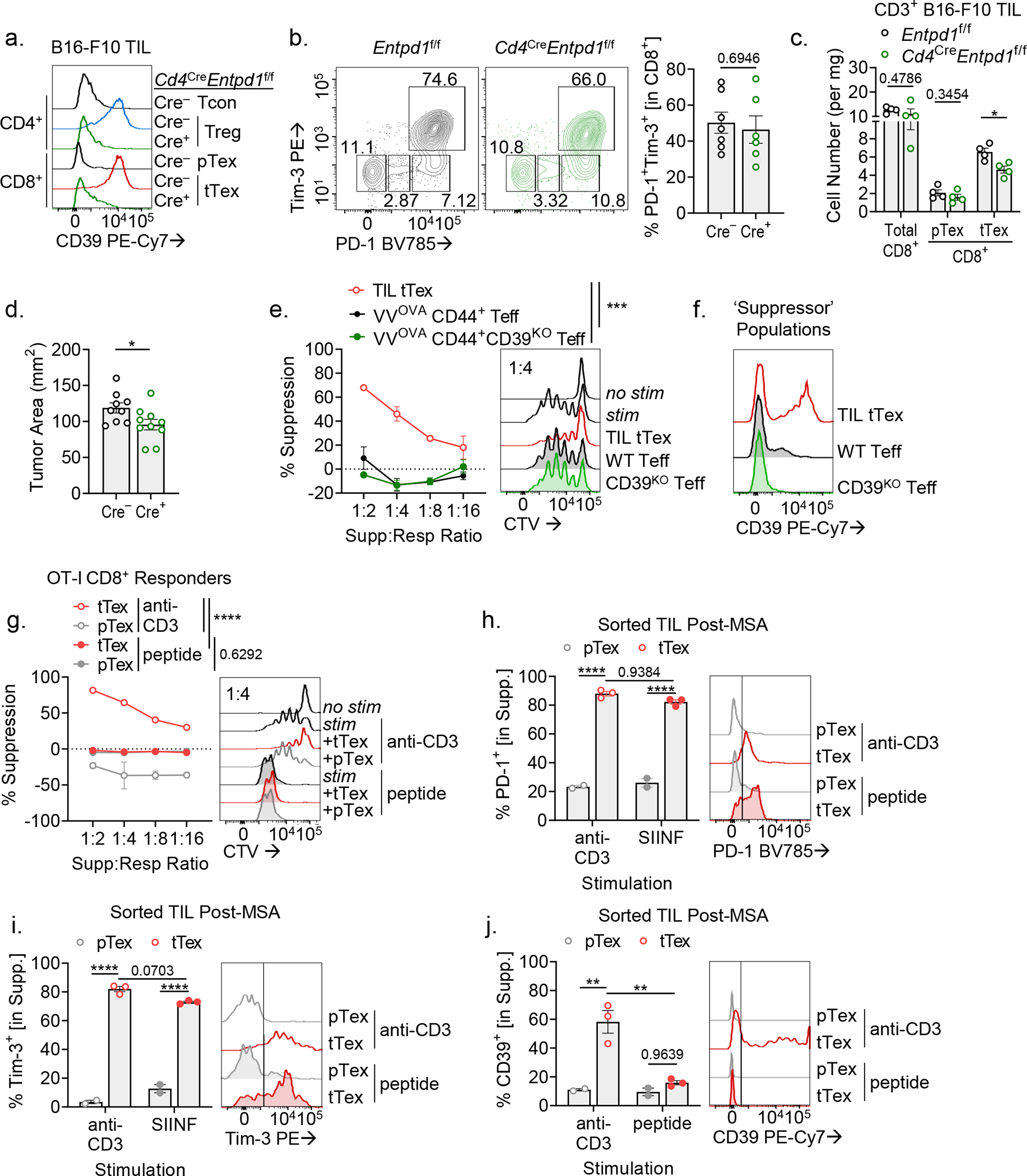
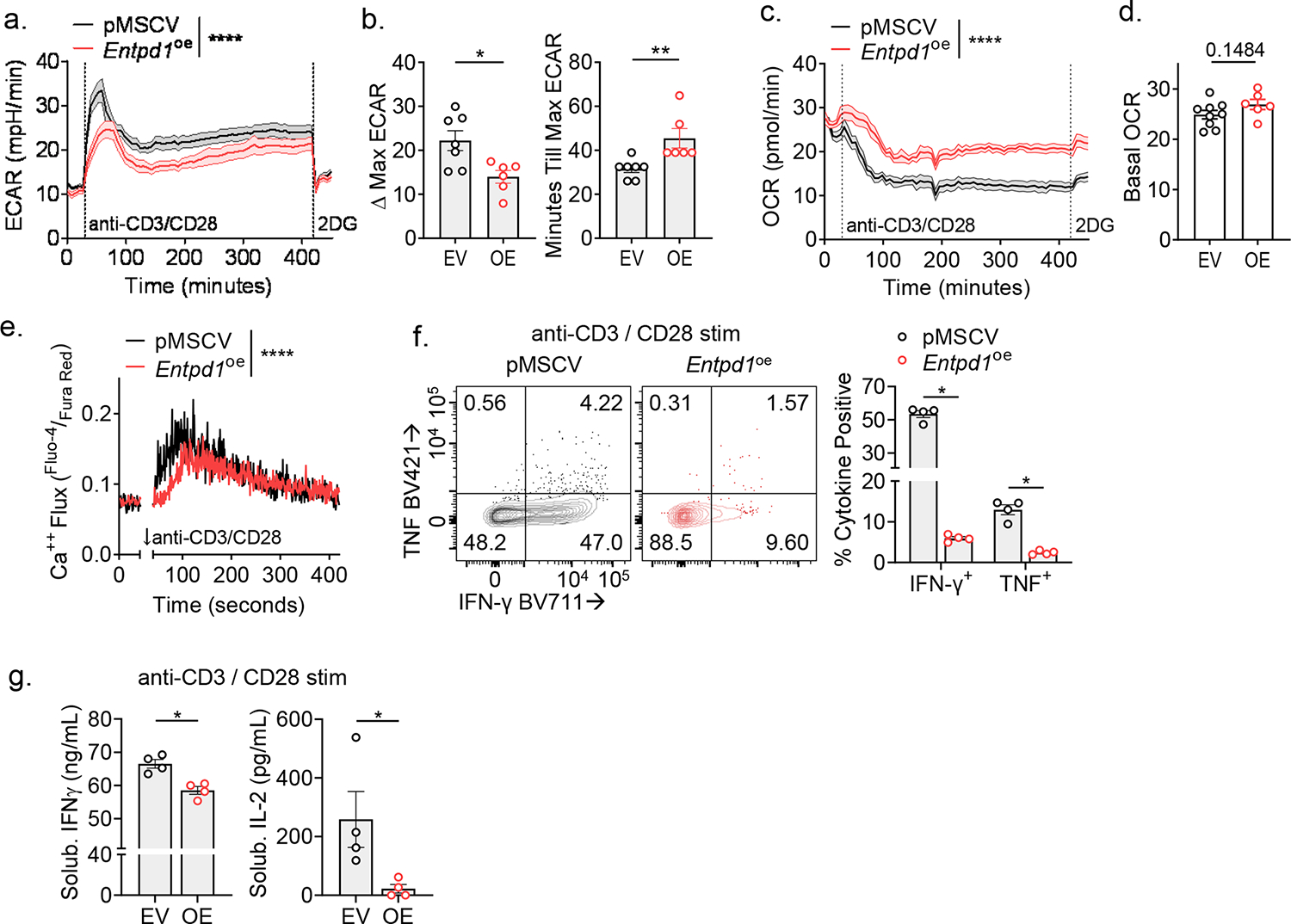
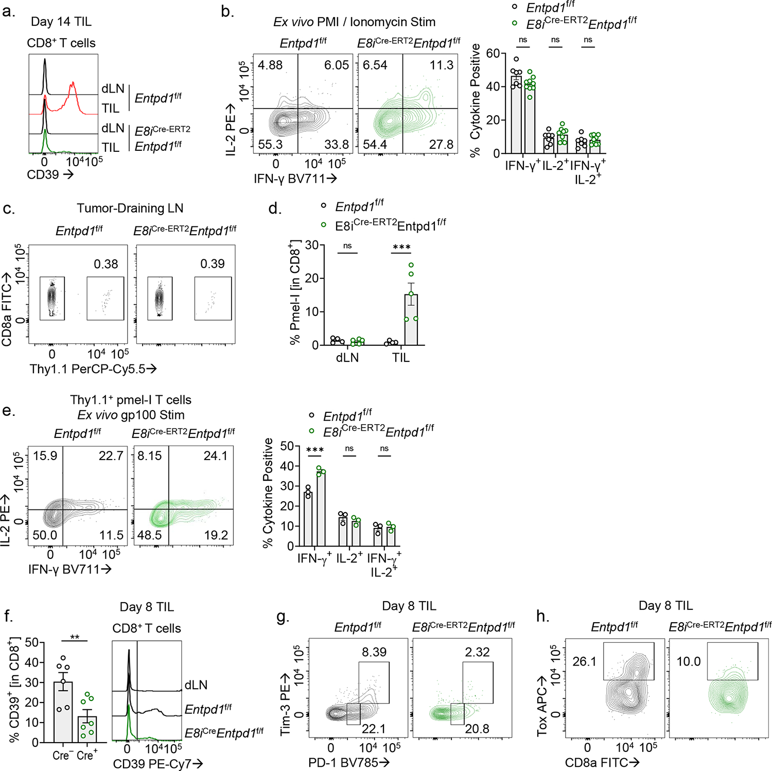
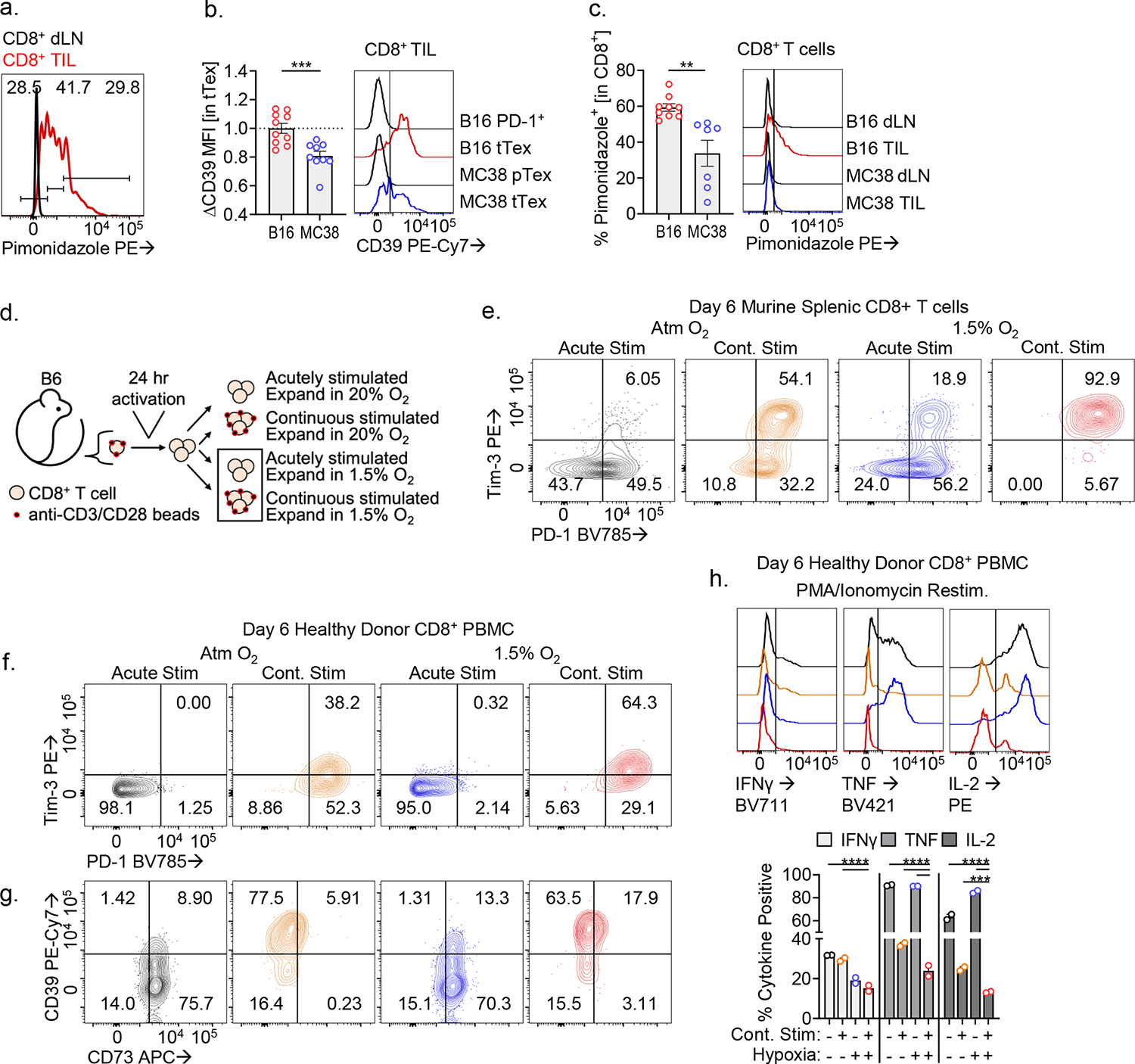
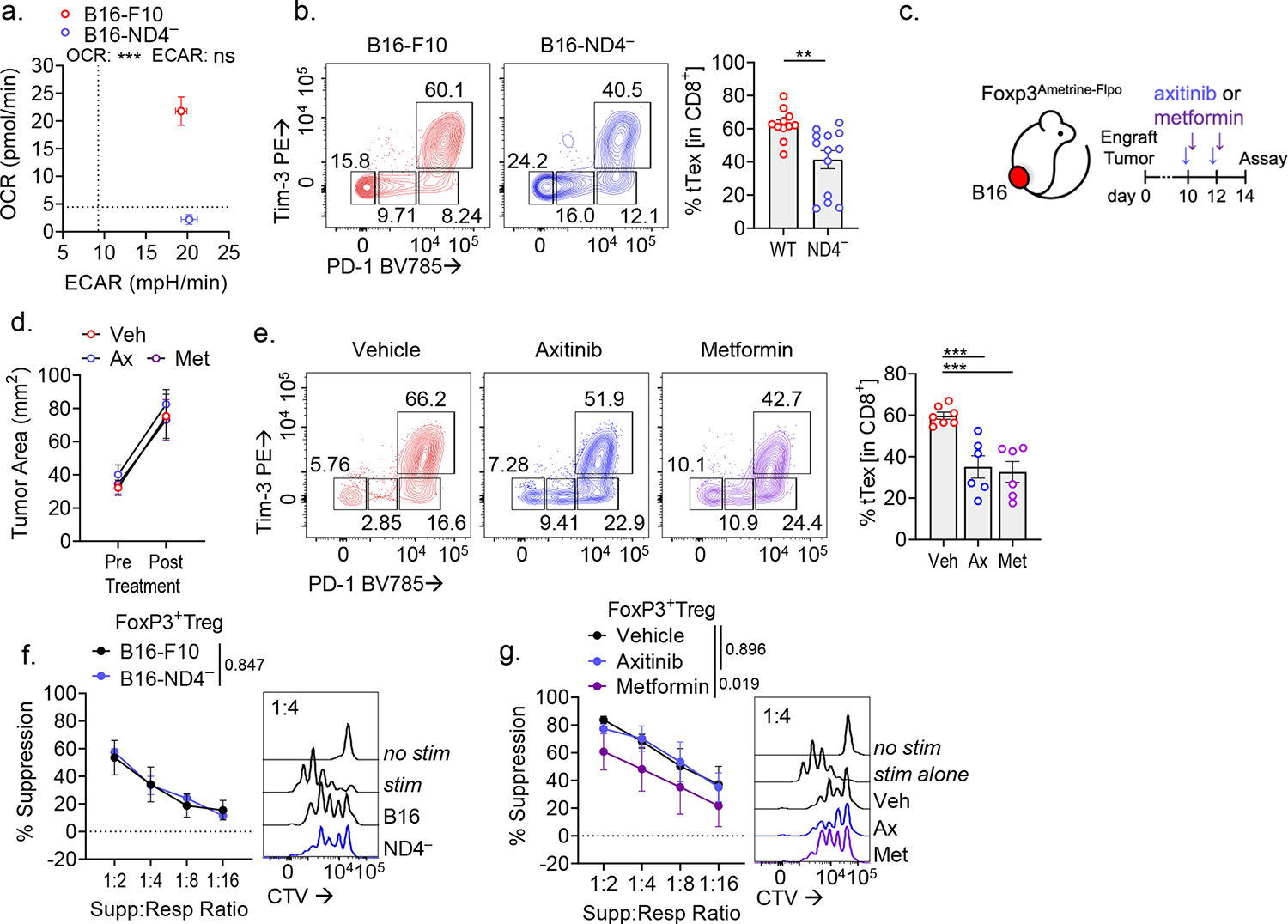
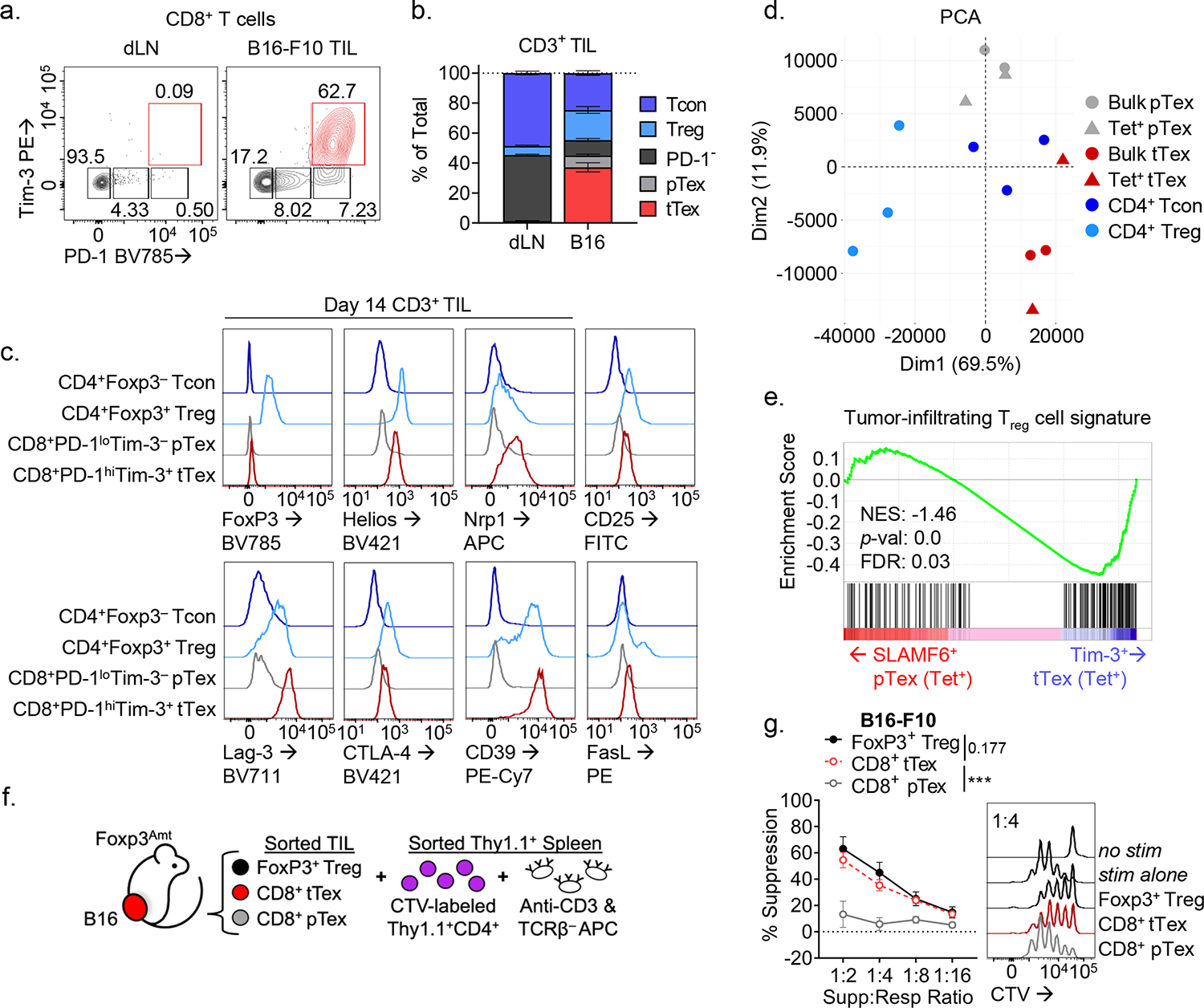
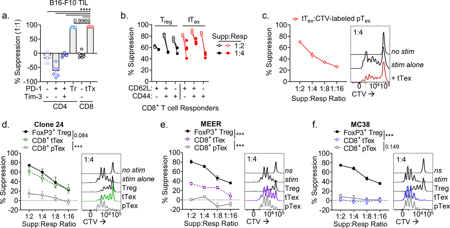
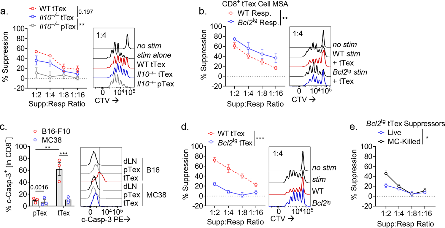
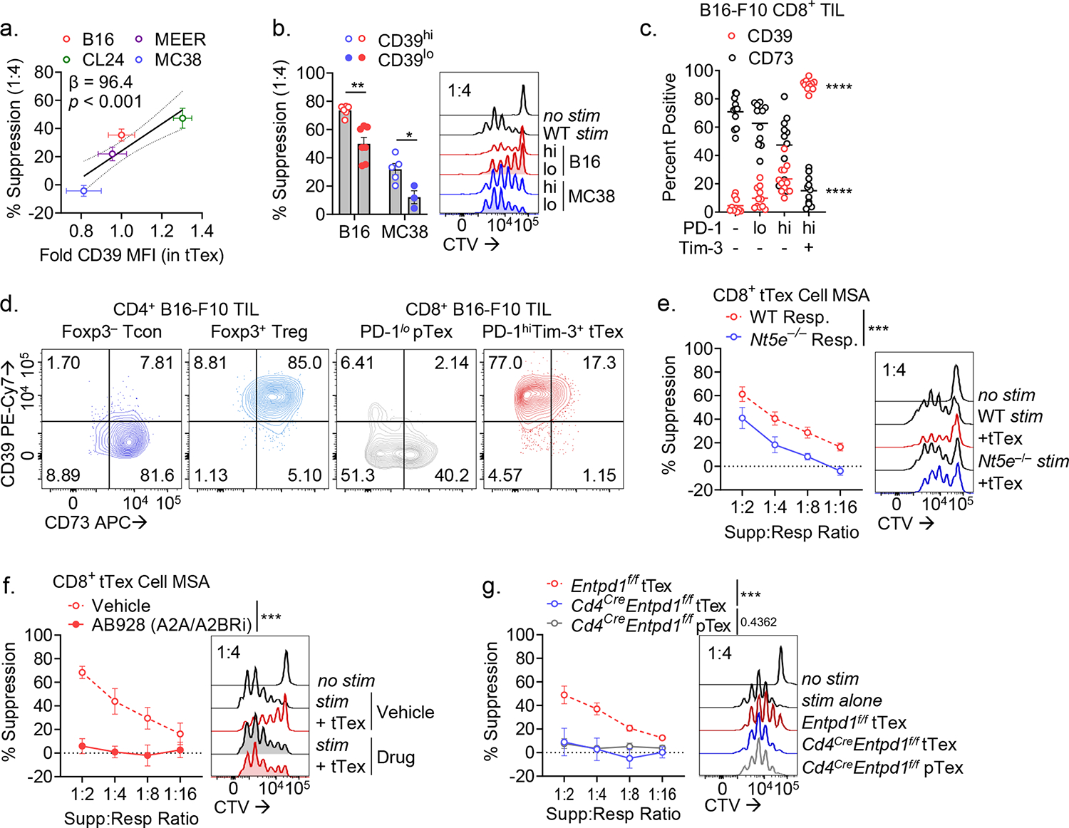
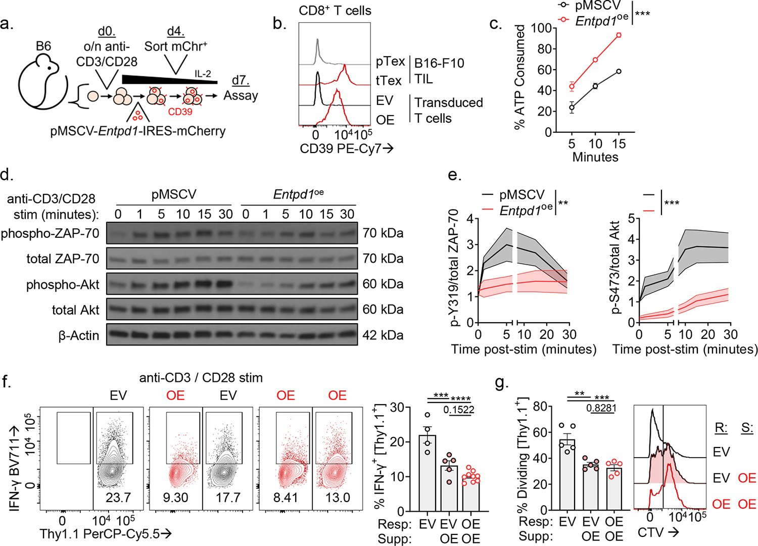
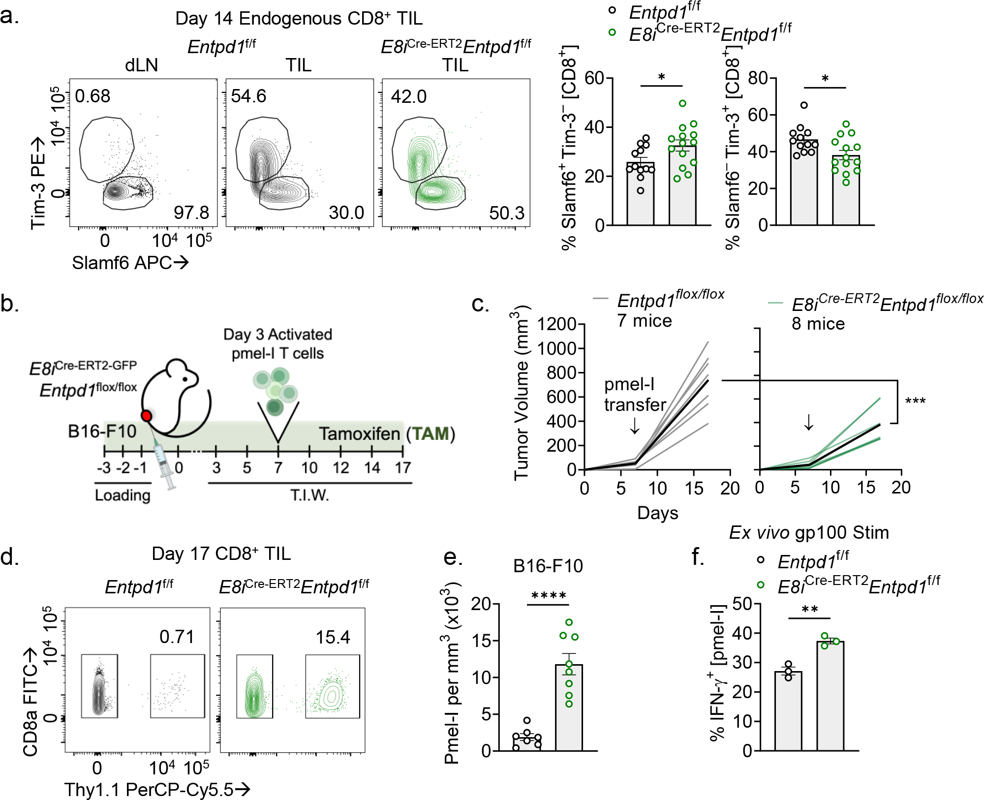
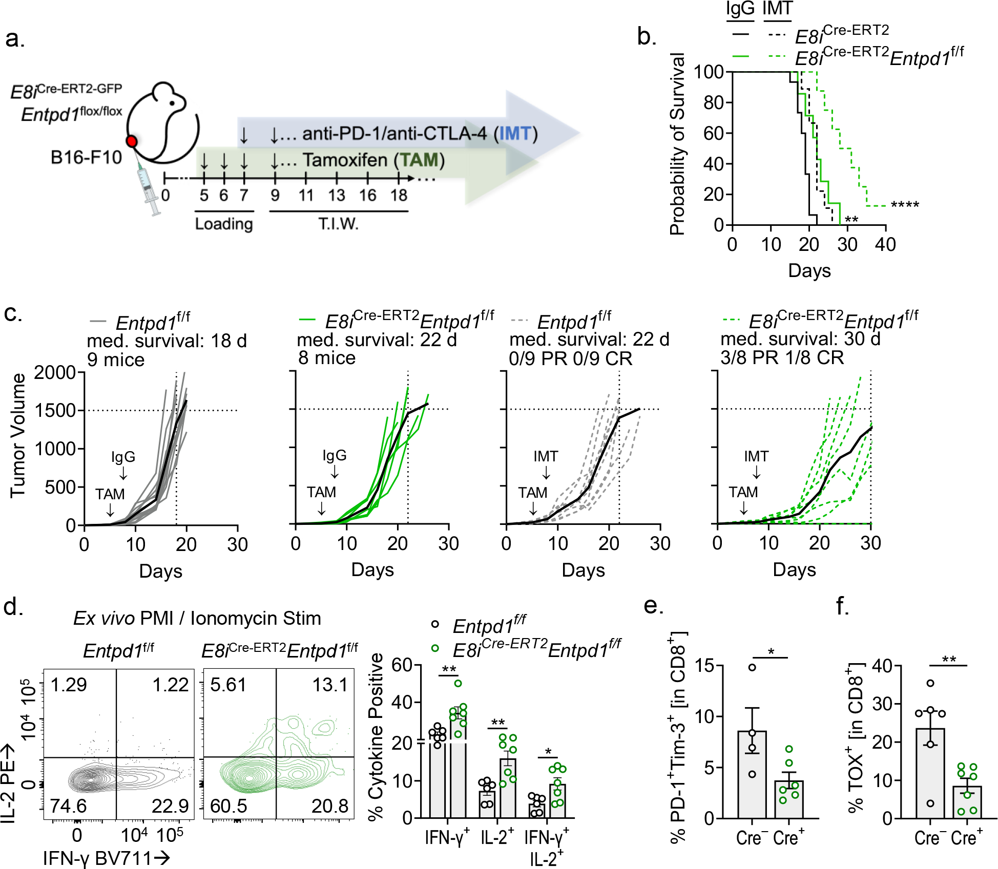
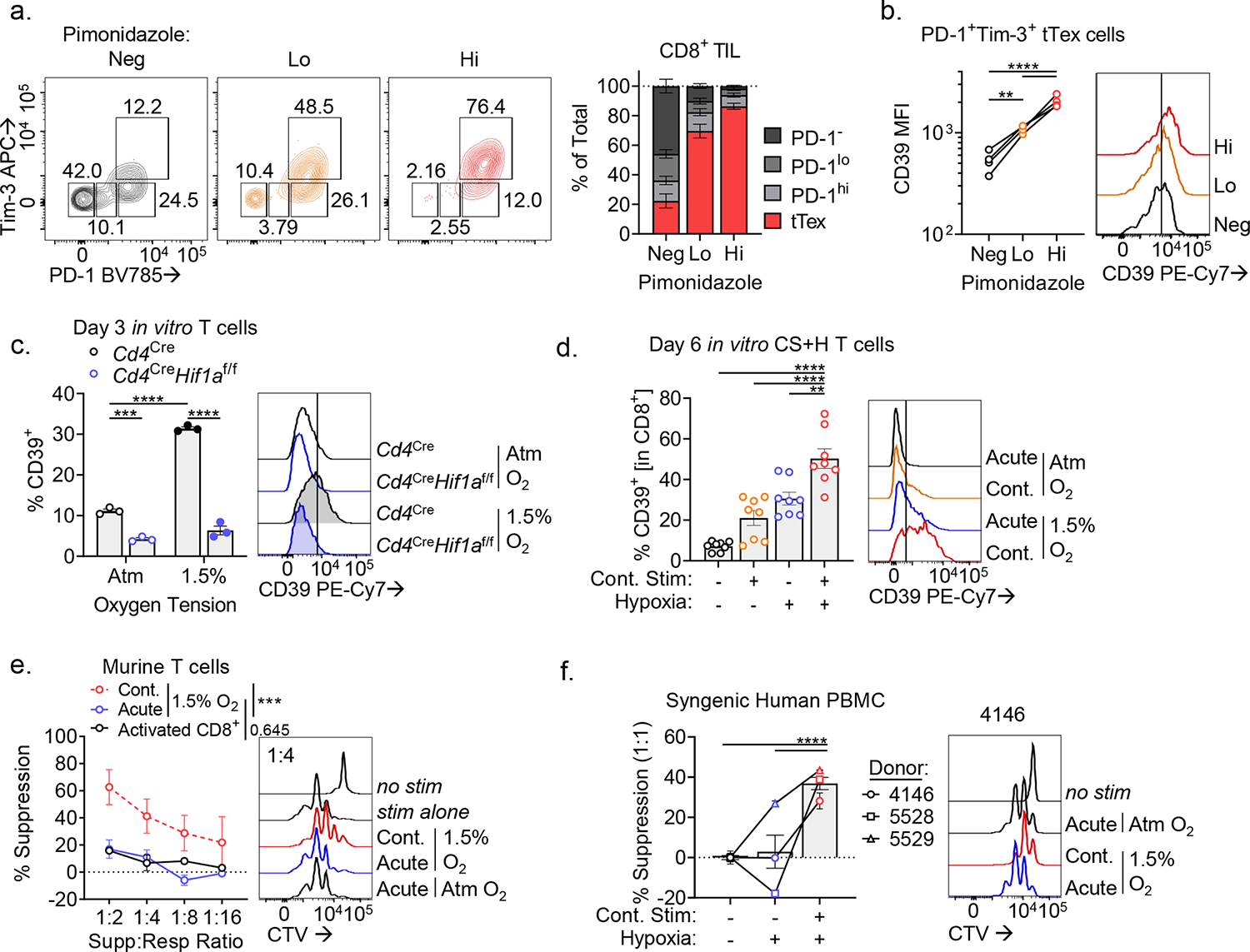
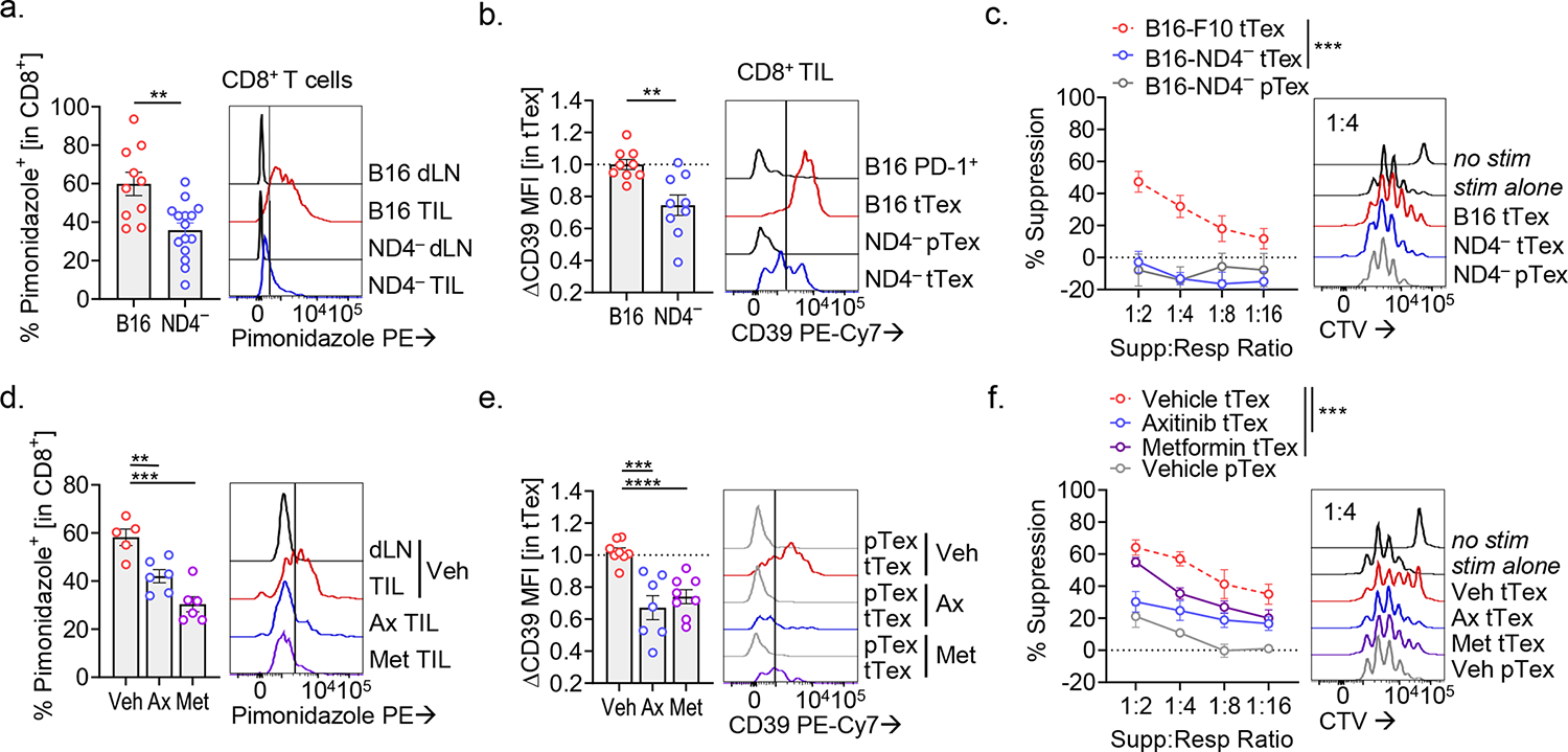
CD8+ T cells are critical for elimination of cancer cells. Factors within the tumor microenvironment (TME) can drive these cells to a hypofunctional state known as exhaustion. The most terminally exhausted T (tTex) cells are resistant to checkpoint blockade immunotherapy and might instead limit immunotherapeutic efficacy. Here we show that intratumoral CD8+ tTex cells possess transcriptional features of CD4+Foxp3+ regulatory T cells and are similarly capable of directly suppressing T cell proliferation ex vivo. tTex cell suppression requires CD39, which generates immunosuppressive adenosine. Restricted deletion of CD39 in endogenous CD8+ T cells resulted in slowed tumor progression, improved immunotherapy responsiveness and enhanced infiltration of transferred tumor-specific T cells. CD39 is induced on tTex cells by tumor hypoxia, thus mitigation of hypoxia limits tTex suppression. Together, these data suggest tTex cells are an important regulatory population in cancer and strategies to limit their generation, reprogram their immunosuppressive state or remove them from the TME might potentiate immunotherapy.


Conflict of interest statement

COMPETING INTERESTS STATEMENT

A.V.M. is currently an employee of Novasenta. G.M.D. declares competing financial interests and has submitted patents targeting exhausted T cells that are licensed or pending and is entitled to a share in net income generated from licensing of these patent rights for commercial development. G.M.D. consults for and/or is on the scientific advisory board of BlueSphere Bio, Century Therapeutics, Nanna Therapeutics, Novasenta, Pieris Pharmaceuticals, and Western Oncolytics/Kalivir; has grants from bluebird bio, Novasenta, Pfizer, Pieris Pharmaceuticals, TCR2, and Western Oncolytics/Kalivir; G.M.D. owns stock in Novasenta, BlueSphere Bio, and RemplirBio. The remaining authors have no competing interests.

Figures

Extended Data Figure 1.
Extended Data Figure 1.. PD-1hiTim-3+ terminally exhausted CD8+ T cells are a numerically dominant in tumor and express numerous Treg cell-associated effector molecules.
(A) Gating strategy for isolating CD8+ T cells in B16-F10 tumor or tumor-draining lymph nodes. (B) Quantified distribution of inhibitory receptor expression on CD8+ TIL from Fig. 1A. (C) Total cell numbers per size-matched B16-F10 tumor or draining lymph node. (D) Quantification from Fig. 1C. Mean fluorescence intensity (MFI) of CD4+Foxp3+ Treg cell-associated genes among tumor infiltrating T cell populations. Statistics are one-way ANOVA (C,D) with *p<0.05, **p<0.01, ***p<0.001 and ****p<0.0001
Extended Data Figure 2.
Extended Data Figure 2.. Tumor infiltrating CD8+ T cells upregulate Treg cell signature upon terminal differentiation.
(A) Gene set enrichment analysis (GSEA) of tumor infiltrating Treg cell signature on bulk SLAMF6+ progenitor and Tim-3+ terminally exhausted CD8+ T cell transcripts from Miller et al. (B,C) Heatmap displaying DESeq2 of log2 normalized transcript expression of genes from the tumor infiltrating Treg cell signature gene set in (B) tetramer+ or (C) bulk progenitor and terminally exhausted CD8+ T cells. Values are transformed log2 (TPM) scaled to row. (D) Heatmap of log2 normalized DESeq2-defined differentially expressed genes (DEG) between progenitor and terminally exhausted CD8+ T cells.
Extended Data Figure 3.
Extended Data Figure 3.. Quantification of tumor sizes and TIL populations from preclinical models with diverse sensitivity to immunotherapy.
(A) Tumor sizes from MSA experiments in Fig. 1 and 2 (B) (F) Average calculated percent suppression at 1:4 suppressor to responder ratio from all replicate experiments in Fig. 1 and supplementary Fig. 3R. (G) Percent of CD8+PD-1+Tim-3+ tTexh cells in various murine tumor models. Bivariant plots and histograms are representative of ≥3 experiments. Statistics are one-way ANOVA (A-C) with *p<0.05, **p<0.01, ***p<0.001 and ****p<0.0001.
Extended Data Figure 4.
Extended Data Figure 4.. tTex cells do not suppress via IL-10 secretion or direct cytotoxicity.
(A,B) Day 14 B16-F10 tumor sizes from Il10−/− experiments and matched CD8+ T cell infiltrate. (C) Suppression assay of B16-F10 infiltrating tTex cells co-cultured with isotype IgG or neutralizing IL-10 antibody at 2.5 or 5.0 μg/mL. (D) Gating strategy for calculation of cell viability in suppression assay populations. (E) Calculated averages of ‘responder’ or ‘APC’ population cell viability per dilution series of ‘suppressor’ T cells from Fig. 1G. (F) Cell viability of responder T cell populations from Fig. 3B. (G) Suppression assay of B16-F10 infiltrating tTex cells co-cultured with CTV-labeled responding T cells with either live or Mytomycin C (MC)-killed T cell-depleted splenocytes, or anti-CD3/anti-CD28 bound microbeads. (H) Cell viability of ‘suppressor’ populations from Fig. 1G. (I) Cell viability of ‘suppressor’ populations from Fig. 3D,E. Statistics are Mann-Whitney (A,B,F,H), linear regression (C), and one-way ANOVA (G,I) with *p<0.05, **p<0.01, ***p<0.001 and ****p<0.0001.
Extended Data Figure 5.
Extended Data Figure 5.. tTex cells suppress through CD39-mediated extracellular ATP depletion and adenosine production.
(A,B) Human CD8+ T cell populations from five melanoma biopsies from treatment-naïve patients and corresponding CD39 expression. (C) Sorting strategy for Fig. 4B. (D) Percent CD8+ T cells expressing CD39 in various tissues in a B16-F10 tumor-bearing C57/BL6 mice. (E,F) Suppression assays of B16-F10 infiltrating CD4+ Treg cells, co-cultured (E) with activated CTV-labeled C57/BL or Nt5e responding T cells or (F) with DMSO vehicle or A2AR/A2BR small-molecule inhibitor AB928 at 3 μg/mL. Statistics are one-way ANOVA (B), and linear regression (E,F) with *p<0.05, **p<0.01, ***p<0.001 and ****p<0.0001.
Extended Data Figure 6.
Extended Data Figure 6.. Cd4-driven Cre recombinase expression efficiently deletes CD39 on CD8+ TIL.
(A,B) TIL analysis of CD39 and inhibitory expression in CD8+ T cells from Cd4CreEntpd1f/f mice. (C) Cell counts per milligram of tumor mass from experiments in Extended Fig. 6A,B. (D) Tumor areas at day 14 from experiments in Fig. 2F. (E) Suppression assay of Cd4CreEntpd1f/f Thy1.1+CD44+ OT-I Teff cells isolated from day 8 of acute VacciniaOVA infection. (F) CD39 expression on TIL tTex cells or VacciniaOVA OT-I T cells in suppression assay co-cultures. (G) Suppression assay of B16-F10-derived CD8+ tTex cells co-cultured with OT-I TCR transgenic CD8+ T cells. Culture were stimulated with either anti-CD3 antibodies as before, or OT-I specific peptide, SIIFEKL. (H-J) Flow cytometric analysis of TIL following MSA from Extended Data Fig. 6G. Statistics are Mann-Whitney (B-D,H-J) and linear regression (E,G) with *p<0.05, **p<0.01, ***p<0.001 and ****p<0.0001.
Extended Data Figure 7.
Extended Data Figure 7.. Enforced CD39 expression on CD8+ T cells limits metabolic reprogramming via disruption of TCR signaling.
(A) Extracellular acidification rate (ECAR) in in-Seahorse activation of rested CD8+ T cells stability transduced with pMSCV or pMSCV-Entpd1. (B) Delta-maximal ECAR quantified by basal ECAR – maximal ECAR; minutes till maximal ECAR per sample well. (C) Oxygen consumption rate (OCR) of cells from Extended Data Fig. 7C with (D) quantified basal OCR. (E) Intracellular calcium flux in day 7 transduced T cells restimulated in a calcium-buffered solution with anti-CD3/anti-CD28-bound microbeads in the presence of calcium indicators, Fluo-4 and Fura Red. (F) Cytokine production of transduced T cells following 24-hour anti-CD3/anti-CD28-bound microbeads stimulation. (G) ELISA of supernatants from repeat experiments of Extended Data Fig. 7F. Statistics are Mann-Whitney (B,D,G) and one-way ANOVA (E,F) with *p<0.05, **p<0.01, ***p<0.001 and ****p<0.0001.
Extended Data Figure 8.
Extended Data Figure 8.. E8i-driven Cre recombinase expression efficiently deletes CD39 on CD8+ TIL.
(A) CD39 expression in day 14 B16-F10 tumor infiltrating CD8+ T cells from Entpd1f/f or E8iCre-ERT2Entpd1f/f mice. (B) Cytokine production following PMA/Ionomycin re-simulation of day 14 CD8+ TIL. (C,D) Infiltration of adoptively transferred pmel-I T cells into tumor-draining lymph nodes (dLN; C) and (D) quantification of pmel-I percentages in a representative experiment. (E) Ex vivo cytokine production of pmel-I T cells following gp100 re-stimulation. (F) TIL analysis of CD39 expression in CD8+ T cells from E8iCre-ERT2Entpd1f/f mice treated for three consecutive days with tamoxifen. (G,H) Representative flow cytometry plots from Fig. 7E,F. Statistics are one-way ANOVA (B,D,E) and Mann-Whitney (F) with *p<0.05, **p<0.01, ***p<0.001 and ****p<0.0001.
Extended Data Figure 9.
Extended Data Figure 9.. Tumor hypoxia enforces CD39 expression on tTex cells.
(A) Histogram overlay displaying hypoxia exposure in CD8+ dLN and TIL from B16-F10 tumors. (B) CD39 staining in exhausted T cells from B16-F10 or MC38, and (C) Pimonidazole staining of bulk TIL. (D) Continuous Activation under Hypoxia (CS+H) assay. In brief, naïve T cells are activated for 24 hours, then split into treatment groups of removal (acute stimulation) or continued presence (continuous stimulation) of anti-CD3/anti-CD28-bound microbeads and cultured under atmospheric oxygen tensions (~20% O2) or tumor hypoxic conditions (1.5% O2) for 5 days. (E) Inhibitory receptor staining in murine day 6 CS+H cells. (F-H) Validation of humanized CS+H assay with healthy donor PBMC-derived CD8+ T cells via staining of (F) inhibitory receptors and (G) enzymes of adenosine metabolism. (H) 24-hour PMA/Ionomycin restimulation of human CS+H cells. Statistics are Mann-Whitney (B,C), one-way ANOVA (H) with *p<0.05, **p<0.01, ***p<0.001 and ****p<0.0001.
Extended Data Figure 10.
Extended Data Figure 10.. Tumor hypoxia mitigation as a therapeutic target to lessen tTex cell-mediated suppression.
(A) Extracellular flux analysis showing validation of mitochondrial respiration knockdown (via OCR measurement) in B16ND4− tumor cells versus parental B16-F10. (B) PD-1 and Tim-3 staining in TIL from day 14 wild-type B16-F10 or B16ND4− tumors. (C) Schematic of treatment plan for therapeutic alleviation of tumor hypoxia. (D) Tumor sizes at treatment initiation and sacrifice in Axitinib/metformin experiments. (E) PD-1 and Tim-3 staining in TIL from Axitinib/metformin experiments in Extended Data Fig. 9D. (F) Suppression assays of CD4+Foxp3+ Treg cells from Fig. 9C. (G) Suppression assays of CD4+Foxp3+ Treg cells from Fig. 9F. Statistics are Mann-Whitney (B), one-way ANOVA (E) and linear regression (F,G) with *p<0.05, **p<0.01, ***p<0.001 and ****p<0.0001
Figure 1.
Figure 1.. PD-1hiTim-3+Foxp3 terminally exhausted CD8+ T cells predominate in solid tumors and adopt a suppressive signature that correlates with immunotherapy response.
(A) CD8+ tumor-infiltrating T cells from day 14 B16-F10 murine melanoma tumor and tumor draining lymph nodes (dLN). (B) Total CD3+ T cells from B16-F10 TIL and dLN with quantified fold change. (C) Expression of CD4+Foxp3+ Treg cell-associated genes among tumor infiltrating T cell populations. (D) Principal component analysis (PCA) plot of tumor-infiltrating T cells from RNAseq mined from Magnuson et al. (2018) and Miller et al. (2019). (E) Gene set enrichment analysis (GSEA) of tumor infiltrating Treg cell signature on (B) tetramer+ SLAMF6+ progenitor and Tim-3+ terminally exhausted CD8+ T cell transcripts from Miller et al. (F) Model of suppression assay and (G) results from sorted B16-F10 melanoma TIL. Statistics are linear regression (G) with *p<0.05, **p<0.01, and ***p<0.001.
Figure 2.
Figure 2.. Suppressive functionality in tTex cells can act on multiple target cells and is environment-dependent.
(A) CD4+ and CD8+ TIL populations from B16-F10 tumors stratified by inhibitory receptor expression and utilized as the ‘suppressor’ group in ex vivo suppression assay. (B) Suppression assay utilizing diverse responder populations from tumor draining lymph nodes and co-cultured with PD-1hiTim-3+ tTex cells or Foxp3+ Treg cells. (C) Proliferation dye labeled pTex cells co-cultured with tTex cells in repeat suppression assays. (D-F) Suppression assay with TIL from (D) Ptenf/fBRafLSL.V600ETyr2Cre.ER–derived melanoma clone, dubbed Clone 24, (E) PD-1-resistant MEER head and neck carcinoma, or (F) PD-1-sensitive MC38 adenocarcinoma. Statistics are one-way ANOVA (A) and linear regression (D-F) with *p<0.05, **p<0.01, ***p<0.001 and ****p<0.0001.
Figure 3.
Figure 3.. CD8+ tTex cell ex vivo suppression is associated by tTex cell-intrinsic apoptosis.
(A) Suppression assay of B16-F10 TIL from wild-type C57/BL6 or Il10−/− mice. (B) Suppression assay of B16-F10-derived tTex cells co-cultured with with activated C57/BL6 or Bcl2tg responding T cells. (C) Cleaved caspase-3 staining in CD8+ T cells from B16-F10 or MC38 tumors and draining lymph nodes (dLN). (D) Suppression assay of B16-F10 TIL from C57/BL6 or Bcl2tg mice. (E) Repeat experiments from Fig. 4D with live or Mytomycin C (MC)-killed tTex cells. Statistics are linear regression (A,B,D,E) and one-way ANOVA (C) with *p<0.05, **p<0.01, ***p<0.001 and ****p<0.0001.
Figure 4.
Figure 4.. tTex cells suppress through CD39-mediated extracellular ATP depletion and adenosine production.
(A) Pearson correlation of percent tTex cell-mediated suppression cell in 1:4 ratio in suppression assay versus fold change in CD39 MFI in tTex cells between murine tumor lines. (B) Repeat experiments from Fig. 1G and 2F, in which tTex cells from B16-F10 and MC38 tumors were stratified by CD39 expression. (C) Percent CD8+ T cell populations from B16-F10 expressing CD39 or CD73. (D) Representative bivariant plots of B16-F10 infiltrating T cell populations. (E) Suppression assay of B16-F10 TIL co-cultured with with activated CTV-labeled C57/BL or Nt5e– ‘responding’ T cells. (F) Suppression assay of B16-F10 infiltrating CD8+ tTex cells co-cultured with DMSO vehicle or A2AR/A2BR small-molecule inhibitor AB928 at 3 μg/mL. (F) MSA of B16-F10 TIL from Entpd1f/f or Cd4CreEntpd1f/f mice. Statistics are linear regression (A,E-G) and one-way ANOVA (B,C) with *p<0.05, **p<0.01, ***p<0.001 and ****p<0.0001.
Figure 5.
Figure 5.. Enforced CD39 expression in Teff cells inhibits effector functions in neighboring CD39 T cells.
(A) Model of activated T cells transduced with pMSCV-Entpd1-IRES-mCherry and then rested for seven days prior to assay. (B) CD39 expression in day 7 transduced T cells or day 14 B16-F10 TIL. (C) Percent of spiked in ATP consumed in transduced T cells. (D) Western Blot of phosphorylation events following of anti-CD3/anti-CD28-coated microbead stimulation in transduced T cells and (E) quantified band intensity normalized to t = 0 in empty vector T cells. (F) Cytokine production of transduced T cells in 24-hour co-culture experiments following anti-CD3/anti-CD28-coated microbead stimulation. (G) Division on CTV-labeled transduced T cells over 72 hours. Statistics are linear regression (C) and one-way ANOVA (E,F,G) with *p<0.05, **p<0.01, ***p<0.001 and ****p<0.0001.
Figure 6.
Figure 6.. Restricted deletion of CD39 on endogenous CD8+ tTex cells reduces suppression of newly infiltrating T cells.
(A) Tim-3 and Slamf6 staining on CD8+ T cells from day 14 B16-F10 tumors. (B) Schematic of adoptive cell therapy protocol. Entpd1f/f or E8iCre-ERT2Entpd1f/f mice were treated with tamoxifen (TAM) starting three days prior to B16-F10 tumor inoculation then continued thrice weekly thereafter. 3–5 million activated pmel-I T cells were transferred retro-orbitally on day 7. (C) Tumor growth from adoptive T cell transfer experiment. (D,E) Percent infiltration and absolute numbers of adoptively transferred pmel-I T cells into B16-F10 tumor. (F) IFN-gamma production in pmel-I T cells restimulated ex vivo with gp100. Statistics are Mann-Whitney (A,E,F), and two-way ANOVA (C) with *p<0.05, **p<0.01, ***p<0.001 and ****p<0.0001.
Figure 7.
Figure 7.. tTex cell-restricted deletion of CD39 invigorates immune responses in an aggressive preclinical model.
(A) Schematic of experimental design. TAM treatment began at day 5 post-tumor inoculation then continued thrice weekly thereafter; immunotherapy (IMT) treatment began on day 8 and was administered thrice weekly. (B,C) Tumor growth over time and survival in Entpd1f/f or E8iCre-ERT2Entpd1f/f mice bearing B16-F10 with or without combination immunotherapy. (D) CD8+ TIL restimulated with PMA and Ionomycin at day 8 of tumor growth, day 3 of TAM treatment. (E,F) PD-1 and Tim-3, or TOX expression in CD8+ T cells on day 8 of tumor growth, day 3 of TAM treatment. Statistics are Log-Rank test (B), one-way ANOVA (D), and Mann-Whitney (E,F) with *p<0.05, **p<0.01, ***p<0.001 and ****p<0.0001.
Figure 8.
Figure 8.. In vitro exposure to continuous TCR stimulation under hypoxic conditions yields exhausted-like, suppressive CD8+ T cells.
(A) PD-1 and Tim-3 staining in Pimonidazole fractions–negative, low, and high. (B) CD39 staining in PD-1+Tim-3+ tTex cells from Pimonidazole fractions. (C) Percent of CD8+ T cells expressing CD39 following 72-hour stimulation at atmospheric or tumor hypoxic conditions (1.5% O2). (D) CD39 staining of day 6 CS+H cells. (E) Proliferation of CTV-labeled murine CD4+ T cells in MSA experiments with CS+H cells. (F) Proliferation of three human healthy donor CTV-labeled CD8+ T cells in MSA experiments with syngeneic human CS+H cells. Statistics are one-way ANOVA (B-D,F), and linear regression (E) with *p<0.05, **p<0.01, ***p<0.001 and ****p<0.0001.
Figure 9.
Figure 9.. Genetic or pharmacologic mitigation of hypoxia reduces the CD39-dependent suppressive phenotype in tTex cells.
(A) Histogram overlay displaying Pimonidazole staining in intratumoral CD8+ T cells from parental B16-F10 or Ndufs4 (B16ND4−) tumors and draining lymph nodes (dLN). (B) CD39 staining intratumoral T cells B16-F10 or B16ND4− tumors. (C) Biological replicate suppression assays utilizing B16-F10 or B16ND4− TIL. (D) Pimonidazole staining in intratumoral CD8+ T cells infiltrating B16-F10 tumors or draining lymph nodes following two treatments of anti-hypoxia agents Axitinib (Ax) and metformin (Met). (E) CD39 staining in intratumoral T cells from various treatment groups. (F) Biological replicate suppression assays of B16-F10 TIL following two treatments of either vehicle, Axitinib, or metformin. Statistics are Mann-Whitney (A,B), linear regression (C,F), and one-way ANOVA (D,E), with *p<0.05, **p<0.01, ***p<0.001 and ****p<0.0001.

Comment in

References

    1. Blank CU et al. Defining “T cell exhaustion”. Nat. Rev. Immunol. 19, 665–674 (2019). - PMC - PubMed
    1. Im SJ et al. Defining CD8+ T cells that provide the proliferative burst after PD-1 therapy. Nature 537, 417–421 (2016). - PMC - PubMed
    1. Miller BC et al. Subsets of exhausted CD8+ T cells differentially mediate tumor control and respond to checkpoint blockade. Nat. Immunol. 20, 326–336 (2019). - PMC - PubMed
    1. Bruni D, Angell HK & Galon J The immune contexture and Immunoscore in cancer prognosis and therapeutic efficacy. Nat. Rev. Cancer 20, 662–680 (2020). - PubMed
    1. Sade-Feldman M et al. Defining T Cell States Associated with Response to Checkpoint Immunotherapy in Melanoma. Cell 175, 998–1013.e20 (2018). - PMC - PubMed

Publication types