Case Reports
. 2022 Dec 7:29:101772.
doi: 10.1016/j.ajoc.2022.101772. eCollection 2023 Mar.

Posterior placoid-like maculopathy and macular hole associated with vitamin A deficiency

Affiliations
Case Reports

Posterior placoid-like maculopathy and macular hole associated with vitamin A deficiency

Eric W Lai et al. Am J Ophthalmol Case Rep. .

Abstract

Purpose: To report a case of bilateral posterior placoid-like maculopathy and a macular hole associated with vitamin A deficiency.

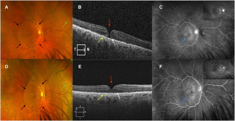
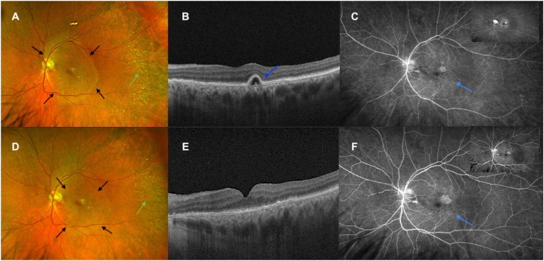
Observations: A 72-year-old male presented with nyctalopia and progressive vision loss in both eyes. Examination and multimodal imaging were consistent with posterior placoid-like maculopathy bilaterally and a macular hole in the right eye. A workup for infectious, inflammatory, and paraneoplastic etiologies revealed a severely low serum vitamin A level. Two months after initiation of vitamin A repletion, there was improvement in best-corrected Snellen visual acuity as well as macular hole closure. A diagnosis of posterior placoid-like maculopathy in the setting of vitamin A deficiency (VAD) was made.

Conclusions and importance: VAD should be considered when symmetric posterior pole placoid-like lesions are observed and other, more common etiologies have been ruled out.

Keywords: Hepatobiliary disease; Macular hole; Posterior placoid maculopathy; Syphilitic retinitis; Vitamin A deficiency.


Conflict of interest statement

The following authors have no financial disclosures: EWL, RD, BKD, SAS.

Figures

Fig. 1
Fig. 1
Fundus photo, OCT macula, and FA of the right eye at presentation (A–C) compared to two months post-vitamin A supplementation (D–F). Fundus photo demonstrates well-circumscribed, placoid-like lesion (A, black arrows) in posterior pole as well as peripheral yellow-white lesions (A, green arrow) with improvement after vitamin A supplementation (D, corresponding line & arrow). OCT macula shows a stage 2 full-thickness macular hole with trace CME (B, red arrow), as well as diffuse outer layer attenuation (B, white arrow), ELM disruption, ellipsoid zone loss, and RPE mottling and thickening (B, yellow arrow). After vitamin A supplementation, the OCT findings significantly improved, and the macular hole resolved (E, corresponding arrows). FA in the early phase, displayed in the upper right hand corner, and late phase, in the central picture, shows disc leakage (C, gray arrow) with stippled hyperfluorescence and staining throughout posterior pole (C, navy arrow), with less distinct margins and less disc leakage after supplementation was initiated (F, corresponding arrows). (For interpretation of the references to color in this figure legend, the reader is referred to the Web version of this article.)
Fig. 2
Fig. 2
Fundus photo, OCT macula, and FA of the left eye at presentation (A–C) compared to two months post-vitamin A supplementation (D–F) show similar findings and improvements as described in Fig. 1. OCT macula shows a serous pigment epithelium detachment (B, blue arrow) that improved with supplementation (E). (For interpretation of the references to color in this figure legend, the reader is referred to the Web version of this article.)
Fig. 3
Fig. 3
Fundus autofluorescence photography of the right eye (A) and left eye (C) at presentation compared to two months post-vitamin A supplementation (B, D) shows improvement in hyperautoflourescence in the placoid-like lesion.

References

    1. Chao J.R., Khurana R.N., Fawzi A.A., Reddy H.S., Rao N.A. Syphilis: reemergence of an old adversary. Ophthalmology. 2006;113(11):2074–2079. - PubMed
    1. Eandi C.M., Neri P., Adelman R.A., Yannuzzi L.A., Cunningham E.T., Jr., International Syphilis Study Group Acute syphilitic posterior placoid chorioretinitis: report of a case series and comprehensive review of the literature. Retina. 2012;32(9):1915–1941. - PubMed
    1. Neri P., Pichi F. Acute syphilitic posterior placoid chorioretinitis: when the great mimicker cannot pretend any more; new insight of an old acquaintance. J Ophthalmic Inflamm Infect. 2022 Feb 22;12(1):9. - PMC - PubMed
    1. DeVience E.X., Schechet S.A., Carney M., et al. Syphilitic retinitis presentations: punctate inner retinitis and posterior placoid chorioretinitis. Int Ophthalmol. 2021;41(1):211–219. - PubMed
    1. Wells J., Wood C., Sukthankar A., Jones N.P. Ocular syphilis: the re-establishment of an old disease. Eye (Lond). 2018;32(1):99–103. - PMC - PubMed

Publication types

LinkOut - more resources