. 2022 Nov;83(6):1312-1326.
doi: 10.3348/jksr.2021.0166. Epub 2022 Mar 21.

Added Value of the Sliding Sign on Right Down Decubitus CT for Determining Adjacent Organ Invasion in Patients with Advanced Gastric Cancer

Added Value of the Sliding Sign on Right Down Decubitus CT for Determining Adjacent Organ Invasion in Patients with Advanced Gastric Cancer

Kyutae Jeon et al. J Korean Soc Radiol. 2022 Nov.

Abstract

Purpose: To investigate the added value of right down decubitus (RDD) CT when determining adjacent organ invasion in cases of advanced gastric cancer (AGC).

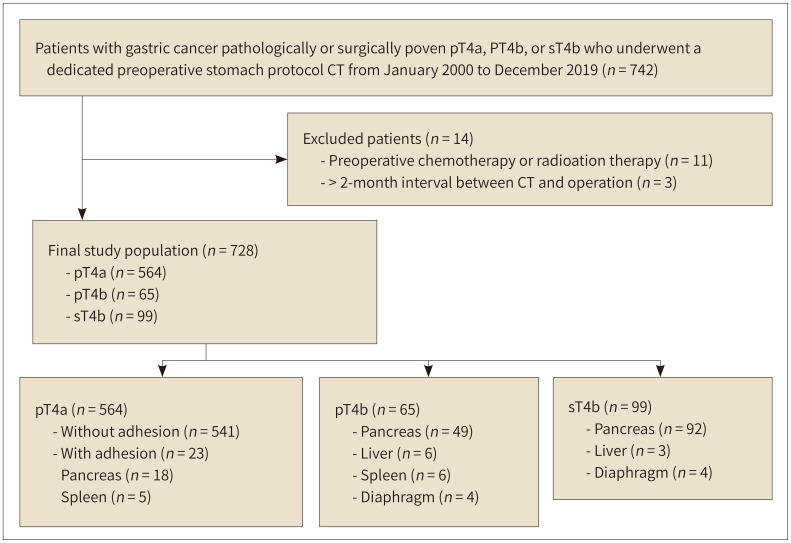
Materials and methods: A total of 728 patients with pathologically confirmed T4a (pT4a), surgically confirmed T4b (sT4b), or pathologically confirmed T4b (pT4b) AGCs who underwent dedicated stomach-protocol CT, including imaging of the left posterior oblique (LPO) and RDD positions, were included in this study. Two radiologists scored the T stage of AGCs using a 5-point scale on LPO CT with and without RDD CT at 2-week intervals and recorded the presence of "sliding sign" in the tumors and adjacent organs and compared its incidence of appearance.

Results: A total of 564 patients (77.4%) were diagnosed with pT4a, whereas 65 (8.9%) and 99 (13.6%) patients were diagnosed with pT4b and sT4b, respectively. When RDD CT was performed additionally, both reviewers deemed that the area under the curve (AUC) for differentiating T4b from T4a increased (p < 0.001). According to both reviewers, the AUC for differentiating T4b with pancreatic invasion from T4a increased in the subgroup analysis (p < 0.050). Interobserver agreement improved from fair to moderate (weighted kappa value, 0.296-0.444).

Conclusion: RDD CT provides additional value compared to LPO CT images alone for determining adjacent organ invasion in patients with AGC due to their increased AUC values and improved interobserver agreement.

목적: 진행성 위암의 인접 장기 침범을 결정함에 있어 우측와위 CT의 추가적 가치를 살펴보았다.

대상과 방법: 병리학적으로 입증된 T4a (p4a), 외과적 그리고 병리학적으로 입증된 T4b (sT4b, pT4b) 위암 환자 중 좌후사위 및 우측와위 자세가 포함된 프로토콜의 CT를 촬영한 환자 총 728명이 포함되었다. 2명의 영상의학과 전문의가 2주 간격으로 각각 우측와위 CT 없이, 우측와위 CT와 함께 좌후사위 CT를 분석하여 5점 척도를 사용하여 T 병기를 평가하고 종양과 인접 장기 사이의 “미끄러짐 징후”의 존재를 기록했다.

결과: 564명의 환자(77.4%)가 pT4a로 진단되었다. 65명(8.9%)과 99명(13.6%)의 환자가 각각 pT4b, sT4b로 진단되었다. 좌후사위 CT 단독 분석에 비하여 우측와위 CT가 추가되었을 때, T4b와 T4a를 구별하기 위한 곡선 아래 면적(area under the curve; 이하 AUC) 값이 두 검토자 모두에서 유의하게 증가했다(Ps < 0.001). 하위집단분석에서 T4a와 췌장을 침범한 T4b 위암을 구별하기 위한 AUC 값 역시 두 검토자 모두에서 증가했다(Ps < 0.050). 관찰자 간 일치도 역시 향상되었다(가중 카파 계수, 0.296–0.444).

결론: 진행위암에서 인접 장기 침범을 판단함에 있어, 우측와위 CT가 추가되었을 때 좌후사위 CT 단독 분석에 비해 더 높은 AUC 값과 관찰자 간 일치도를 보임으로써 추가적 가치가 있었다.

Keywords: Multidetector Computed Tomography; Neoplasm Invasiveness; Pancreas; Stomach Neoplasms; Task Performance and Analysis.


Conflict of interest statement

Conflicts of Interest: The authors have no potential conflicts of interest to disclose.

Figures

Fig. 1
Fig. 1. The flow chart for patients’ enrollment.
pT4a = pathologically confirmed T4a, pT4b = pathologically confirmed T4b with adjacent organ invasion, sT4b = surgically confirmed T4b with adjacent organ invasion in the surgical field
Fig. 2
Fig. 2. “Sliding sign” in the right down decubitus position CT.
A, B. An illustration (A) and corresponding CT image (B) in the left posterior oblique position reveal a tumor (arrow) at the posterior wall of the gastric body is widely abutting to the pancreas body. The fat plane between the tumor and the pancreas is obliterated (arrowheads). C. This illustration shows that the stomach and tumor, which are located in the peritoneal cavity, exhibit free movement in a right down decubitus position, whereas the pancreas is fixed as the pancreas is fixedly located at the retroperitoneum during the position change. Resultantly, there is a “sliding sign” (arrow) between the gastric tumor and the pancreas body. D, E. An illustration (D) and corresponding CT image (E) in the right down decubitus position show that a fat plane (asterisks in D and arrowheads in E) newly appears between the tumor (arrow) and the pancreas (P), suggesting that the tumor does not invade the pancreas. The right down decubitus CT is rotated 90 degrees clockwise so as to show it in a familiar orientation (thick blue arrow in D).
Fig. 3
Fig. 3. A 64-year-old female with advanced gastric cancer with a pathologically proven T4a stage.
A. Contrast-enhanced axial CT obtained in a left posterior oblique position reveals a diffuse low-attenuating wall thickening (arrows) at the gastric antrum. There is a broad attachment (arrowheads) between the tumor and pancreas neck at the far distal portion of the tumor, and a flat plane (arrowheads) between them was obliterated. Therefore, both radiologists scored 4 (probably T4b) with pancreas invasion. B. CT perfomed in a right down debucitus reveals a sliding sign between the gastric tumor (arrow) and pancreas neck (P). A linear fat plane (arrowheads) between the tumor (arrow) and pancreas neck (P) is newly visualized. Both radiologists recorded the presence of sliding signs and scored 1 (definitely T4a) without adjacent organ invasion. The patient subsequently underwent a subtotal gastrectomy. There is no evidence of adjacent organ invasion in the surgical field. Moderately differentiated adenocarcinoma with pT4aN0 stage is finally diagnosed via histopathology.
Fig. 4
Fig. 4. A 21-year old female with advanced gastric cancer and pathologically proven liver invasion (pT4a) with adhesion to the pancreas.
A. This contrast-enhanced axial CT image obtained in a left posterior oblique position shows a focal low-attenuating wall thickening (arrows) at the posterior wall of the gastric high body. The tumor is broadly abutted (arrowheads) to the pancreas tail (P). Two radiologists scored the tumor as 3 (possibly T4b) with pancreas invasion. B. A delayed phase CT image taken in a right down debucitus reveals that the gastric tumor (arrows) is still abutted (arrowheads) to the tail of the pancreas and there is no sliding sign. Therefore, both radiologists recorded the absence of sliding sign and scored 4 (probably T4b) with pancreas invasion. The patient underwent surgery. The tumor is broadly attached to the pancreas tail in the surgical field. Therefore, both distal pancreatectomy and total gastrectomy, is performed. C. This photograph of the cut surface of the gross specimen shows focal wall thickening of the stomach (arrow). The pancreatic tail (P) is also attached to the gastric tumor. However, the microscopic image showed that tumor did not invade the pancreas capsule or parenchyma (not shown). Therefore, the final histopathologic stage is pT4a. This case shows that simple adhesion by the tumor can cause false interpretation of sliding signs.
Fig. 5
Fig. 5. A 70-year old male with advanced gastric cancer and pathologically proven pancreas invasion (pT4b).
A. This image obtained using contrast-enhanced axial CT in a left posterior oblique position shows diffuse low-attenuating wall thickening (arrows) with punctate calcifications at the gastric angle to antrum. The posterior portion of the tumor broadly is abutted to the pancreas body. The fat plane (arrowhead) between the tumor and pancreas body is obliterated at the far distal portion of the tumor. Therefore, both radiologists scored the tumor as 3 (possibly T4b) with pancreas invasion. B. This CT image obtained after position change to right down decubitus, shows that the gastric tumor (arrows) and pancreas body (P) are still abutted (arrowhead) and there is no sliding sign. Both radiologists recorded the absence of sliding sign and scored 5 (definitely T4b) with pancreas invasion. The patient underwent a palliative total gastrectomy. A suspicious invasion of the pancreas body is present in the surgical field. Pancreas invasion by poorly cohesive carcinoma was proven using final histopathology.
Fig. 6
Fig. 6. The ROC curves for two radiologists with and without RDD CT images.
A, B. These panels show the ROC curves for all the patients drafted by the reviewers. When RDD CT images are provided in addition to the LPO CT images, the AUC values obtained by both radiologists for differentiating T4b from T4a increased significantly from 0.667 to 0.917 for reviewer 1 (A) and 0.850 to 0.894 for reviewer 2 (B) (p < 0.001). C, D. These panels show the ROC curves of reviewers for patients with pancreatic invasion. When RDD CT images are provided in addition to LPO CT images, AUC values for differentiating T4b with pancreatic invasion from T4a obtained by both reviewers increased significantly (from 0.654 to 0.893 for reviewer 1, p < 0.001; from 0.786 to 0.814 for reviewer 2, p = 0.035). AUC = areas under the curve, LPO = left posterior oblique, RDD = right down decubitus, ROC = receiver operating characteristic

References

    1. Sung H, Ferlay J, Siegel RL, Laversanne M, Soerjomataram I, Jemal A, et al. Global cancer statistics 2020: GLOBOCAN estimates of incidence and mortality worldwide for 36 cancers in 185 countries. CA Cancer J Clin. 2021;71:209–249. - PubMed
    1. Parkin DM, Bray F, Ferlay J, Pisani P. Global cancer statistics, 2002. CA Cancer J Clin. 2005;55:74–108. - PubMed
    1. Guideline Committee of the Korean Gastric Cancer Association (KGCA), Development Working Group & Review Panel. Korean practice guideline for gastric cancer 2018: an evidence-based, multi-disciplinary approach. J Gastric Cancer. 2019;19:1–48. - PMC - PubMed
    1. Kweon SS. Updates on cancer epidemiology in Korea, 2018. Chonnam Med J. 2018;54:90–100. - PMC - PubMed
    1. De Vita F, Giuliani F, Galizia G, Belli C, Aurilio G, Santabarbara G, et al. Neo-adjuvant and adjuvant chemotherapy of gastric cancer. Ann Oncol. 2007;18 Suppl 6:vi120–vi123. - PubMed