. 2022 Dec 12:2022:6987806.
doi: 10.1155/2022/6987806. eCollection 2022.

Comprehensive Assessment of Biomolecular Interactions of Morpholine-Based Mixed Ligand Cu(II) and Zn(II) Complexes of 2,2'-Bipyridine as Potential Anticancer and SARS-CoV-2 Agents: A Synergistic Experimental and Structure-Based Virtual Screening

Affiliations

Comprehensive Assessment of Biomolecular Interactions of Morpholine-Based Mixed Ligand Cu(II) and Zn(II) Complexes of 2,2'-Bipyridine as Potential Anticancer and SARS-CoV-2 Agents: A Synergistic Experimental and Structure-Based Virtual Screening

Karunganathan Sakthikumar et al. Bioinorg Chem Appl. .

Abstract

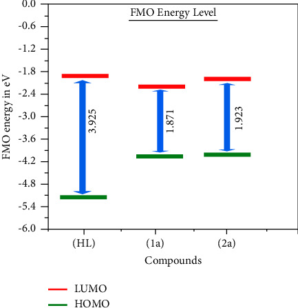
A new class of pharmacologically active mixed-ligand complexes (1a-2a) [MII(L)2 (bpy)], where L = 2-(4-morpholinobenzylideneamino)phenol), bpy = 2,2'-bipyridine, MII = Cu (1a), and Zn (2a), were assigned an octahedral geometry by analytical and spectral measurements. Gel electrophoresis showed that complex (1a) demonstrated the complete DNA cleavage mediated by H2O2. The overall DNA-binding constants observed from UV-vis, fluorometric, hydrodynamic, and electrochemical titrations were in the following sequence: (1a) > (2a) > (HL), which suggests that the complexes might intercalate DNA, a possibility that is further supported by the biothermodynamic characteristics. The binding constant results of BSA by electronic absorption and fluorometric titration demonstrate that complex (1a) exhibits the highest binding effectiveness among others, which means that all compounds could interact with BSA through a static approach, additionally supported by FRET measurements. Density FunctionalTheory (DFT) and molecular docking calculations were relied on to unveil the electronic structure, reactivity, and interacting capability of all substances with DNA, BSA, and SARS-CoV-2 main protease (Mpro). These observed binding energies fell within the following ranges: -7.7 to -8.6, -7.2 to -10.2, and -6.7 to -8.2 kcal/mol, respectively. The higher reactivity of the complexes compared to free ligand is supported by the Frontier MolecularOrbital (FMO) theory. The in vitro antibacterial, cytotoxic, and radical scavenging characteristics revealed that complex (1a) has the best biological efficacy compared to others. This is encouraged because all experimental findings are closely correlated with the theoretical measurements.


Conflict of interest statement

The authors declare that there are no conflicts of interest in this work.

Figures

Scheme 1
Scheme 1
The suggested geometry of complexes (1a-2a) [MII(L)2(bpy)].
Figure 1
Figure 1
Ethidium bromide displacement assay: gel electrophoresis demonstrates the DNA cleavage property in the H2O2 environment for the following substances: Lane: 1 DNA alone + H2O2; lane: 2 ligand (HL) + DNA + H2O2; lane: 3 complex (1a) + DNA + H2O2; lane: 4 complex (2a) + DNA + H2O2. Raw data for electrophoretic gels and blots were also enclosed in the electronic supplementary information file.
Figure 2
Figure 2
Increasing concentrations of CT-DNA were present while the ligand (HL) and its complexes (1a-2a) were measured for their absorption spectra in a Tris-HCl buffer solution at 25°C. Arrows depict the changes in absorbance that occur as CT-DNA concentration is increased, and another arrow with isosbestic points denotes that equilibrium between DNA and complexes has been achieved.
Figure 3
Figure 3
DNA thermal denaturation profile at 260 nm in the absence and presence of compounds in 5 mM Tris-HCl/50 mM NaCl buffer pH = 7.2, [DNA]/[Complex] = 1 (R).
Figure 4
Figure 4
Derivative melting curve for DNA thermal denaturation at 260 nm in the absence and presence of compounds in 5 mM Tris-HCl/50 mM NaCl buffer pH = 7.2, [DNA]/[Complex] = 1 (R).
Figure 5
Figure 5
Fluorescence quenching curve of ethidium bromide bound DNA in the presence of ligand (HL) and complexes (1a-2a).
Figure 6
Figure 6
The overlap of UV-vis spectra of ligand (HL) and mixed ligand complexes (1a-2a) (acceptor) at 336–337 nm with fluorescence emission spectrum of BSA (donor) at 350 nm.
Figure 7
Figure 7
Bovine serum albumin's UV-visible titration spectra at 25°C in a Tris-HCl buffer at a pH of 7.2 in the absence and presence of rising amounts of test substances. Arrow shows the changes in absorbance upon increasing the substance concentration.
Figure 8
Figure 8
The optimized geometries for the free ligand (HL) and its complexes (1a-2a).
Figure 9
Figure 9
FMO of the free ligand (HL) and its complexes (1a-2a).
Figure 10
Figure 10
FMO energy level diagram by DFT computation for all compounds.
Figure 11
Figure 11
Molecular electrostatic potential (MEP) maps of the free ligand (HL) and associated complexes (1a-2a). Plots generated at the 0.002 isosurface value.
Figure 12
Figure 12
Best docking poses of guest molecules inside the active site of the BSA protein.
Figure 13
Figure 13
Best docking pose of guest molecules inside the active site of 3CLPro.
Figure 14
Figure 14
Best binding pose of our guest molecules in the CT-DNA double helix.
Figure 15
Figure 15
Agar disc diffusion technique histogram comparing the antibacterial effects of all substances.
Figure 16
Figure 16
The evaluation of the anticancer properties of ligand (HL) and its complexes (1a-2a) against cancer cell lines and normal cell lines in comparison to the standard medication cisplatin (CP). Error limits ±2.5–5.0% (P ≤ 0.05).

References

    1. Utthra P. P., Raman N. Probing the potency of triazole tethered Schiff base complexes and the effect of substituents on their biological attributes. International Journal of Biological Macromolecules . 2018;116:194–207. - PubMed
    1. Kumar G., Devi S., Johari R., Kumar D. Synthesis, spectral characterization and antimicrobial evaluation of Schiff base Cr (III), Mn (III) and Fe (III) macrocyclic complexes. European Journal of Medicinal Chemistry . 2012;52:269–274. - PubMed
    1. Sung H., Ferlay J., Siegel R. L., et al. Global cancer statistics 2020: GLOBOCAN estimates of Incidence and mortality worldwide for 36 cancers in 185 Countries. CA: A Cancer Journal for Clinicians . 2021;71(3):209–249. doi: 10.3322/caac.21660. - DOI - PubMed
    1. Abu-Surrah A., Kettunen M. Platinum group Antitumor chemistry: design and development of new anticancer drugs Complementary to cisplatin. Current Medicinal Chemistry . 2006;13(11):1337–1357. doi: 10.2174/092986706776872970. - DOI - PubMed
    1. Gao A., Wu Y., Yu J., et al. Synthesis and anticancer activity of two highly water-soluble and ionic Pt(IV) complexes as prodrugs for Pt(II) anticancer drugs. RSC Medicinal Chemistry . 2022;13(5):594–598. doi: 10.1039/d2md00004k. - DOI - PMC - PubMed

LinkOut - more resources