

Laparoscopic resection of a rare diaphragmatic haemangioma: a case report
- PMID: 36545868
- PMCID: PMC9793033
- DOI: 10.1177/03000605221140688
Laparoscopic resection of a rare diaphragmatic haemangioma: a case report
Abstract
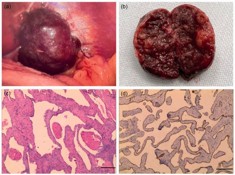
This case report describes the laparoscopic resection of a rare diaphragmatic haemangioma. A 45-year-old male patient was diagnosed incidentally with a left subphrenic mass by computed tomography. Laparoscopic left subphrenic mass excision was performed under general anaesthesia. A phrenic haemangioma was confirmed by postoperative pathology. Tumours originating in the diaphragm are rare, with only approximately 200 cases reported in the past century. The diaphragmatic tumour was determined to be primary because intraoperative imaging showed that the tumour was relatively isolated and had no obvious relationship with the surrounding tissues and organs.
Keywords: Laparoscopic resection; diaphragmatic haemangioma.
Conflict of interest statement
The authors declare that there are no conflicts of interest.
Figures

References
-
- Mordant P, Le Pimpec-Barthes F, Gangi A, et al.. Tumours of the diaphragm. In: Kuzdzal J. (ed) ESTS Textbook of Thoracic Surgery. Cracow, Poland: Medycyna Praktyczna, 2015, pp.199–208.
-
- Baldes N, Schirren J. Primary and Secondary Tumors of the Diaphragm. Thorac Cardiovasc Surg 2016; 64: 641–646. - PubMed
-
- Kono R, Terasaki H, Fujimoto K, et al.. Venous hemangioma arising from the diaphragm: a case report of computed tomography and magnetic resonance imaging findings. J Thorac Imaging 2006; 21: 231–234. - PubMed
-
- Gagnier JJ, Kienle G, Altman DG, et al.. The CARE guidelines: consensus-based clinical case reporting guideline development. Headache 2013; 53: 1541–1547. - PubMed
-
- Burcharth F, Agger P. Singultus: a case of hiccup with diaphragmatic tumour. Acta Chir Scand 1974; 140: 340–341. - PubMed
Publication types
MeSH terms
LinkOut - more resources
Full Text Sources
Medical
Miscellaneous
