. 2023 Mar 8;72(1):59-69.
doi: 10.33549/physiolres.934975. Epub 2022 Dec 22.

Prenatal exposure to acrylamide differently affected the sex ratio, aromatase and apoptosis in female adult offspring of two subsequent generations

Affiliations

Prenatal exposure to acrylamide differently affected the sex ratio, aromatase and apoptosis in female adult offspring of two subsequent generations

M Arafah et al. Physiol Res. .

Abstract

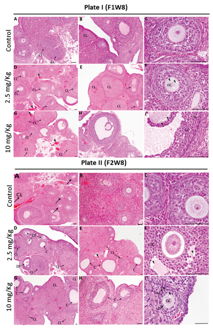
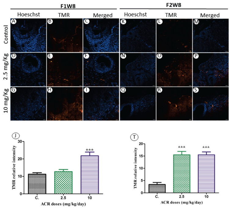
In the present study, we investigated the effect of acrylamide (ACR) exposure during pregnancy on the ovary of female adult offspring of two subsequent generations. Sixty-day-old Wistar albino female rats were given different doses of ACR (2.5 and 10 mg/kg/day) from day 6 of pregnancy until giving birth. Females from the first generation (AF1) were fed ad libitum, and thereafter, a subgroup was euthanized at 8 weeks of age and ovary samples were obtained. The remaining females were maintained until they reached sexual maturity (50 days old) and then treated in the same way as the previous generation to obtain the second generation of females (AF2). The histopathological examination indicated a high frequency of corpora lutea along with an increased number of antral follicles that reached the selectable stage mainly at a dose of 2.5 mg/kg/day. Interestingly, ACR exposure significantly increased the mRNA levels of CYP19 gene and its corresponding CYP19 protein expression in AF1 females. The TUNEL assay showed a significantly high rate of apoptosis in stromal cells except for dose of 2.5 mg/kg/day. However, in AF2 females, ACR exposure significantly increased the number of degenerating follicles and cysts while the number of growing follicles was reduced. Moreover, in both ACR-treated groups, estradiol-producing enzyme CYP19A gene and its corresponding protein were significantly reduced, and an excessive apoptosis was produced. We concluded that the ovarian condition of AF1 females had considerable similarity to the typical early perimenopausal stage, whereas that of AF2 females was similar to the late perimenopausal stage in women.


Conflict of interest statement

Conflict of Interest

There is no conflict of interest.

Figures

Fig. 1
Fig. 1
(A) The sex ratio of offspring in the first generation AF1 compared to that in control CF1. No significant difference between the control CF1 and other treatment groups. (B) The sex ratio of offspring in second generation AF2 compared to that in control CF2. ACR (10 mg/kg/day) caused significant elevation, while the dose of ACR (2.5 mg/kg/day) did not show any different from the control CF2. Values are means ± S.E.M. ** P<0.001.
Fig. 2
Fig. 2
Plate I: Histological sections from 8-week-old females of the first generation (F1W8) from treated and control groups. Ovaries from control (CF1) displayed normal growing follicles and few corpora lutea (CL) (arrows) (A–C). In ovaries from females AF1, proportional increase of the number of corpora lutea (CL) with vacuolations and degenerative luteal cells (arrows), blood vessel congestion and vacuolated oocyte (OC) (arrowheads) can be observed with the dose (2.5 mg/kg) (D–F). Rising number of corpora lutea (CL) (arrows), blood congestion and pyknosis in granulosa cells (GC) (arrowheads) can be seen in the dose 10 mg/kg (G–I). Scale bar=60 μm. Plate II: Histological evaluation of ovary structure in 8-week-old females of the second generation (F2W8). Ovaries from control (A–C) CF2 show normal growing follicles and few corpora lutea (CL) (arrows). AF2 females treated with the dose (2.5 mg/kg) showed an increased number of corpora lutea (CL), degenerated follicles (DF) and cysts (C) (arrows), reduction in growing follicles. Congestion of blood vessels and vacuolation of granulosa cell’s layers (arrowheads) can also been seen (D–F). In (10 mg/kg) ovary showing rising in cysts (C) and corpora lutea (CL) number (arrows). Oocytes (OC) vacuolation, irregular thinning zona pellucida (ZP) and pyknosis in the granulosa cells nuclei (arrowheads) (G–I). Scale bar=60 μm.
Fig. 3
Fig. 3
The ovarian CYP19 localization and expression in 8-week-old female offspring from the first (F1W8) and the second (F2W8) generation compared to control. F1W8: Stained sections from control CF1 (A–C), AF1 females treated with the dose 2.5 mg/kg (D–F) and 10 mg/kg (G–I). F2W8: Stained sections from AF2 females treated with the dose 2.5 mg/kg (J–L) and 10 mg/kg (M–O). Sections of ovary was performed by immunofluorescence using specific CYP19 antibody stained with FITC (green), cell nuclei are stained with Hoechst (blue). Scale bar=20 μm. The relative CYP19 intensity in 8-week-old AF1 females and AF2 females compared with control females are presented in the graphs (P) and (R), respectively. Effect of ACR on the mRNA expression level of the CYP19 gene in 8-week-old AF1 females (Q) and in 8-week-old AF2 females (S) compared with control females. Values are means ± S.E.M. * P<0.05; ** P<0.001; *** P<0.0001.
Fig. 4
Fig. 4
The TUNEL reaction in 8-week-old female offspring from the first (F1W8) and second (F2W8) generations compared to control. Stained sections from control CF1 (A–C), AF1 females treated with the dose 2.5 mg/kg (D–F) and the dose 10 mg/kg (G–I). (J) The TMR relative intensity in 8-week-old AF1 females compared with control CF1. The TUNEL reaction in 8-week-old females of control CF2 (K–M), AF2 females treated with the dose 2.5 mg/kg (N–P) and the dose 10 mg/kg (Q–S). Sections of ovary apoptosis was performed by confocal images of follicles with TUNEL fluorescence in both granulosa and oocyte nuclei stained in (red) TMR, cell nuclei are stained with Hoechst (blue). Scale bar=50 μm. The TMR relative intensity in 8-week-old AF2 females compared with control CF2 (T). Values are means ± S.E.M. *** P<0.0001.

References

    1. Silvestris E, Lovero D, Palmirotta R. Nutrition and female fertility: an interdependent correlation. Fron Endocrinol (Lausanne) 2019;10:346. doi: 10.3389/fendo.2019.00346. - DOI - PMC - PubMed
    1. Hanson B, Johnstone E, Dorais J, Silver B, Peterson CM, Hotaling J. Female infertility, infertility-associated diagnoses, and comorbidities: a review. J Assist Reprod Genet. 2017;34:167–177. doi: 10.1007/s10815-016-0836-8. - DOI - PMC - PubMed
    1. Jalouli M, Mofti A, Elnakady YA, Nahdi S, Feriani A, Alrezaki A, Sebei K, et al. Allethrin promotes apoptosis and autophagy associated with the oxidative stress-related PI3K/AKT/mTOR signaling pathway in developing rat ovaries. Int J Mol Sci. 2022;23:6397. doi: 10.3390/ijms23126397. - DOI - PMC - PubMed
    1. Tarko A, Štochmal’ová A, Hrabovszká S, Vachanová A, Harrath AH, Aldahmash W, Grossman R, Sirotkin AV. Potential protective effect of puncture vine (Tribulus terrestris, L.) against xylene toxicity on bovine ovarian cell functions. Physiol Res. 2022;71:249. doi: 10.33549/physiolres.934871. - DOI - PMC - PubMed
    1. Harrath AH, Alrezaki A, Mansour L, Alwasel SH, Palomba S. Food restriction during pregnancy and female offspring fertility: adverse effects of reprogrammed reproductive lifespan. J Ovarian Res. 2017;10:1–9. doi: 10.1186/s13048-017-0372-x. - DOI - PMC - PubMed