. 2023 Feb;16(2):271-279.
doi: 10.1002/aur.2875. Epub 2022 Dec 22.

Increased resting-state thalamocortical functional connectivity in children and young adults with autism spectrum disorder

Affiliations

Increased resting-state thalamocortical functional connectivity in children and young adults with autism spectrum disorder

Bengi Baran et al. Autism Res. 2023 Feb.

Abstract

There is converging evidence that abnormal thalamocortical interactions contribute to attention deficits and sensory sensitivities in autism spectrum disorder (ASD). However, previous functional MRI studies of thalamocortical connectivity in ASD have produced inconsistent findings in terms of both the direction (hyper vs. hypoconnectivity) and location of group differences. This may reflect, in part, the confounding effects of head motion during scans. In the present study, we investigated resting-state thalamocortical functional connectivity in 8-25 year-olds with ASD and their typically developing (TD) peers. We used pre-scan training, on-line motion correction, and rigorous data quality assurance protocols to minimize motion confounds. ASD participants showed increased thalamic connectivity with temporal cortex relative to TD. Both groups showed similar age-related decreases in thalamic connectivity with occipital cortex, consistent with a process of circuit refinement. Findings of thalamocortical hyperconnectivity in ASD are consistent with other evidence that decreased thalamic inhibition leads to increase and less filtered sensory information reaching the cortex where it disrupts attention and contributes to sensory sensitivity. This literature motivates studies of mechanisms, functional consequences, and treatment of thalamocortical circuit dysfunction in ASD.

Keywords: MRI; autism spectrum disorder; functional connectivity; resting state; sensory sensitivity; thalamus.


Figures

Figure 1.
Figure 1.. Thalamic seed analyses of thalamocortical functional connectivity.
Left: Statistical map of the group differences in the connectivity of the thalamus with the cortex, displayed on the template brain at pFWE-corrected ≤ .05. Greater connectivity in ASD is depicted in blue. There were no regions of significantly greater connectivity in TD. Right: Dot plot of averaged thalamocortical connectivity in the group difference cluster: blue dots represent TD, red dots represent ASD, black bars represent group means
Figure 2.
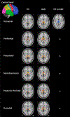
Figure 2.. Cortical seed analyses of thalamocortical functional connectivity.
The cortex was parcellated into six non-overlapping seeds. Statistical maps of thalamic connectivity of each seed are displayed on the template brain for TD and ASD groups separately. The third column corresponds to the statistical map of group differences displayed on the template brain at pFWE-corrected ≤ .05. Greater connectivity in ASD is depicted in blue. There were no regions of significantly greater connectivity in TD. (ns: non-significant)
Figure 3.
Figure 3.. Age effects in thalamocortical functional connectivity (cortical seed analysis).
(A) Statistical map of the main effect of age on the connectivity of the occipital cortex seed with the thalamus displayed on the template brain at p FWE-corrected ≤ .05. Negative relationship is depicted in blue, there were no regions of age-related increases in thalamocortical connectivity or any significant age-relations with the rest of the cortical seeds. (B) Scatterplot of the relations between occipital thalamocortical connectivity and age in the age effect mask: blue dots and regression lines represent TD, red dots regression lines represent ASD, black line represents the regression line for the main effect of age.

References

    1. Al-Aidroos N, Said CP, Turk-Browne NB (2012). Top-down attention switches coupling between low-level and high-level areas of human visual cortex. Proc Natl Acad Sci U S A 109, 14675–80 doi:10.1073/pnas.1202095109 - DOI - PMC - PubMed
    1. Alkonyi B, Juhász C, Muzik O, Behen ME, Jeong JW, Chugani HT (2011). Thalamocortical connectivity in healthy children: asymmetries and robust developmental changes between ages 8 and 17 years. AJNR Am J Neuroradiol 32, 962–9 doi:10.3174/ajnr.A2417 - DOI - PMC - PubMed
    1. Arnold Anteraper S, Triantafyllou C, Sawyer AT, Hofmann SG, Gabrieli JD, Whitfield-Gabrieli S (2014). Hyper-connectivity of subcortical resting-state networks in social anxiety disorder. Brain Connect 4, 81–90 doi:10.1089/brain.2013.0180 - DOI - PubMed
    1. Ayub R, et al. (2021). Thalamocortical connectivity is associated with autism symptoms in high-functioning adults with autism and typically developing adults. Transl Psychiatry 11, 93 doi:10.1038/s41398-021-01221-0 - DOI - PMC - PubMed
    1. Baran B, et al. (2019). Increased Thalamocortical Connectivity in Schizophrenia Correlates With Sleep Spindle Deficits: Evidence for a Common Pathophysiology. Biol Psychiatry Cogn Neurosci Neuroimaging 4, 706–714 doi:10.1016/j.bpsc.2019.04.012 - DOI - PMC - PubMed

Publication types