. 2022 Dec 1;13(4):269.
doi: 10.3390/jfb13040269.

Evaluation of an Injectable Biphasic Calcium Sulfate/Hydroxyapatite Cement for the Augmentation of Fenestrated Pedicle Screws in Osteoporotic Vertebrae: A Biomechanical Cadaver Study

Affiliations

Evaluation of an Injectable Biphasic Calcium Sulfate/Hydroxyapatite Cement for the Augmentation of Fenestrated Pedicle Screws in Osteoporotic Vertebrae: A Biomechanical Cadaver Study

Xinggui Tian et al. J Funct Biomater. .

Abstract

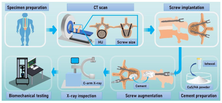
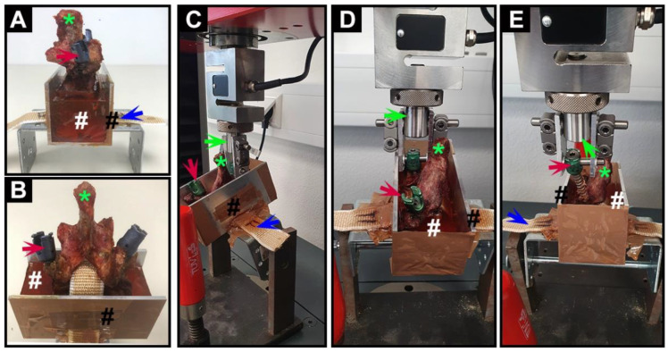
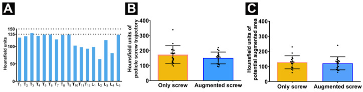
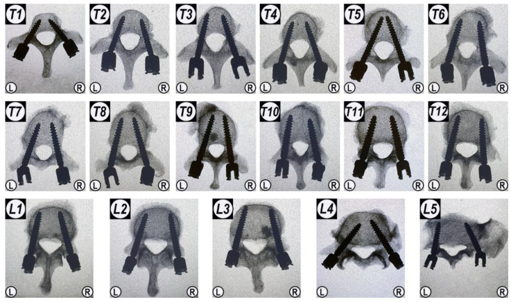
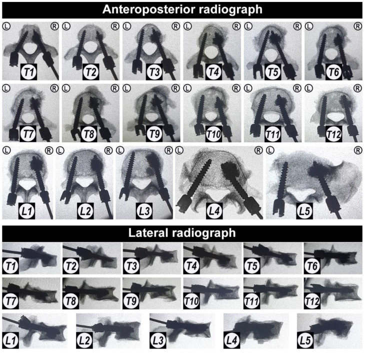
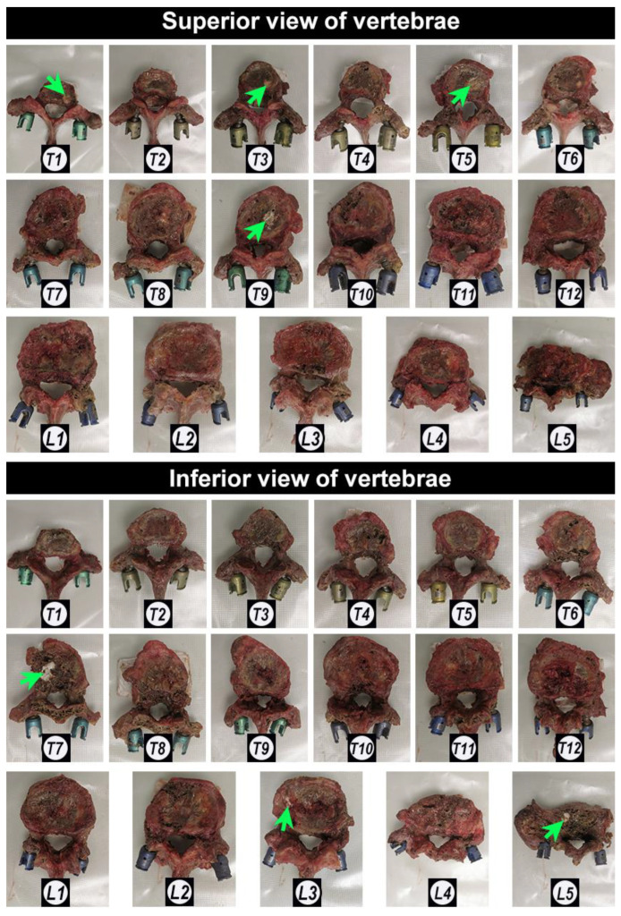
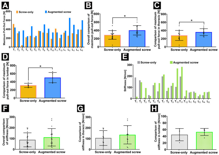
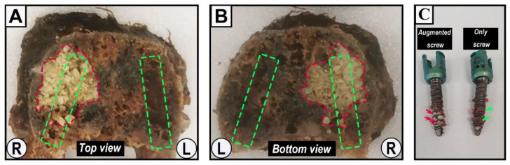
Cement augmentation of pedicle screws is one of the most promising approaches to enhance the anchoring of screws in the osteoporotic spine. To date, there is no ideal cement for pedicle screw augmentation. The purpose of this study was to investigate whether an injectable, bioactive, and degradable calcium sulfate/hydroxyapatite (CaS/HA) cement could increase the maximum pull-out force of pedicle screws in osteoporotic vertebrae. Herein, 17 osteoporotic thoracic and lumbar vertebrae were obtained from a single fresh-frozen human cadaver and instrumented with fenestrated pedicle screws. The right screw in each vertebra was augmented with CaS/HA cement and the un-augmented left side served as a paired control. The cement distribution, interdigitation ability, and cement leakage were evaluated using radiographs. Furthermore, pull-out testing was used to evaluate the immediate mechanical effect of CaS/HA augmentation on the pedicle screws. The CaS/HA cement presented good distribution and interdigitation ability without leakage into the spinal canal. Augmentation significantly enhanced the maximum pull-out force of the pedicle screw in which the augmented side was 39.0% higher than the pedicle-screw-alone side. Therefore, the novel biodegradable biphasic CaS/HA cement could be a promising material for pedicle screw augmentation in the osteoporotic spine.

Keywords: biomaterial; biomechanical; calcium sulfate/hydroxyapatite; cement; osteoporosis; pedicle screw augmentation.


Conflict of interest statement

L.L. is a board member of Ortho Cell, Australia and BONESUPPORT AB, Sweden. L.L., M.T., and D.B.R. hold stocks in Moroxite AB, Sweden. The authors declare that they have no other competing interests pertaining to this study.

Figures

Figure 1
Figure 1
Flow chart summarizing the methodology. After the specimens’ acquisition, computed tomography (CT) scan was performed to determine that the specimen met the experimental criteria and to determine the parameters of the pedicle screws. Pedicle screws were implanted and augmented with CaS/HA cement, and then a pull-out experiment was performed to evaluate the effect of the augmentation. Abbreviations: HU: Hounsfield units; CaS/HA: calcium sulfate/hydroxyapatite.
Figure 2
Figure 2
Photographs of vertebra fixation and biomechanical testing process. (A) Lateral view and (B) anterior view of a vertebra (*) fixed in the customized metal container (black #) filled with epoxy resin (white #); (C) lateral view; and (D) anterior view of the mechanical testing process of pedicle screws; and (E) the picture of pedicle screws being successfully pulled out from the vertebra. Red arrows indicate the pedicle screws; blue arrows indicate the fixed band; green arrows indicate the piston of the testing machine.
Figure 3
Figure 3
Quantification of radiodensity in CT scans: (A) Radiodensity in Hounsfield units (HU) for all vertebral bodies T1–T12 and L1–L5; (B) Radiodensity values (in HU) of pedicle screw trajectory comparing augmented screw side and only screw side; and (C) the HU between the potential augmented area and contralateral area. Data are presented as mean ± SD. The two horizontal dotted lines in panel A represent the critical values for diagnosing osteoporosis by HU in previous literature [29].
Figure 4
Figure 4
The anteroposterior X-ray images of all vertebrae after the fenestrated pedicle screw implantation prior to augmentation with CaS/HA material. “L” and “R” indicate the left and right side; T1–T12 and L1–L5 indicate the thoracic and lumbar position of the vertebrae.
Figure 5
Figure 5
The anteroposterior radiograph and the lateral radiograph of the fenestrated pedicle screws augmented with CaS/HA cement. “L” and “R” indicate the left and right side; T1–T12 and L1–L5 indicate the thoracic and lumbar position of the vertebrae.
Figure 6
Figure 6
Photographs of the appearance of all the vertebrae after cement injection. Green arrows indicate the CaS/HA cement. T1–T12 and L1–L5 indicate the thoracic and lumbar position of the vertebrae.
Figure 7
Figure 7
(AD) The maximum pull-out force in N for screws pulled out from the cement-augmented screw side and screw-only side; (A,B) overall comparison; (C) comparison for thoracic vertebrae; and (D) lumbar comparison of screw only vs. augmented screw. (EH) Stiffness in N/mm of vertebrae comparison between the cement-augmented screw side and screw-only side; (E,F) overall; (G) thoracic; and (H) lumbar comparison. Data are presented as mean ± SD, * p < 0.05.
Figure 8
Figure 8
Representative specimens of vertebrae and fenestrated pedicle screws after pull-out testing. (A) The top section; and (B) the bottom section of vertebra L2 after axial cross-section along the midline of the pedicle. The red area of irregular shape indicates the CaS/HA cement-augmented area, while the rectangular area marked by the green dashed lines indicates the rough zone of the pedicle screw trajectory. “L” and “R” indicate the respective orientations. (C) A pair of pedicle screws (6.5/30 mm) from T1 after the pull-out experiment. Red arrows indicate remnants of CaS/HA cement; green arrows indicate remnants of the trabecular bone tissue.

References

    1. Krenzlin H., Foelger A., Mailänder V., Blase C., Brockmann M., Düber C., Ringel F., Keric N. Novel Biodegradable Composite of Calcium Phosphate Cement and the Collagen I Mimetic P-15 for Pedicle Screw Augmentation in Osteoporotic Bone. Biomedicines. 2021;9:1392. doi: 10.3390/biomedicines9101392. - DOI - PMC - PubMed
    1. Reginster J.Y., Burlet N. Osteoporosis: A still increasing prevalence. Bone. 2006;38((Suppl. 1)):4–9. doi: 10.1016/j.bone.2005.11.024. - DOI - PubMed
    1. Park S.B., Chung C.K. Strategies of spinal fusion on osteoporotic spine. J. Korean Neurosurg. Soc. 2011;49:317–322. doi: 10.3340/jkns.2011.49.6.317. - DOI - PMC - PubMed
    1. Xie F., Zhou B., Wang J., Liu T., Wu X., Fang R., Kang Y., Dai R. Microstructural properties of trabecular bone autografts: Comparison of men and women with and without osteoporosis. Arch. Osteoporos. 2018;13:18. doi: 10.1007/s11657-018-0422-z. - DOI - PubMed
    1. Paré P.E., Chappuis J.L., Rampersaud R., Agarwala A.O., Perra J.H., Erkan S., Wu C. Biomechanical evaluation of a novel fenestrated pedicle screw augmented with bone cement in osteoporotic spines. Spine (Phila Pa 1976) 2011;36:E1210–E1214. doi: 10.1097/BRS.0b013e318205e3af. - DOI - PubMed

LinkOut - more resources