. 2022 Dec 1;13(4):271.
doi: 10.3390/jfb13040271.

TiO2/HA and Titanate/HA Double-Layer Coatings on Ti6Al4V Surface and Their Influence on In Vitro Cell Growth and Osteogenic Potential

Affiliations

TiO2/HA and Titanate/HA Double-Layer Coatings on Ti6Al4V Surface and Their Influence on In Vitro Cell Growth and Osteogenic Potential

Michalina Ehlert et al. J Funct Biomater. .

Abstract

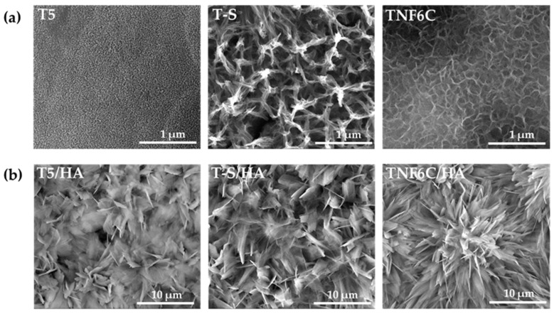
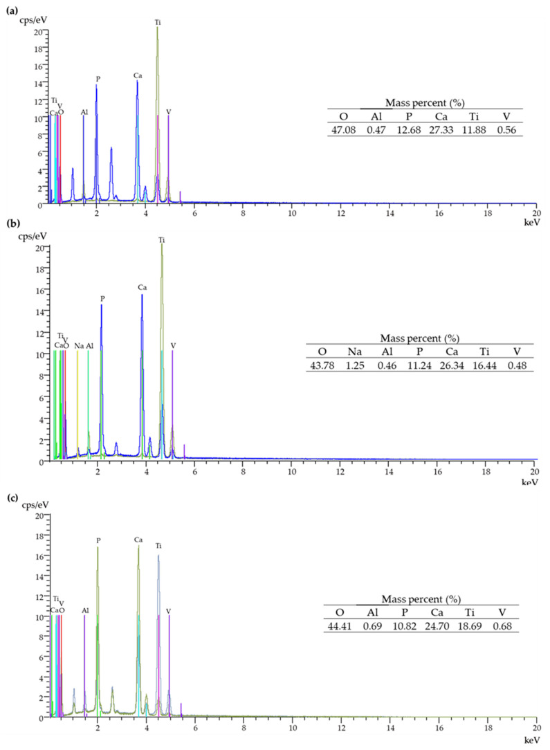
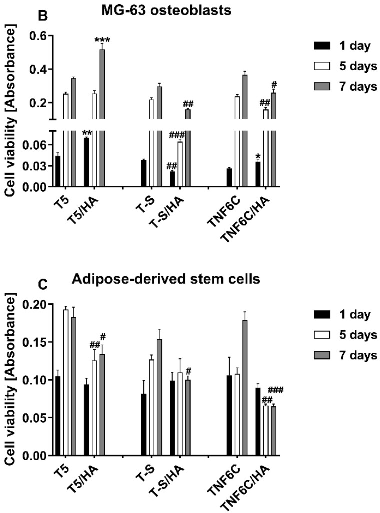
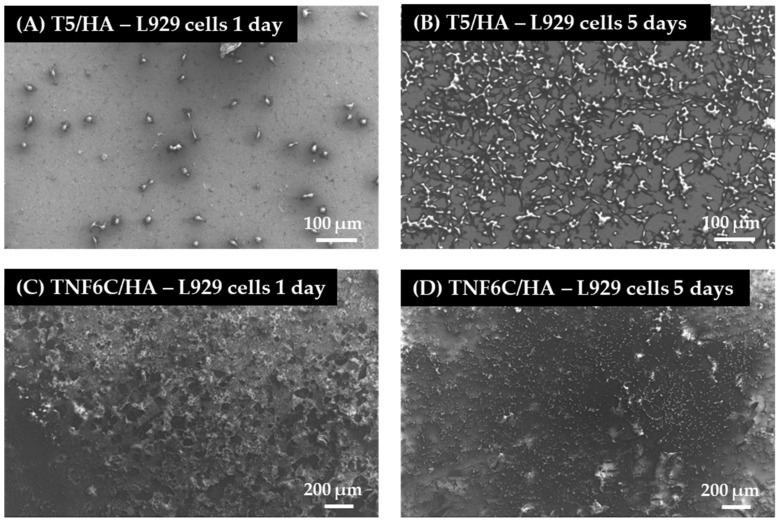
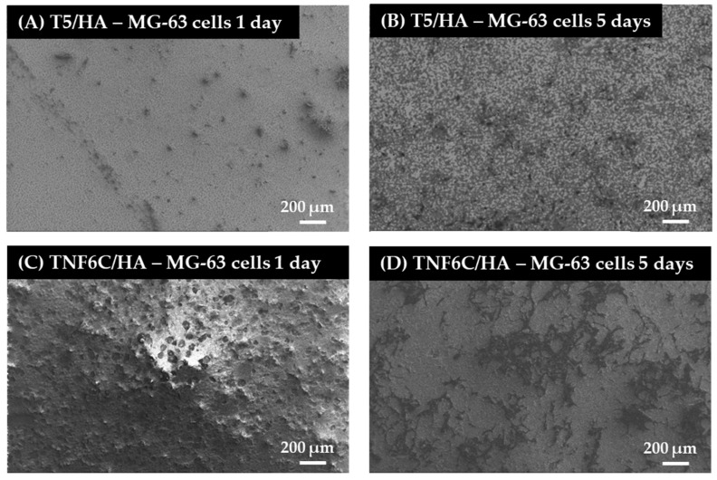
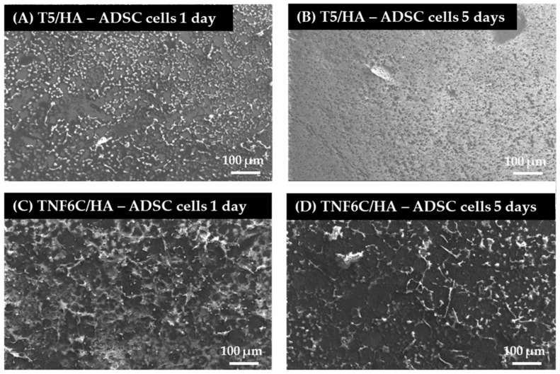
Hydroxyapatite (HA) layers are appropriate biomaterials for use in the modification of the surface of implants produced inter alia from a Ti6Al4V alloy. The issue that must be solved is to provide implants with appropriate biointegration properties, enabling the permanent link between them and bone tissues, which is not so easy with the HA layer. Our proposition is the use of the intermediate layer ((IL) = TiO2, and titanate layers) to successfully link the HA coating to a metal substrate (Ti6Al4V). The morphology, structure, and chemical composition of Ti6Al4V/IL/HA systems were characterized by scanning electron microscopy (SEM), X-ray diffraction (XRD), and energy-dispersive X-ray spectrometry (EDS). We evaluated the apatite-forming ability on the surface of the layer in simulated body fluid. We investigated the effects of the obtained systems on the viability and growth of human MG-63 osteoblast-like cells, mouse L929 fibroblasts, and adipose-derived human mesenchymal stem cells (ADSCs) in vitro, as well as on their osteogenic properties. Based on the obtained results, we can conclude that both investigated systems reflect the physiological environment of bone tissue and create a biocompatible surface supporting cell growth. However, the nanoporous TiO2 intermediate layer with osteogenesis-supportive activity seems most promising for the practical application of Ti6Al4V/TiO2/HA as a system of bone tissue regeneration.

Keywords: adipose-derived mesenchymal stem cells; antimicrobial activity; biointegration; cathodic electrodeposition; hydroxyapatite; titanate nanolayers; titanium dioxide.


Conflict of interest statement

The authors declare no conflict of interest. The funders had no role in the design of the study; in the collection, analyses, or interpretation of data; in the writing of the manuscript; or in the decision to publish the results.

Figures

Figure 1
Figure 1
Scheme to produce systems with a hydroxyapatite layer.
Figure 2
Figure 2
SEM images of the surface morphology of the T5, T-S, TNF6C (a), T5/HA, T-S/HA, and TNF6C/HA (b) samples.
Figure 3
Figure 3
EDS spectra and quantitative data of the T5/HA (a), T-S/HA (b), and TNF6C/HA (c) systems.
Figure 4
Figure 4
SEM images of the (a) T5/HA, (b) T-S/HA, and (c) TNF6C/HA samples after immersing in SBF for 1–4 weeks.
Figure 5
Figure 5
Weight gain for the samples with hydroxyapatite layer after immersing in SBF for 1–4 weeks.
Figure 6
Figure 6
XRD patterns of T-S/HA, T5/HA and TNF6C/HA samples after immersing in SBF for four weeks. (hkl) for CaTiO3 are marked in violet. S is assigned to the sodium titanate. Ti is assigned to the Ti6Al4V substrate (TiO2 anatase phase (A)).
Figure 7
Figure 7
The viability of L929 fibroblasts (A), MG-63 osteoblasts (B) and adipose-derived stem cells (C) cultured on the scaffolds (nanoporous TiO2 (T5), titanate (T-S) and nanofibrous TiO2 (TNF6C)) coated or not with a hydroxyapatite layer (HA) evaluated using MTT assays after one, five and seven days. The presented data are from four independent experiments. Asterisks and hash marks show statistical differences between the scaffolds coated with HA and the samples without HA at the appropriate time. Asterisks show differences when cell viability measured for the samples with HA was greater compared with the specimens without HA (*** p < 0.001, ** p < 0.01, * p < 0.05). Hash marks denote differences when absorbance values noticed for the scaffolds with HA was lower than the samples not covered with HA (### p < 0.001, ## p < 0.01, # p < 0.05).
Figure 7
Figure 7
The viability of L929 fibroblasts (A), MG-63 osteoblasts (B) and adipose-derived stem cells (C) cultured on the scaffolds (nanoporous TiO2 (T5), titanate (T-S) and nanofibrous TiO2 (TNF6C)) coated or not with a hydroxyapatite layer (HA) evaluated using MTT assays after one, five and seven days. The presented data are from four independent experiments. Asterisks and hash marks show statistical differences between the scaffolds coated with HA and the samples without HA at the appropriate time. Asterisks show differences when cell viability measured for the samples with HA was greater compared with the specimens without HA (*** p < 0.001, ** p < 0.01, * p < 0.05). Hash marks denote differences when absorbance values noticed for the scaffolds with HA was lower than the samples not covered with HA (### p < 0.001, ## p < 0.01, # p < 0.05).
Figure 8
Figure 8
SEM images of L929 fibroblasts growing on the scaffolds coated with hydroxyapatite layer (HA) (nanoporous (T5/HA) and nanofibrous TiO2 (TNF6C/HA) for one and five days. The type of specimens and culture time are indicated in the figures.
Figure 9
Figure 9
Micrographs from SEM presenting MG-63 osteoblasts cultured on the nanoporous and nanofibrous TiO2 scaffolds coated with hydroxyapatite layer (T5/HA and TNF6C/HA, respectively). The type of samples and culture time are described in the figures.
Figure 10
Figure 10
SEM micrographs that present adipose-derived mesenchymal stem cells (ADSCs) growing on the surface of the nanoporous and nanofibrous TiO2 specimens coated with hydroxyapatite layer (T5/HA and TNF6C/HA, respectively). The type of specimens and culture time are indicated in the figures.
Figure 11
Figure 11
Determination of calcium deposit formation in the extracellular matrix of MG-63 osteoblasts (A) and human adipose-derived mesenchymal stem cells (B) evaluated after one, five and seven days using Alizarin Red S staining. Asterisks denote differences between Alizarin staining determined for the samples with HA and without HA (*** p < 0.001, ** p < 0.01; * p < 0.05).
Figure 11
Figure 11
Determination of calcium deposit formation in the extracellular matrix of MG-63 osteoblasts (A) and human adipose-derived mesenchymal stem cells (B) evaluated after one, five and seven days using Alizarin Red S staining. Asterisks denote differences between Alizarin staining determined for the samples with HA and without HA (*** p < 0.001, ** p < 0.01; * p < 0.05).
Figure 12
Figure 12
Determination of ALP activity in MG-63 osteoblasts (A) and adipose-derived mesenchymal stem cells (B) evaluated after one, five and seven days of culture on selected scaffolds. Asterisks and hash marks show statistical differences between the scaffolds with HA and without HA at the appropriate culture time. Asterisks present differences when ALP activity measured for the samples with HA was higher compared with the specimens without HA (* p < 0.05). Hash marks indicate differences when ALP activity noticed for the scaffolds with HA was lower than the samples without HA (### p < 0.001).

References

    1. Jain P., Kathuria H., Dubey N. Advances in 3D Bioprinting of Tissues/Organs for Regenerative Medicine and in-Vitro Models. Biomaterials. 2022;287:121639. doi: 10.1016/j.biomaterials.2022.121639. - DOI - PubMed
    1. Chan B.P., Leong K.W. Scaffolding in Tissue Engineering: General Approaches and Tissue-Specific Considerations. Eur. Spine J. 2008;17:467–479. doi: 10.1007/s00586-008-0745-3. - DOI - PMC - PubMed
    1. Olson J.L., Atala A., Yoo J.J. Tissue Engineering: Current Strategies and Future Directions. Chonnam Med. J. 2011;47:1–13. doi: 10.4068/cmj.2011.47.1.1. - DOI - PMC - PubMed
    1. Todros S., Todesco M., Bagno A. Biomaterials and Their Biomedical Applications: From Replacement to Regeneration. Processes. 2021;9:1949. doi: 10.3390/pr9111949. - DOI
    1. Apablaza J.A., Días F.J., Sánchez K.G., Navarro P., Venegas C., Fuentes R. Analysis of the Chemical Composition and Morphological Characterization of Tissue Osseointegrated to a Dental Implant after 5 Years of Function. Int. J. Mol. Sci. 2022;23:8882. doi: 10.3390/ijms23168882. - DOI - PMC - PubMed

LinkOut - more resources