. 2022 Dec 5;13(4):276.
doi: 10.3390/jfb13040276.

Healing Patterns of Non-Collagenated Bovine and Collagenated Porcine Xenografts Used for Sinus Floor Elevation: A Histological Study in Rabbits

Affiliations

Healing Patterns of Non-Collagenated Bovine and Collagenated Porcine Xenografts Used for Sinus Floor Elevation: A Histological Study in Rabbits

Yuhei Miyauchi et al. J Funct Biomater. .

Abstract

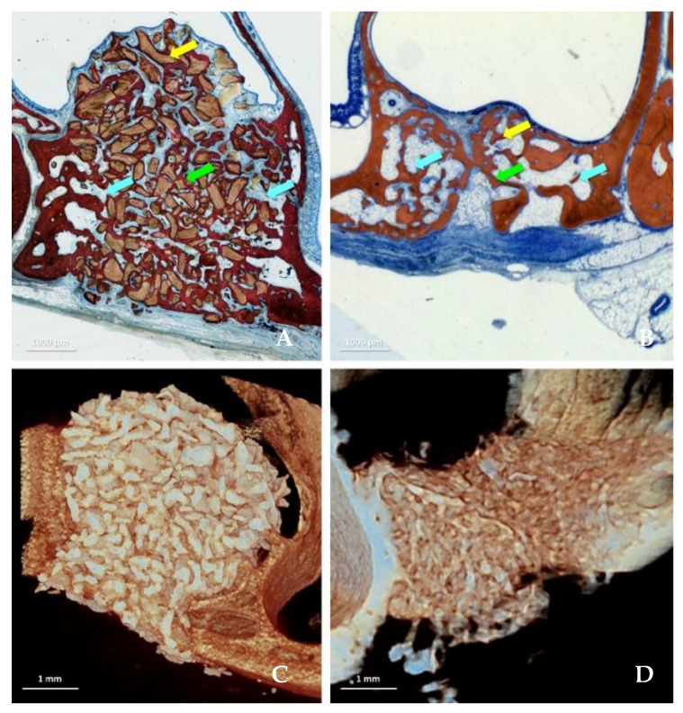
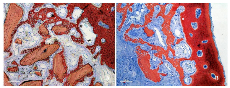
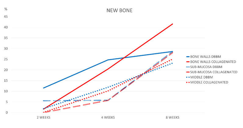
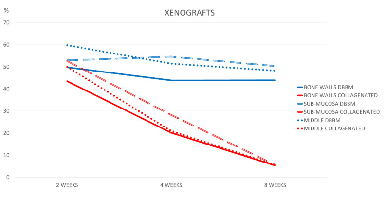
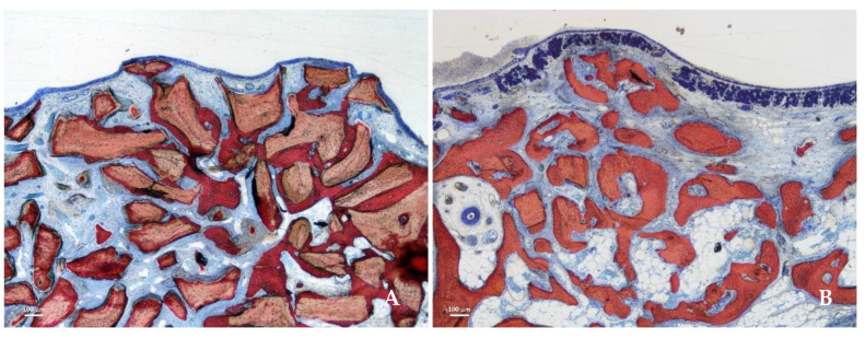
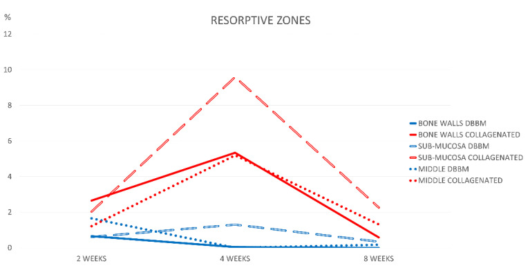
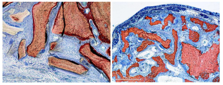
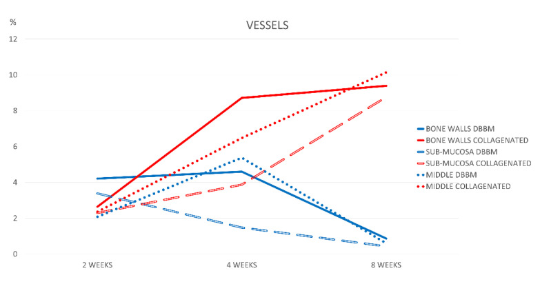
Objective: To compare healing of collagenated and non-collagenated xenografts used for maxillary sinus floor elevation. Materials and Methods: Two different xenografts were used: deproteinized bovine bone (DBBM group) and collagenated corticocancellous porcine bone (collagenated group). Healing was studied after 2, 4, and 8 weeks. The loss of dimensions of the elevated area and the percentages of new bone, xenograft remnants, osteoclastic zones, vessels, inflammatory infiltrates, and soft tissues were analyzed. Three regions were evaluated: close to the bone walls (bone wall region), subjacent the sinus mucosa (submucosa region), and the center of the elevated area (middle region). The primary variables were the percentage of new bone and xenograft remnants. Results: Between 2 and 8 weeks, the elevated areas showed a reduction of 16.3% and 52.2% in the DBBM and collagenated groups, respectively (p < 0.01 between the two areas after 8 weeks). After 8 weeks, the highest content of new bone was observed in the bone wall region, which was higher in the collagenated group than in the DBBM group (41.6% and 28.6%, respectively; p < 0.01). A similar quantity of new bone was found between the two groups in other regions. A higher percentage of vessels in all regions evaluated (p < 0.01) and soft tissue in the sub-mucosa region (p < 0.05) was found in the collagenated group than in the DBBM group. Conclusions: The present study showed that both xenografts allowed new bone formation. In comparison with the non-collagenated xenograft, the collagenated xenograft underwent higher resorption, resulting in greater shrinkage of the elevated space after sinus lifting and a higher content of new bone in the regions close to the bone walls. Clinical relevance: In this study, the region adjacent to the bone wall showed the highest new bone content. This region resembles the base of the sinus, closest to the sinus floor and walls, and is the most important region from a clinical point of view because it is where the implant will be installed. Residues of the biomaterial remained after 8 weeks of healing. Other reports have shown that these biomaterial residues may interfere with the integration of implants.

Keywords: animal model; bone formation; bone regeneration; bone resorption; bone substitutes; sinus floor augmentation.


Conflict of interest statement

The authors declare no conflict of interest.

Figures

Figure 1
Figure 1
Healing after 8 weeks in the DBBM (A,C) and collagenated (B,D) groups illustrated in histological (A,B) and microCT images (C,D). The tissues were evaluated in three different regions: close to the sinus bone walls (bone walls region, light blue arrows), subjacent the sinus mucosa (sub-mucosa region, yellow arrow) and in the center of the elevated space (middle region, green arrow). Stevenel’s blue and alizarin red stain.
Figure 2
Figure 2
Photomicrographs of ground sections representing the healing after 2 weeks. New bone was formed from the sinus bone walls and surrounded the neighbor granules. (A) DBBM group. (B) collagenated group. Stevenel’s blue and alizarin red stain.
Figure 3
Figure 3
Graph illustrating percentage bone formation in the various regions examined after 2, 4, and 8 weeks of healing.
Figure 4
Figure 4
Graph illustrating percentage xenograft resorption in the various regions examined after 2, 4, and 8 weeks of healing.
Figure 5
Figure 5
Photomicrographs of ground sections representing the healing after 8 weeks in the submucosa region. Xenograft particles and soft tissues were present in both groups, but in higher percentages in the DBBM group. (A) DBBM group. (B) Collagenated group. Stevenel’s blue and alizarin red stain.
Figure 6
Figure 6
Graph illustrating the percentages of resorptive zones in the various regions examined after 2, 4, and 8 weeks of healing.
Figure 7
Figure 7
Photomicrographs of ground sections showing resorptive zones after 4 weeks. (A) DBBM group. (B) Collagenated group. Stevenel’s blue and alizarin red stain.
Figure 8
Figure 8
Graph illustrating the percentages of vessels in the various regions examined after 2, 4, and 8 weeks of healing.

References

    1. Buser D., Dula K., Hess D., Hirt H.P., Belser U.C. Localized ridge augmentation with autografts and barrier membranes. Periodontology 2000. 1999;19:151–163. doi: 10.1111/j.1600-0757.1999.tb00153.x. - DOI - PubMed
    1. Pjetursson B.E., Tan W.C., Zwahlen M., Lang N.P. A systematic review of the success of sinus floor elevation and survival of implants inserted in combination with sinus floor elevation. J. Clin. Periodontol. 2008;35((Suppl. S8)):216–240. doi: 10.1111/j.1600-051X.2008.01272.x. - DOI - PubMed
    1. Lundgren S., Cricchio G., Hallman M., Jungner M., Rasmusson L., Sennerby L. Sinus floor elevation procedures to enable implant placement and integration: Techniques, biological aspects and clinical outcomes. Periodontology 2000. 2017;73:103–120. doi: 10.1111/prd.12165. - DOI - PubMed
    1. Kawakami S., Botticelli D., Nakajima Y., Sakuma S., Baba S. Anatomical analyses for maxillary sinus floor augmentation with a lateral approach: A cone beam computed tomography study. Ann. Anat. 2019;226:29–34. doi: 10.1016/j.aanat.2019.07.003. - DOI - PubMed
    1. Testori T., Weinstein T., Taschieri S., Wallace S.S. Risk factors in lateral window sinus elevation surgery. Periodontology 2000. 2019;81:91–123. doi: 10.1111/prd.12286. - DOI - PubMed

LinkOut - more resources