. 2022 Dec 19;20(12):787.
doi: 10.3390/md20120787.

Complexes of Cu-Polysaccharide of a Marine Red Microalga Produce Spikes with Antimicrobial Activity

Affiliations

Complexes of Cu-Polysaccharide of a Marine Red Microalga Produce Spikes with Antimicrobial Activity

Nofar Yehuda et al. Mar Drugs. .

Abstract

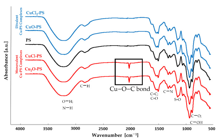
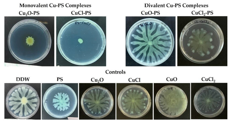
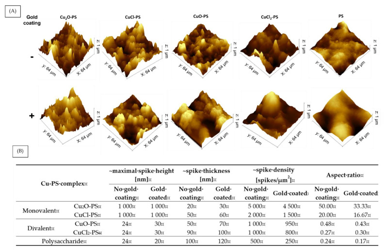
Metal-polysaccharides have recently raised significant interest due to their multifunctional bioactivities. The antimicrobial activity of a complex of Cu2O with the sulfated polysaccharide (PS) of the marine red microalga Porphyridium sp. was previously attributed to spikes formed on the complex surface (roughness). This hypothesis was further examined here using other Cu-PS complexes (i.e., monovalent-Cu2O, CuCl and divalent-CuO, CuCl2). The nanostructure parameters of the monovalent complexes, namely, longer spikes (1000 nm) and greater density (2000-5000 spikes/µm2) were found to be related to the superior inhibition of microbial growth and viability and biofilm formation. When Escherichia coli TV1061, used as a bioluminescent test organism, was exposed to the monovalent Cu-PS complexes, enhanced bioluminescence accumulation was observed, probably due to membrane perforation by the spikes on the surface of the complexes and consequent cytoplasmic leakage. In addition, differences were found in the surface chemistry of the monovalent and divalent Cu-PS complexes, with the monovalent Cu-PS complexes exhibiting greater stability (ζ-potential, FTIR spectra, and leaching out), which could be related to spike formation. This study thus supports our hypothesis that the spikes protruding from the monovalent Cu-PS surfaces, as characterized by their aspect ratio, are responsible for the antimicrobial and antibiofilm activities of the complexes.

Keywords: antimicrobial activities; biomaterials; metal complexes; red microalgae; spike formation; sulfated polysaccharides.


Conflict of interest statement

The authors declare no conflict of interest.

Figures

Figure 1
Figure 1
FTIR transmission spectra of the divalent Cu–PS complexes (blue), the monovalent Cu–PS complexes (red) and the polysaccharide (black). All Cu–PS complexes contained 0.7% polysaccharide (w/v) and 500 ppm copper. To facilitate ease of viewing, the spectra are displaced with respect to the Y axis.
Figure 2
Figure 2
Copper-release profiles from monovalent and divalent Cu–PS complexes. The copper concentration was determined in distilled water. Data represent the average values of three independent experiments. All the Cu–PS complexes contained 0.7% polysaccharide (w/v) and 500 ppm copper. Copper concentration was measured using a SPECTRO ARCOS ICP-OES analyzer.
Figure 3
Figure 3
Effect of the monovalent and divalent Cu–PS complexes on (A) the inhibition of growth and (B) the cell viability of a fungus (Candida albicans), Gram-negative bacteria (Acinetobacter baumannii, Pseudomonas aeruginosa, and Escherichia coli), and Gram-positive bacteria (Staphylococcus aureus and Bacillus subtilis). All the Cu−PS complexes contained 0.07% (w/v) polysaccharide and 30 ppm copper. For the growth inhibition experiments, the control was the absorbance of the relevant growth medium with only the bacterium or the fungus (see the experimental section). The microbial cultures were incubated with shaking in 96-well plates at 37 °C for 14 h for each bacterial species or 48 h for C. albicans. Each sample was plated on an agar plate of the relevant medium after serial dilution and incubated overnight at 37 °C. CFUs were counted the following morning and were assessed vs. untreated cells. Values are the means ± standard error of mean (SEM)of three independent experiments performed in triplicate. All of the results were significantly different from their relative controls (ANOVA; p < 0.05).
Figure 4
Figure 4
Effect of monovalent and divalent Cu–PS complexes on the swarming motility of P. aeruginosa PA14. Top row: Motility of P. aeruginosa PA14 in the presence of Cu–PS complexes. Bottom row: Controls. The bacteria were inoculated into the center of each plate consisting of M9 solidified with 0.5% (w/v) Difco agar and containing 0.1% of the relevant Cu–PS complex. Surface coverage was assessed after 24 h of growth at 37 °C. All of the Cu–PS complexes contained 0.7% polysaccharide (w/v) and 500 ppm copper. For the control treatments, the copper concentration in the copper-containing plates was 500 ppm, and the PS plate contained 0.7% Porphyridium sp. polysaccharide (w/v).
Figure 5
Figure 5
Effect of monovalent and divalent Cu–PS complexes on P. aeruginosa PA14 biofilm formation. Forty-eight hours after inoculation, biofilm formation was assessed by CLSM on (A) an untreated surface, (B) a surface pre-coated with Porphyridium sp. polysaccharide, (C,D) surfaces pre-coated with monovalent Cu–PS complexes, and (E,F) surfaces pre-coated with divalent Cu–PS complexes. Viable cells stained green, and dead cells stained red with the BacLight® DEAD/LIVE Kit for scanned areas of ~318 μm × 318 μm. (G) Cell-layer thickness (μm3/μm2) for live and dead cells; values are means ± standard error of mean (SEM). The results are presented for three independent sets of flow-cell experiments, each containing 30 measurements. Significant differences between the groups (p < 0.0001) by two-way ANOVA, followed by Tukey’s test. (H) Ratio of dead-to-live cells (calculated from Figure 5G). All of the Cu–PS complexes contained 0.7% polysaccharide (w/v) and 500 ppm copper.
Figure 6
Figure 6
Surface topography and spike distribution of monovalent and divalent Cu–PS complexes with and without the gold coating. (A) AFM 3D images and (B) spike parameters. 3D images of the Cu–PS complexes and the polysaccharide were manually analyzed using Gwyddion and ImageJ software. Spike thickness and density were calculated manually using ImageJ and the aspect ratio was calculated from the data collected from the AFM images analyzed by Gwyddion and ImageJ software. All of the Cu–PS complexes contained 0.7% polysaccharide (w/v) and 500 ppm copper.
Figure 7
Figure 7
SEM micrographs showing the effect of monovalent and divalent Cu–PS complexes that were surface coated with gold vs. non-coated on P. aeruginosa PA14 biofilm formation. ×10,000, scale bar = 4 µm. All of the Cu–PS complexes contained 0.7% polysaccharide (w/v) and 500 ppm copper. Images of the control (glass surface alone) are presented in Figure S3, Supplementary Materials.
Figure 8
Figure 8
(A) Bioluminescence signal from E. coli TV1061 induced by monovalent and divalent Cu–PS complexes. The bioluminescence was measured in relative light units (RLU) at 490 nm (i.e., the wavelength attributed to bacterial luciferase). (B) Area under the peak showing the leakage of luciferase from the bacterial cells. All of the Cu−PS complexes contained 0.07% (w/v) polysaccharide and 30 ppm copper. In the copper salts (Cu2O, CuCl, CuO, CuCl2), the same pattern of bioluminescence as the PS alone was shown (not shown).
Figure 9
Figure 9
Microbial cell death as a function of the characteristics of the spikes induced on the Cu–PS complexes. (A) Aspect ratios (•) of the spikes of the various Cu–PS complexes. (B) Luminescence of E. coli TV1061 as a function of the aspect ratio (). (C) Microbial growth inhibition as function luminescence (∆) for five different microorganisms: Candida albicans, Acinetobacter baumannii, Pseudomonas aeruginosa, Escherichia coli, Staphylococcus aureus, and Bacillus subtilis. All the Cu−PS complexes contained 0.07% (w/v) polysaccharide and 30 ppm copper. The results presented are calculated from Figure 3 and Figure 6.

References

    1. Yu Y., Shen M., Song Q., Xie J. Biological Activities and Pharmaceutical Applications of Polysaccharide from Natural Resources: A Review. Carbohydr. Polym. 2018;183:91–101. doi: 10.1016/j.carbpol.2017.12.009. - DOI - PubMed
    1. Wang X.Y., Zhang D.D., Yin J.Y., Nie S.P., Xie M.Y. Recent Developments in Hericium Erinaceus Polysaccharides: Extraction, Purification, Structural Characteristics and Biological Activities. Crit. Rev. Food Sci. Nutr. 2018;59:S96–S115. doi: 10.1080/10408398.2018.1521370. - DOI - PubMed
    1. Li X., Jiang F., Liu M., Qu Y., Lan Z., Dai X., Huang C., Yue X., Zhao S., Pan X., et al. Synthesis, Characterization, and Bioactivities of Polysaccharide Metal Complexes: A Review. J. Agric. Food Chem. 2022;70:6922–6942. doi: 10.1021/acs.jafc.2c01349. - DOI - PubMed
    1. Xu Y., Wu Y., Sun P., Zhang F., Linhardt R.J., Zhang A. Chemically Modified Polysaccharides: Synthesis, Characterization, Structure Activity Relationships of Action. Int. J. Biol. Macromol. 2019;132:970–977. doi: 10.1016/j.ijbiomac.2019.03.213. - DOI - PubMed
    1. Lapidot M., Shrestha R.P., Weinstein Y., Arad S. Red Microalgae: From Basic Know-How to Biotechnology. Springer; Berlin/Heidelberg, Germany: 2010. pp. 205–225.

LinkOut - more resources