. 2023 Jan 27;8(79):eade2798.
doi: 10.1126/sciimmunol.ade2798. Epub 2023 Jan 27.

Class switch toward noninflammatory, spike-specific IgG4 antibodies after repeated SARS-CoV-2 mRNA vaccination

Affiliations

Class switch toward noninflammatory, spike-specific IgG4 antibodies after repeated SARS-CoV-2 mRNA vaccination

Pascal Irrgang et al. Sci Immunol. .

Abstract

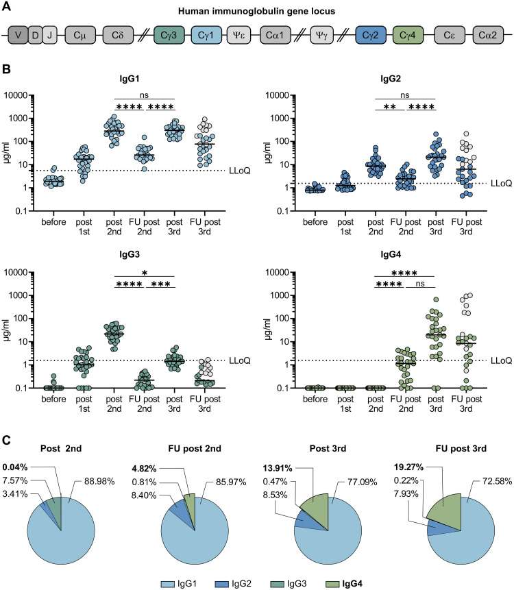
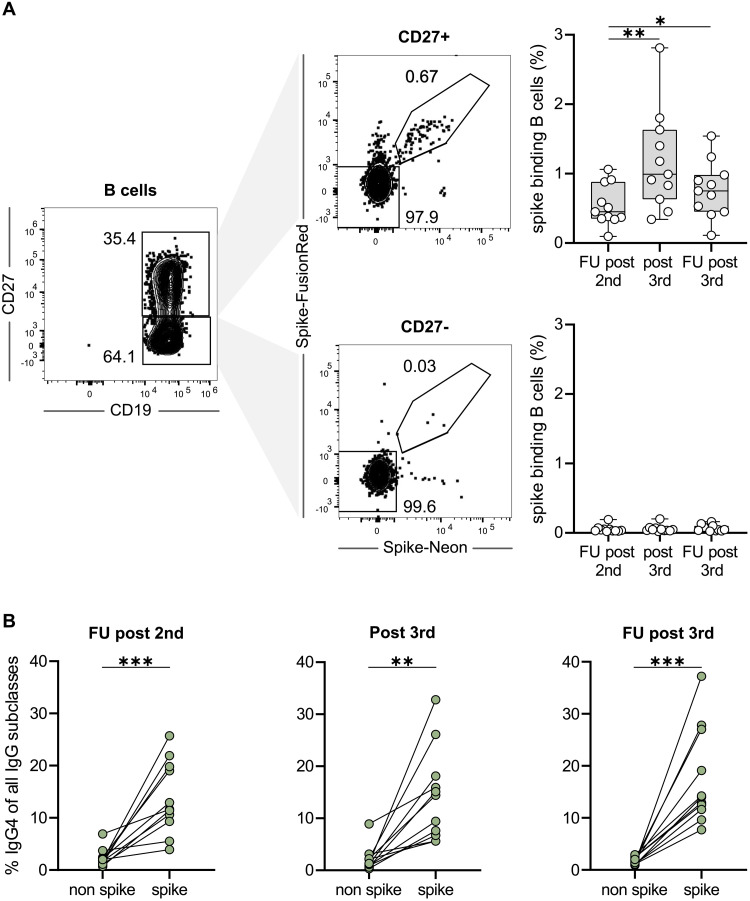
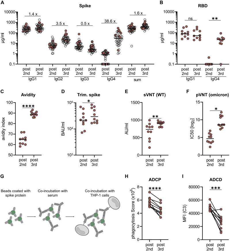
RNA vaccines are efficient preventive measures to combat the severe acute respiratory syndrome coronavirus 2 (SARS-CoV-2) pandemic. High levels of neutralizing SARS-CoV-2 antibodies are an important component of vaccine-induced immunity. Shortly after the initial two mRNA vaccine doses, the immunoglobulin G (IgG) response mainly consists of the proinflammatory subclasses IgG1 and IgG3. Here, we report that several months after the second vaccination, SARS-CoV-2-specific antibodies were increasingly composed of noninflammatory IgG4, which were further boosted by a third mRNA vaccination and/or SARS-CoV-2 variant breakthrough infections. IgG4 antibodies among all spike-specific IgG antibodies rose, on average, from 0.04% shortly after the second vaccination to 19.27% late after the third vaccination. This induction of IgG4 antibodies was not observed after homologous or heterologous SARS-CoV-2 vaccination with adenoviral vectors. Single-cell sequencing and flow cytometry revealed substantial frequencies of IgG4-switched B cells within the spike-binding memory B cell population [median of 14.4%; interquartile range (IQR) of 6.7 to 18.1%] compared with the overall memory B cell repertoire (median of 1.3%; IQR of 0.9 to 2.2%) after three immunizations. This class switch was associated with a reduced capacity of the spike-specific antibodies to mediate antibody-dependent cellular phagocytosis and complement deposition. Because Fc-mediated effector functions are critical for antiviral immunity, these findings may have consequences for the choice and timing of vaccination regimens using mRNA vaccines, including future booster immunizations against SARS-CoV-2.


Figures

Fig. 1.
Fig. 1.. Longitudinal analyses of vaccine induced antibody response.
(A) Schematic representation of the human immunoglobulin heavy chain gene locus. 5′ of each functional C-region (except Cδ) switch (S) regions are positioned directing class-switch recombination. Gene segments denoted by ψ resemble pseudogenes (B) 29 volunteers received three doses of the mRNA vaccine Comirnaty as detailed in Table 1. Serum samples were collected at a median of ten days after each vaccination (post first, post second, post third) as well as during follow-up visits at 210 days after the second vaccination (FU second) and 180 days after the third vaccination (FU third). Ten individuals experienced a breakthrough infection in the time frame between post third and FU third (indicated by grey circles). The different IgG subclasses were quantified by flow cytometry using recombinant monoclonal receptor binding domain (RBD)-antibodies as a standard. The median MFI of three negative sera were used to set the background for each subclass. For visualization purposes, all sera with MFI values below the background were set to 0.1 μg/ml. The lowest limit of quantification (LLoQ) is indicated in each graph by a dotted line and represents the lowest amount of the respective standard mAbs, which was detected (1.56 μg/ml for IgG2, IgG3 and IgG4; 5.6 μg/ml for IgG1). Depicted are individual donors and the respective median. n.a. = not analyzed. For the sake of clarity only statistical comparisons between post second, FU post second, and post third are shown irrespective of statistical significance. (C) The proportion of the different IgG subclasses of the total anti-S IgG response is shown for the four last time points. Depicted are the means of each IgG subclass. Over-time comparison within one group was done by Kruskal-Wallis test followed by Dunn’s multiple comparisons test. *p < 0.05, **p < 0.01, ***p < 0.001, ****p < 0.0001, and n.s. indicates not significant.
Fig. 2.
Fig. 2.. Longitudinal monitoring of class-switching in SARS-CoV-2-specific memory B cells.
PBMCs from 11 volunteers of cohort 1 were analyzed for the IgG subclass contribution of spike-binding memory B cells at the indicated time points (follow-up post second, 10 days post third and follow-up post third). (A) Flow cytometric gating for CD19-postive and then either CD27-positive or negative B cells (left panel), binding simultaneously to recombinant Spike-Neon and Spike-FusionRed proteins (middle panel). Percentages of spike-binding cells among CD27-positive and negative B cells are summarized on the right panels. (B) Pairwise comparison of the contribution of IgG4 subclasses on spike-binding memory B cells versus non-binding memory B cells at three different time points. Percentages were calculated from IgG4 binding cells and the sum of cells of all 4 IgG subclasses. * p < 0.05, ** p < 0.01, *** p < 0.001; paired t-Test.
Fig. 3.
Fig. 3.. Comparison of functional antibody responses after two or three mRNA vaccinations.
From a second cohort of 38 vaccinees having received three immunizations with Comirnaty (see Table 1, cohort 2), we selected ten persons to characterize the vaccine-induced antibody profile (indicated by filled circles). Only one individual in this cohort showed a positive nucleocapsid serology, despite no anamnestic clinical breakthrough infection, and was not included in the smaller sub-cohort used for functional testing. Paired serum samples collected after the second (post second) or the third (post third) vaccination were analyzed. The IgG subclass distribution measured in the flow cytometric assay and the sum of all IgG are shown for the whole cohort (A). The amounts of RBD-specific IgG1 and IgG4 (B) and the avidity (C) were determined by ELISA. For avidity measurements, sera were normalized according to total anti-S IgG levels and equal amounts of specific IgG were used. A fully automated CLIA assay was used to measure antibodies binding to trimeric spike protein. Antibody levels were quantified according to the WHO International Reference standard and given as BAU/ml (D). The neutralizing capacity was determined in a surrogate VNT against WT (E) and in a pseudotype VNT against the Omicron VOC (F). Antibody-dependent phagocytosis by the monocytic THP-1 cell line (G) was analyzed by using either monoclonal RBD antibodies of the different subclasses (Fig. S7) or the paired sera. The phagocytosis score is calculated as follows: % of THP-1 bead-positive x mean fluorescence intensity of bead-positive (H). Antibody-dependent complement deposition was analyzed on spike-coated microbeads after incubation with the paired sera. C3 deposition was detected by fluorescently labeled antibodies and mean fluorescence intensity of complement-loaded beads are shown (I). Circles represent individual sera and solid lines indicate the median (H, I). Statistical comparison for the two time points were done by a paired T-test. *p < 0.05, **p < 0.01, ***p < 0.001, ****p < 0.0001, and n.s. indicates not significant.

Comment in

References

    1. Meslé M. M., Brown J., Mook P., Hagan J., Pastore R., Bundle N., Spiteri G., Ravasi G., Nicolay N., Andrews N., Dykhanovska T., Mossong J., Sadkowska-Todys M., Nikiforova R., Riccardo F., Meijerink H., Mazagatos C., Kyncl J., McMenamin J., Melillo T., Kaoustou S., Lévy-Bruhl D., Haarhuis F., Rich R., Kall M., Nitzan D., Smallwood C., Pebody R. G., Estimated number of deaths directly averted in people 60 years and older as a result of COVID-19 vaccination in the WHO European Region, December 2020 to November 2021. Euro. Surveill. 26, 2101021 (2021). - PMC - PubMed
    1. Watson O. J., Barnsley G., Toor J., Hogan A. B., Winskill P., Ghani A. C., Global impact of the first year of COVID-19 vaccination: A mathematical modelling study. Lancet Infect. Dis. 22, 1293–1302 (2022). - PMC - PubMed
    1. Polack F. P., Thomas S. J., Kitchin N., Absalon J., Gurtman A., Lockhart S., Perez J. L., Pérez Marc G., Moreira E. D., Zerbini C., Bailey R., Swanson K. A., Roychoudhury S., Koury K., Li P., Kalina W. V., Cooper D., Frenck R. W., Hammitt L. L., Türeci Ö., Nell H., Schaefer A., Ünal S., Tresnan D. B., Mather S., Dormitzer P. R., Şahin U., Jansen K. U., Gruber W. C.; C4591001 Clinical Trial Group , Safety and Efficacy of the BNT162b2 mRNA Covid-19 Vaccine. N. Engl. J. Med. 383, 2603–2615 (2020). - PMC - PubMed
    1. Baden L. R., El Sahly H. M., Essink B., Kotloff K., Frey S., Novak R., Diemert D., Spector S. A., Rouphael N., Creech C. B., McGettigan J., Khetan S., Segall N., Solis J., Brosz A., Fierro C., Schwartz H., Neuzil K., Corey L., Gilbert P., Janes H., Follmann D., Marovich M., Mascola J., Polakowski L., Ledgerwood J., Graham B. S., Bennett H., Pajon R., Knightly C., Leav B., Deng W., Zhou H., Han S., Ivarsson M., Miller J., Zaks T.; COVE Study Group , Efficacy and Safety of the mRNA-1273 SARS-CoV-2 Vaccine. N. Engl. J. Med. 384, 403–416 (2021). - PMC - PubMed
    1. Abu-Raddad L. J., Chemaitelly H., Butt A. A.; National Study Group for COVID-19 Vaccination , Effectiveness of the BNT162b2 Covid-19 Vaccine against the B.1.1.7 and B.1.351 Variants. N. Engl. J. Med. 385, 187–189 (2021). - PMC - PubMed

Publication types

Supplementary concepts