. 2023 Feb 1;134(2):242-252.
doi: 10.1152/japplphysiol.00401.2022. Epub 2022 Dec 22.

Modifying the ICP pulse wave: effects on parenchymal blood flow pulsatility

Affiliations

Modifying the ICP pulse wave: effects on parenchymal blood flow pulsatility

Sara Qvarlander et al. J Appl Physiol (1985). .

Abstract

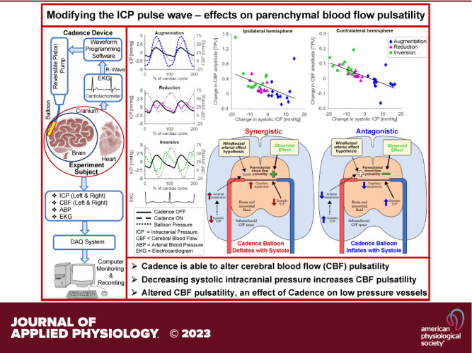
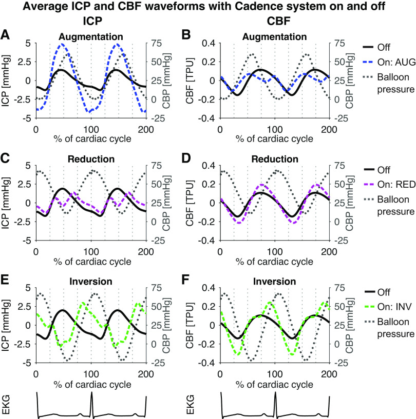
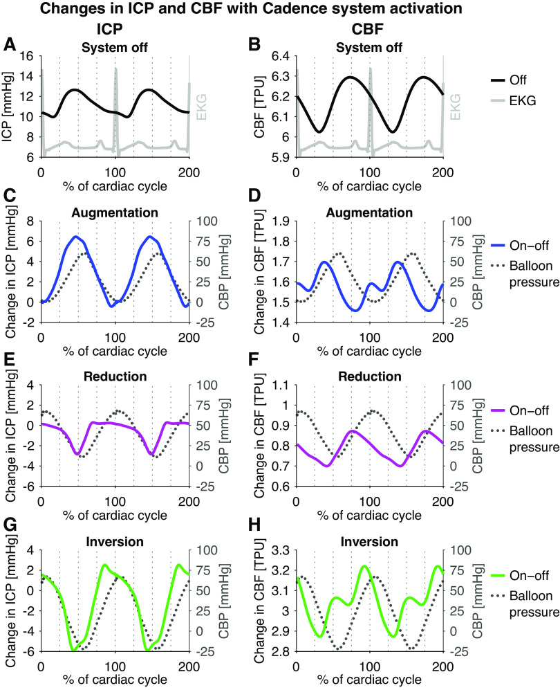
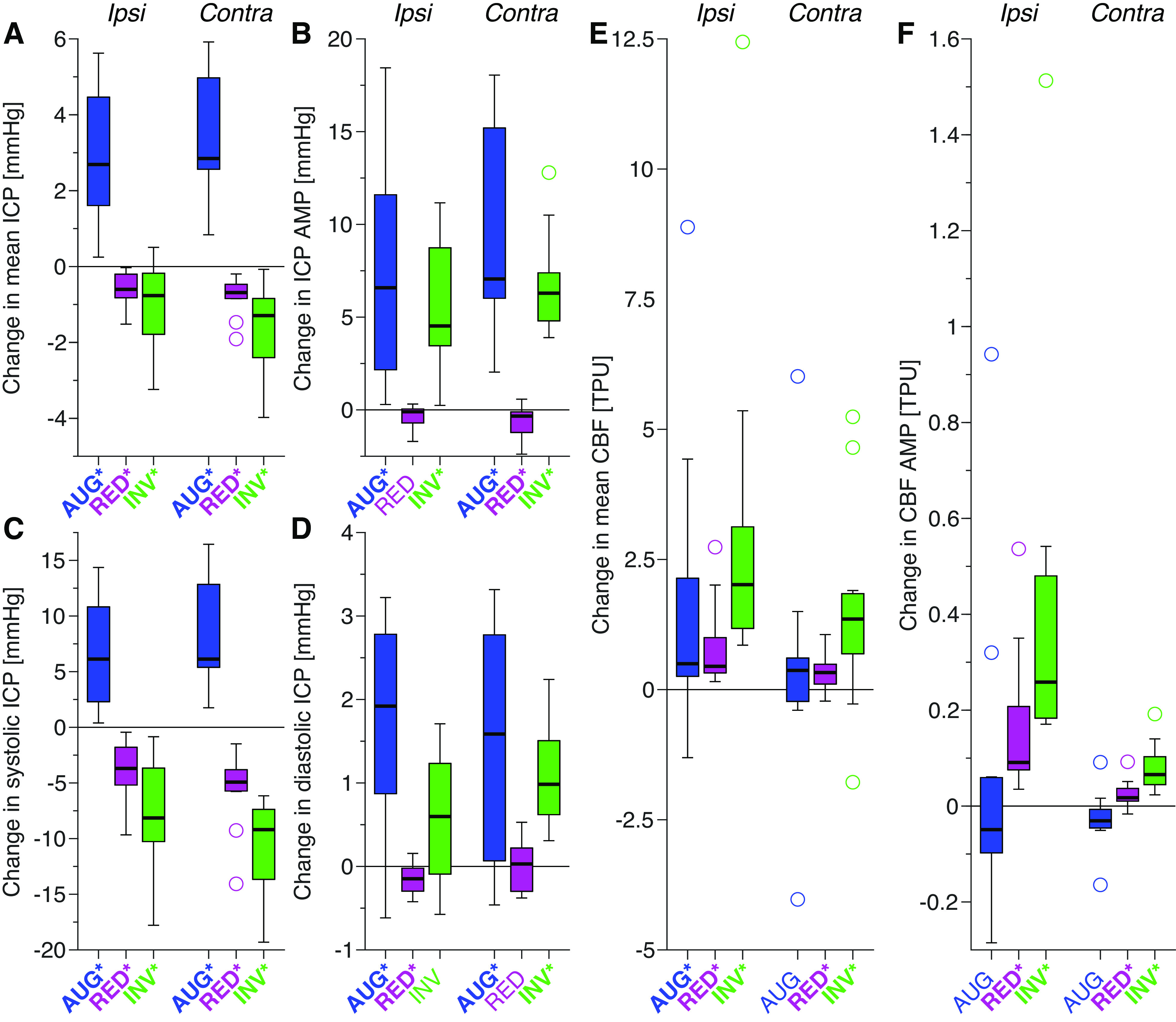
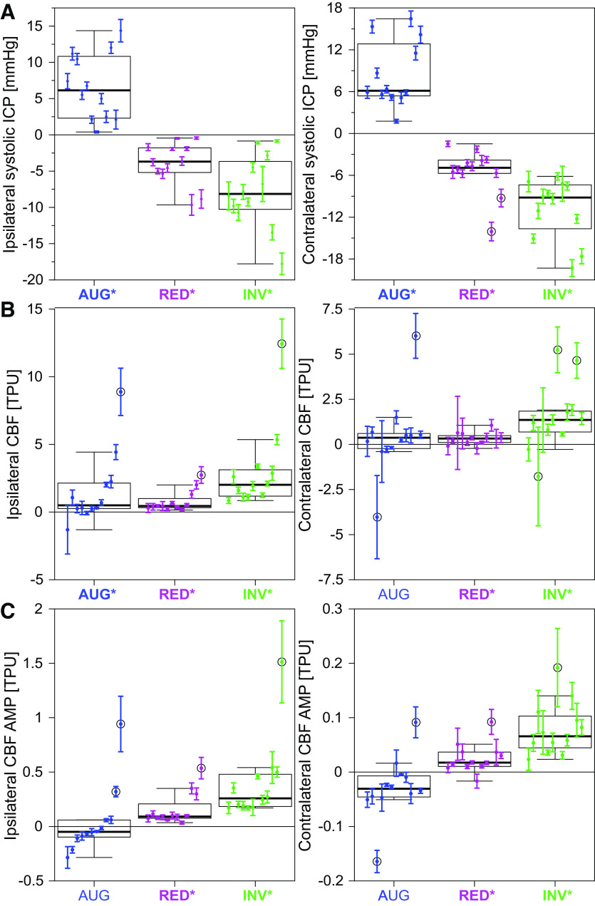
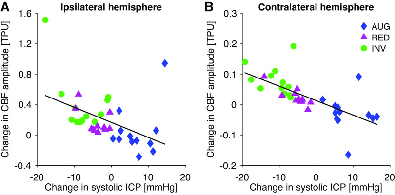
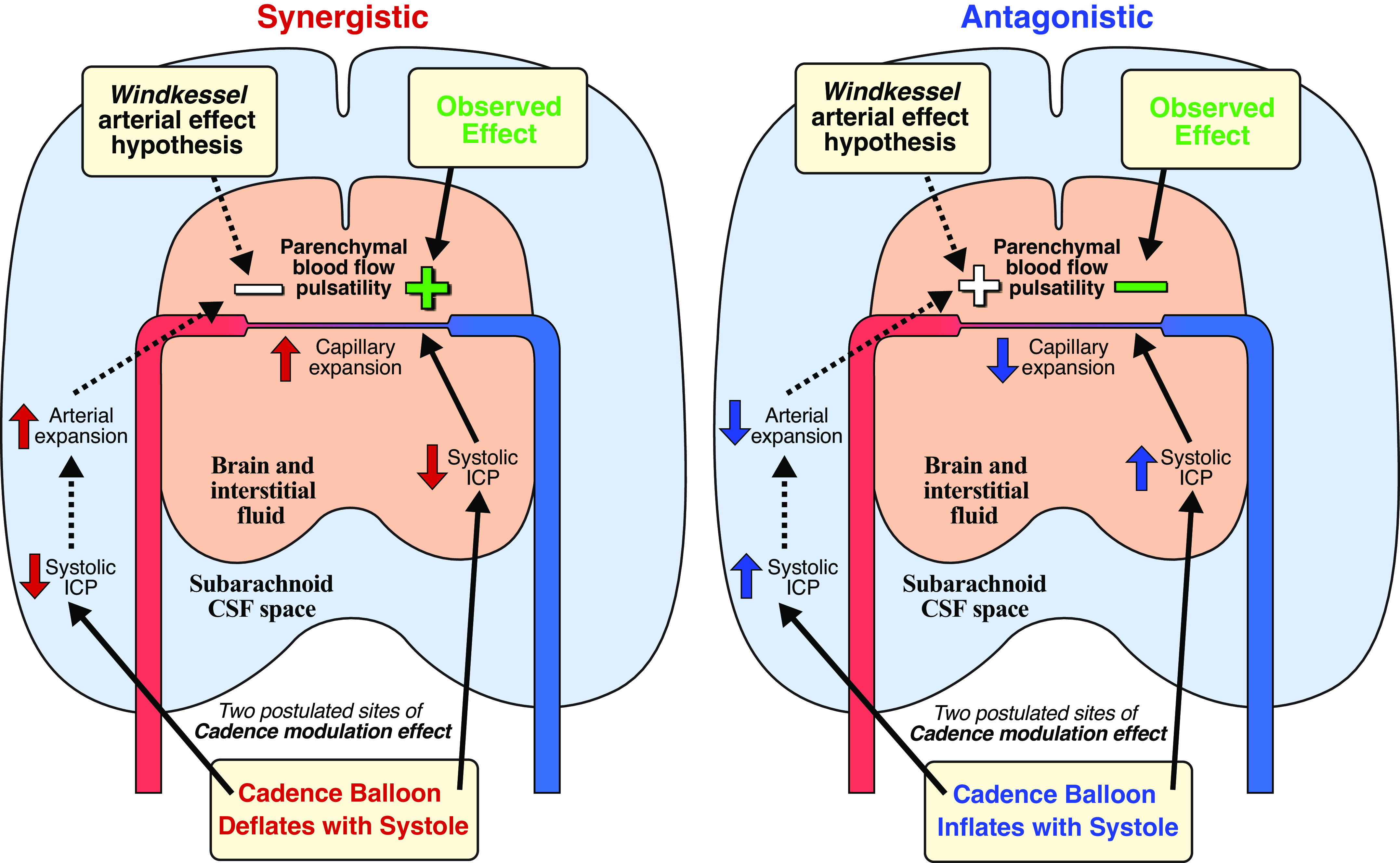
Pulsation of the cerebral blood flow (CBF) produces intercranial pressure (ICP) waves. The aim of this study is to determine whether externally modifying ICP pulsatility alters parenchymal blood flow pulsatility. A cardiac-gated inflatable device was inserted in the lateral epidural space of 12 anesthetized canines (canis familiaris) and used to cause reduction, inversion, and augmentation of the ICP pulse. CBF in each hemisphere was measured using laser Doppler velocimetry. A significant increase in both mean CBF and its amplitude was observed for reduction as well as inversion of the ICP pulse, with larger changes observed for the inversion protocol. Significant increases in the mean CBF were also observed ipsilaterally for the augmentation protocol together with indications of reduced CBF amplitude contralaterally. External alteration of the ICP pulse thus caused significant changes in parenchymal blood flow pulsatility. The inverse relationship between the ICP and CBF amplitude suggests that the changes did not occur via modification of the intracranial Windkessel mechanism. Thus, the effects likely occurred in the low-pressure vessels, i.e., capillaries and/or venules, rather than the high-pressure arteries. Future MRI studies are however required to map and quantify the effects on global cerebral blood flow.NEW & NOTEWORTHY This study demonstrated that external modification of ICP pulsatility, using a cardiac-gated inflatable device implanted epidurally in canines, alters brain tissue blood flow pulsatility. Specifically, decreasing systolic ICP increased blood flow pulsatility in brain tissue. The results suggest that the altered CBF pulsatility is unlikely to depend on modification of the Windkessel effect on the feeding arterial system but was rather an effect directly on tissue and the lower pressure distal vessels.

Keywords: cardiac-gated inflatable device; cerebral blood flow; experimental model; intracranial pressure; intracranial pulsatility.


Conflict of interest statement

M.G.L. and S.M.D. are listed as inventors on two patents regarding the device presented in this paper, but do not have any conflict of interest otherwise. None of the other authors has any conflicts of interest, financial or otherwise, to disclose.

Figures

None
Graphical abstract
Figure 1.
Figure 1.
Averaged ICP (left) and CBF waveforms (right) for the baseline measurements and the three balloon protocols: augmentation (blue, A and B), reduction (magenta, C and D), and inversion (green, E and F). For easier comparison of the original and altered waveforms, the graphs show mean corrected waveforms. Cadence balloon pressure (CBP) is shown as grey dotted lines. The augmentation protocol augmented the typical ICP pulse wave (A), while suppressing the peak of the CBF signal (B). Conversely, the reduction protocol suppressed the systolic peak in ICP (C), while enhancing the CBF pulse wave (D). The inversion protocol inverted the systolic part of the ICP pulse wave (E), enhancing the CBF pulse wave even more than the reduction protocol (F). Interestingly, the onset of systolic increase in ICP occurred slightly earlier with the augmentation protocol, and this behavior was reflected in the CBF pulse wave. AUG, augmentation balloon protocol; CBF, cerebral (parenchymal) blood flow; ICP, intracranial pressure; INV, inversion balloon protocol; RED, reduction balloon.
Figure 2.
Figure 2.
Changes in the average waveform of ICP (left) and CBF (right) resulting from Cadence system activation: EKG (grey) and baseline waveforms (A and B), waveform change with augmentation (C and D, blue), reduction (E and F, magenta), and inversion (G and H, green). Balloon pressure is shown as grey dotted lines. For the inversion and augmentation protocol, two peaks/valleys in the CBF change occurred, while for the reduction protocol only one peak/valley was evident. Though individual data is not shown, it was noted that the double peak was not seen for all canines. CBF, cerebral (parenchymal) blood flow; ICP, intracranial pressure.
Figure 3.
Figure 3.
Box plots of the changes in ICP and CBF parameters for the different balloon protocols: augmentation (blue), reduction (magenta), and inversion (green). The analyzed parameters are mean ICP (A), ICP AMP (B), systolic ICP (C), diastolic ICP (D), mean CBF (E), and CBF AMP (F). Statistically significant changes (P < 0.05) are represented using bold text and * in the x-axis labels (the P value of contralateral CBF AMP for AUG was P = 0.052). The boxplots show the median (line), first and third quartile (box), range (whiskers), and statistical outliers (circles). AMP, amplitude; AUG, augmentation balloon protocol; CBF, cerebral (parenchymal) blood flow; contra, contralateral; CSF, cerebrospinal fluid; ICP, intracranial pressure; INV, inversion balloon protocol; ipsi, ipsilateral; RED, reduction balloon protocol; TPU, tissue perfusion units.
Figure 4.
Figure 4.
Intra-animal variability across the repeated measurements (means ± SD: dots and error bars, 8–12 repetitions) for changes in systolic ICP (top, A), mean CBF (middle, B), and CBF AMP (bottom, C). The data for each hemisphere (ispi-/contralateral) and balloon protocol (augmentation: blue, reduction: magenta, inversion: green) are presented with the individual animals in the same order (left to right according to ipsilateral CBF AMP, AUG). Overlayed are the corresponding box plots for the group, showing median (line), first and third quartile (box), and range (whiskers); statistical outliers are circled. Statistically significant changes on the group level (P < 0.05) are represented using bold text and * in the x-axis labels. Note that the scale of the y-axis is different for each panel. ABP, arterial blood pressure; AMP, amplitude; AUG, augmentation balloon protocol; CBF, cerebral (parenchymal) blood flow; contra, contralateral; CSF, cerebrospinal fluid; ICP, intracranial pressure; INV, inversion balloon protocol; ipsi, ipsilateral; RED, reduction balloon protocol; SD, standard deviation; TPU, tissue perfusion units.
Figure 5.
Figure 5.
Illustration of the correlation between changes in systolic ICP and CBF amplitude for the hemisphere ipsilateral (left, A; R2 = 0.20) and contralateral (right, B; R2 = 0.47) to the Cadence balloon. Each symbol and color illustrate measurements from a different type of protocol: inversion (INV; green circles), reduction (RED; magenta triangles), and augmentation (AUG; blue diamonds). AUG, augmentation balloon protocol; CBF, cerebral (parenchymal) blood flow; ICP, intracranial pressure; INV, inversion balloon protocol; RED, reduction balloon protocol; TPU, tissue perfusion units.
Figure 6.
Figure 6.
Illustration of the two hypothesized mechanisms by which the Cadence device may affect the pulsatility of parenchymal blood flow. One potential mechanism was by facilitating or hindering the arterial Windkessel mechanism, but this was not supported by the data. Thus, it is hypothesized that the relevant effect occurred more locally in the tissue, e.g., by facilitating or hindering capillary expansion. Left: synergistic inflation protocol. Right: antagonistic inflation protocol. CSF, cerebrospinal fluid; ICP, intracranial pressure.

References

    1. Westerhof N, Lankhaar J-W, Westerhof BE. The arterial Windkessel. Med Biol Eng Comput 47: 131–141, 2009. doi:10.1007/s11517-008-0359-2. - DOI - PubMed
    1. Wagshul ME, Eide PK, Madsen JR. The pulsating brain: a review of experimental and clinical studies of intracranial pulsatility. Fluids Barriers CNS 8: 5, 2011. doi:10.1186/2045-8118-8-5. - DOI - PMC - PubMed
    1. Mitchell GF. Effects of central arterial aging on the structure and function of the peripheral vasculature: implications for end-organ damage. J Appl Physiol (1985) 105: 1652–1660, 2008. doi:10.1152/japplphysiol.90549.2008. - DOI - PMC - PubMed
    1. Bateman GA. Pulse wave encephalopathy: a spectrum hypothesis incorporating Alzheimer’s disease, vascular dementia and normal pressure hydrocephalus. Med Hypotheses 62: 182–187, 2004. doi:10.1016/S0306-9877(03)00330-X. - DOI - PubMed
    1. Mitchell GF, Van Buchem MA, Sigurdsson S, Gotal JD, Jonsdottir MK, Kjartansson Ó, Garcia M, Aspelund T, Harris TB, Gudnason V, Launer LJ. Arterial stiffness, pressure and flow pulsatility and brain structure and function: the age, gene/environment susceptibility-Reykjavik Study. Brain 134: 3398–3407, 2011. doi:10.1093/brain/awr253. - DOI - PMC - PubMed

Publication types

LinkOut - more resources