Review
. 2023 Mar 9;141(10):1119-1135.
doi: 10.1182/blood.2022018092.

Metabolism in acute myeloid leukemia: mechanistic insights and therapeutic targets

Affiliations
Review

Metabolism in acute myeloid leukemia: mechanistic insights and therapeutic targets

Sushanta Kumar Mishra et al. Blood. .

Abstract

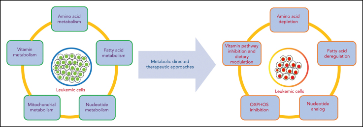
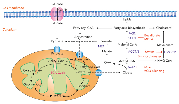
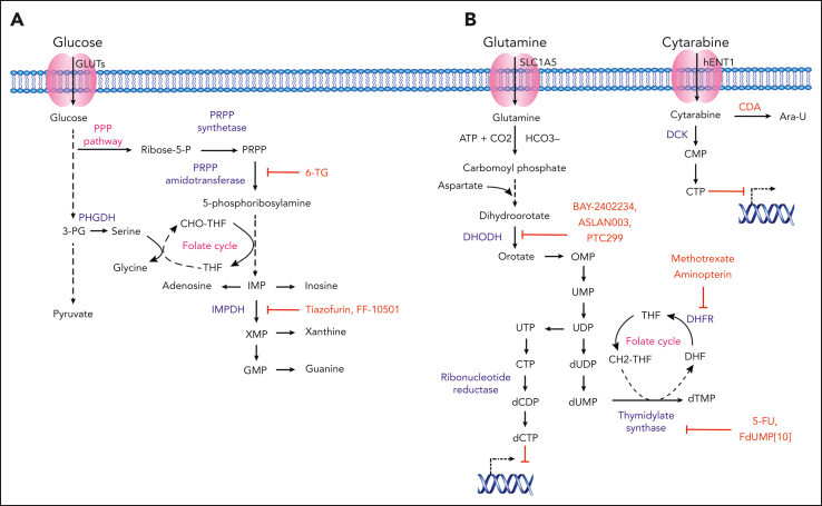
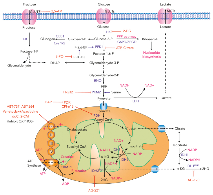
Metabolic rewiring and cellular reprogramming are trademarks of neoplastic initiation and progression in acute myeloid leukemia (AML). Metabolic alteration in leukemic cells is often genotype specific, with associated changes in epigenetic and functional factors resulting in the downstream upregulation or facilitation of oncogenic pathways. Targeting abnormal or disease-sustaining metabolic activities in AML provides a wide range of therapeutic opportunities, ideally with enhanced therapeutic windows and robust clinical efficacy. This review highlights the dysregulation of amino acid, nucleotide, lipid, and carbohydrate metabolism in AML; explores the role of key vitamins and enzymes that regulate these processes; and provides an overview of metabolism-directed therapies currently in use or development.


Conflict of interest statement

Conflict-of-interest disclosure: The authors declare no competing financial interests.

Figures

None
Graphical abstract
Figure 1.
Figure 1.
Amino acid metabolism in AML and relevant therapeutic strategies. Schematic representation showing the cellular uptake and utilization of select amino acids and their downstream intermediates. Enzymes with demonstrated relevance to AML pathophysiology are highlighted in blue. Inhibitory compounds or proteins are highlighted in red. Biological outputs and metabolic pathways are highlighted in pink. ADI, arginine deaminase; ASL, arginosuccinate lyase; ASN, asparagine; ASS1, arginosuccinate synthetase 1; NO, nitric oxide; OAA, oxaloacetic acid; OTC, ornithine transcarbamylase; PSAT1, phosphoserine aminotransferase 1; PSPH, phosphoserine phosphatase; 3-PG, 3-phosphoglycerate; 3-PHP, 3-phosphohydroxypyruvate; 3-PS, 3-phosphoserine; THF, tetrahydrofolate; 5,10-mTHF, 5,10-methylenetetrahydrofolate.
Figure 2.
Figure 2.
FA metabolism in AML and relevant therapeutic strategies. Schematic representation showing the transport of citrate into the cytoplasm, conversion to acetyl-CoA, and generation of FA intermediates through either downstream carboxylation or alternatively through the MVA pathway. Enzymes with demonstrated relevance to AML pathophysiology are highlighted in blue. Chemical inhibitors or gene silencing effects are highlighted in red. ACC, acetyl Co-A carboxylase; ACLY, ATP citrate lyase; DCV, dehydrocurvularin; FASN, fatty acid synthase; HMGCR, HMG-CoA reductase; ME1, malic enzyme 1; OAA, oxaloacetate; SCD1, stearoyl CoA desaturase 1.
Figure 3.
Figure 3.
Nucleotide metabolism in AML and relevant therapeutic strategies. (A) Purine nucleotide biosynthesis in AML. Schematic representation showing purine synthesis through the utilization of glycolytic intermediates. Key enzymes, inhibitory compounds, and metabolic pathways with demonstrated relevance to AML pathophysiology are highlighted in blue, red, and pink, respectively. (B) Pyrimidine nucleotide biosynthesis in AML. Schematic representation showing the key enzymes involved in de novo pyrimidine synthesis. The transport of pyrimidine analog cytarabine and the mechanistic inhibition of DNA synthesis in leukemic cells is represented. Different enzymes, inhibitory compounds, and metabolic pathways with demonstrated relevance to AML pathophysiology are highlighted in blue, red, and pink, respectively. Ara-U, uracil arabinoside; CDP, cytidine diphosphate; CMP, cytarabine monophosphate; CTP, cytidine triphosphate; DHF, dihydrofolate; DHFR, dihydrofolate reductase; DOODH, dihydroorotate dehydrogenase; GMP, guanine monophosphate; IMP, inosine monophosphate; IMPDH, 5′-monophosphate dehydrogenase; OMP, orotidine 5′-monophosphate; 3-PG, 3-phosphoglycerate; PHGDP, 3-phosphoglycerate dehydrogenase; PPP, pentose phosphate pathway; 6-TG; 6-thioguanine; UMP, uridine monophosphate; TS, thymidylate synthase; UDP, uridine diphosphate; UTP, uridine triphosphate; XMP, xanthine monophosphate.
Figure 4.
Figure 4.
Glycolysis and mitochondrial OXPHOS in AML and relevant therapeutic strategies. Schematic representation showing the cellular uptake and downstream utilization of glucose and fructose. Enzymes with demonstrated relevance to AML pathophysiology are highlighted in blue. Chemical and biological inhibitors are highlighted in red. Biological outputs and metabolic pathways are highlighted in pink. 2,5-AM, 2,5-anhydro-D-mannitol; FK, fructose kinase; LDH, lactate dehydrogenase; MCTs, monocarboxylate transporters; PEP, phosphoenolpyruvate.
Figure 5.
Figure 5.
Vitamin metabolism in AML and relevant therapeutic strategies. (A) Regulation of TET enzymes by vitamin C (ascorbate). Schematic representation demonstrating the ascorbate-dependent regulation of the TET enzymes by ascorbate, as well as downstream epigenetic effects in HSCs (multipotent progenitors [MPPs]) and cancer stem cells. (B) Regulation of vitamin B6 (pyridoxal) in AML. Schematic representation of pyridoxal uptake and downstream utilization in AML cells, including a central role for PLP and downstream effector enzymes, ODC1 and GOT2 in leukemic proliferation. Enzymes and inhibitory compounds with demonstrated relevance to AML pathophysiology are highlighted in blue, and red, respectively. BER, base exclusion repair; 5CaC, 5-carboxylcytosine; 5fC, 5-formyl cytosine; DHA, dehydroascorbate; GOT2, glutamic-oxaloacetic transaminase 2; 5mC, 5-methylcytosine; 5hmC, 5-hydroxymethylcytosine; ODC1, ornithine decarboxylase.

References

    1. Siegel RL, Miller KD, Fuchs HE, Jemal A. Cancer statistics, 2022. CA Cancer J Clin. 2022;72(1):7–33. - PubMed
    1. Ferrara F, Schiffer CA. Acute myeloid leukaemia in adults. Lancet. 2013;381(9865):484–495. - PubMed
    1. De Kouchkovsky I, Abdul-Hay M. Acute myeloid leukemia: a comprehensive review and 2016 update. Blood Cancer J. 2016;6(7):e441. - PMC - PubMed
    1. Döhner H, Wei AH, Appelbaum FR, et al. Diagnosis and management of AML in adults: 2022 recommendations from an international expert panel on behalf of the ELN. Blood. 2022;140(12):1345–1377. - PubMed
    1. Khwaja A, Bjorkholm M, Gale RE, et al. Acute myeloid leukaemia. Nat Rev Dis Prim. 2016;2(1):1–22. - PubMed

MeSH terms