. 2023 Jan 11;31(1):18-32.e9.
doi: 10.1016/j.chom.2022.11.015. Epub 2022 Dec 21.

Two distinct gut microbial pathways contribute to meta-organismal production of phenylacetylglutamine with links to cardiovascular disease

Affiliations

Two distinct gut microbial pathways contribute to meta-organismal production of phenylacetylglutamine with links to cardiovascular disease

Yijun Zhu et al. Cell Host Microbe. .

Abstract

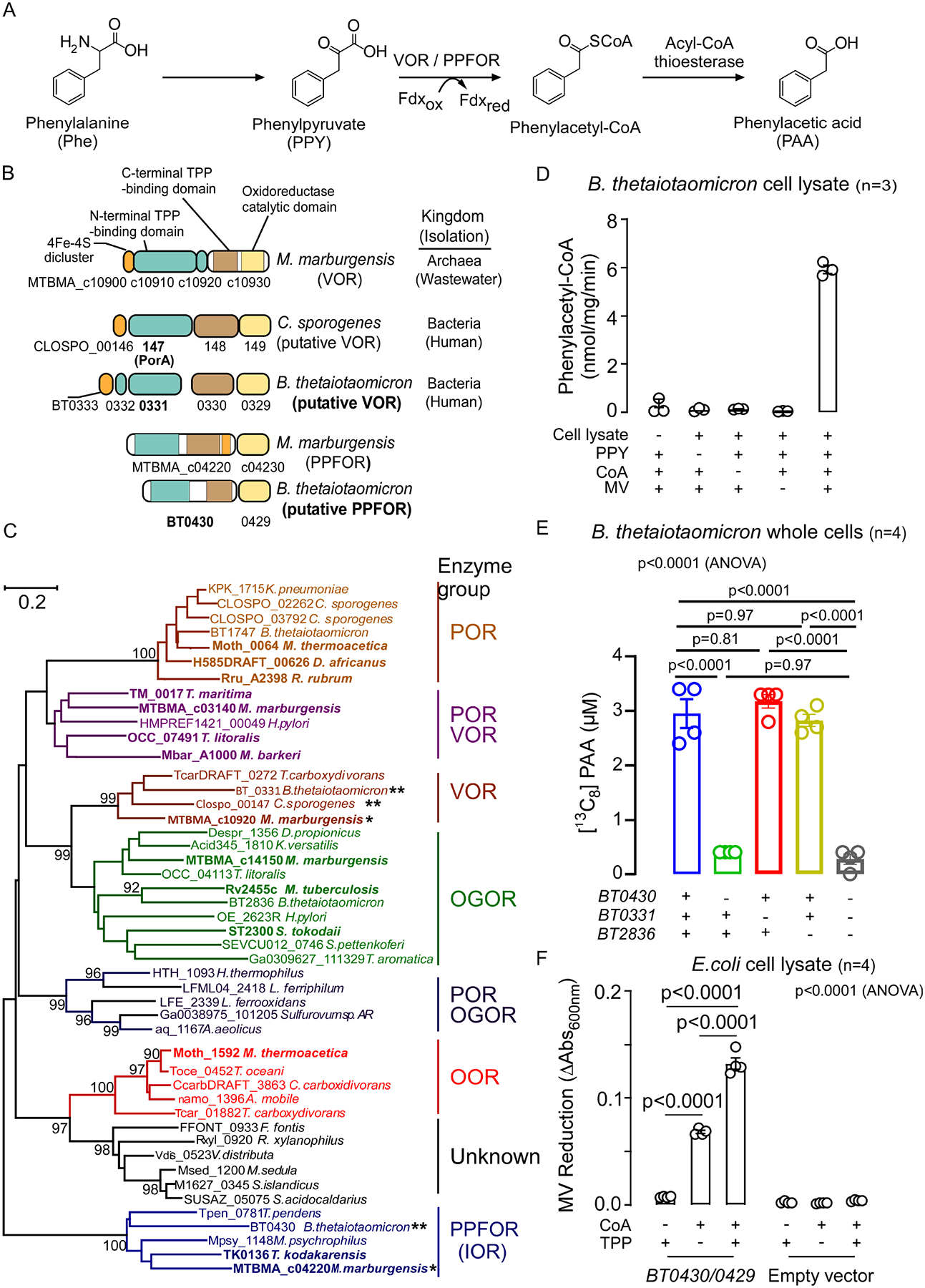
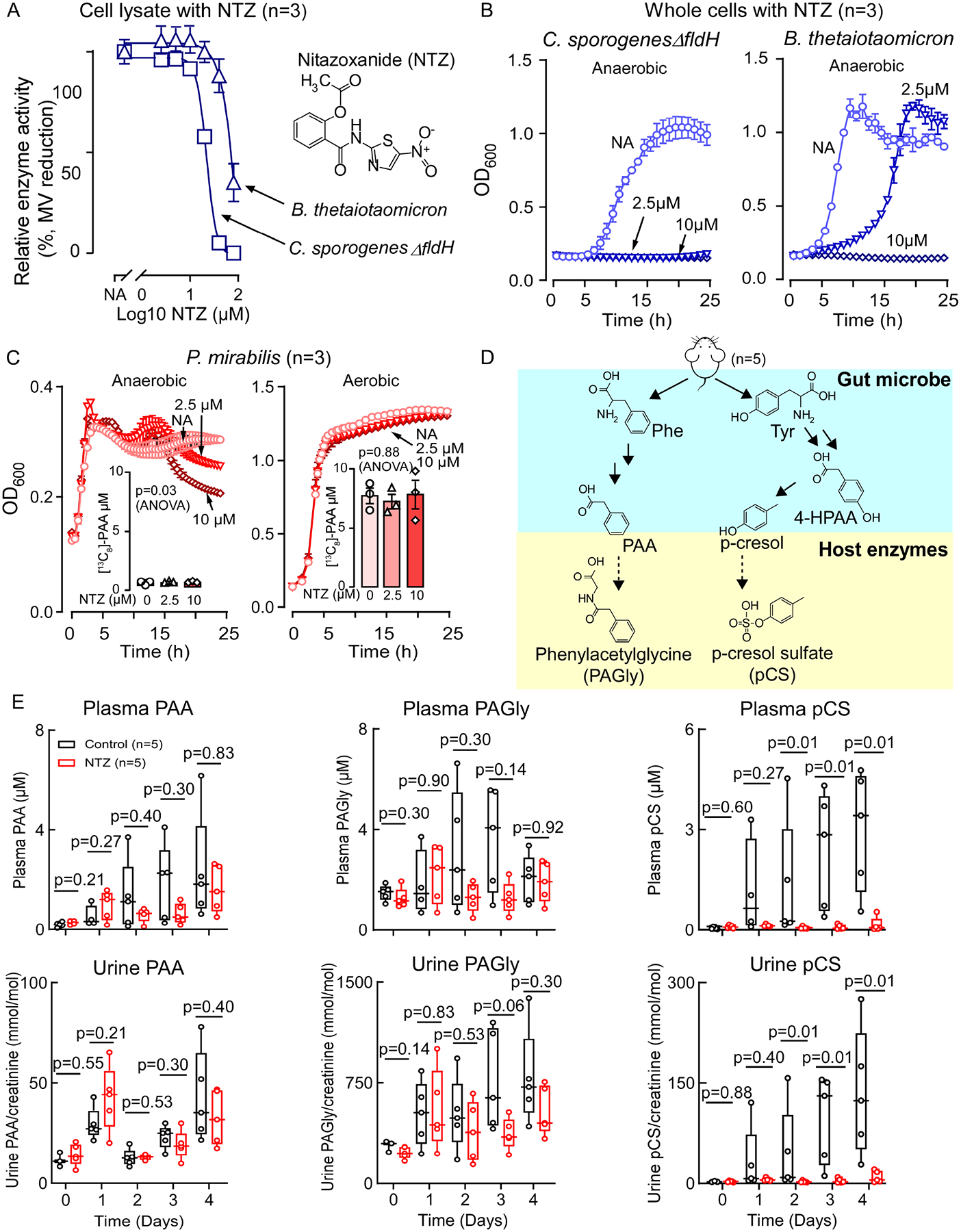
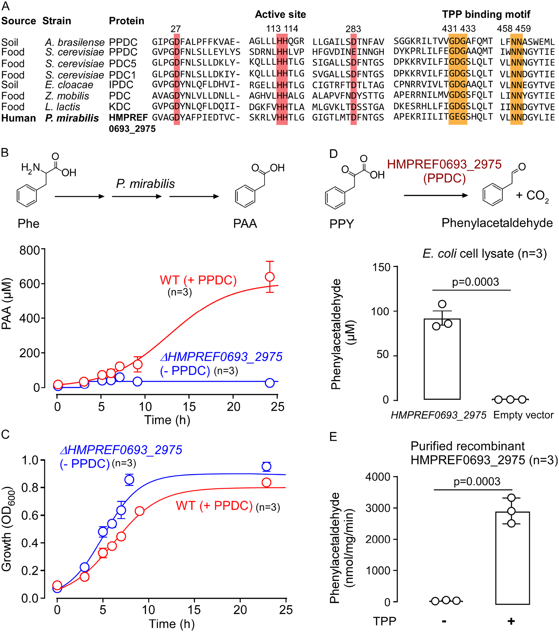
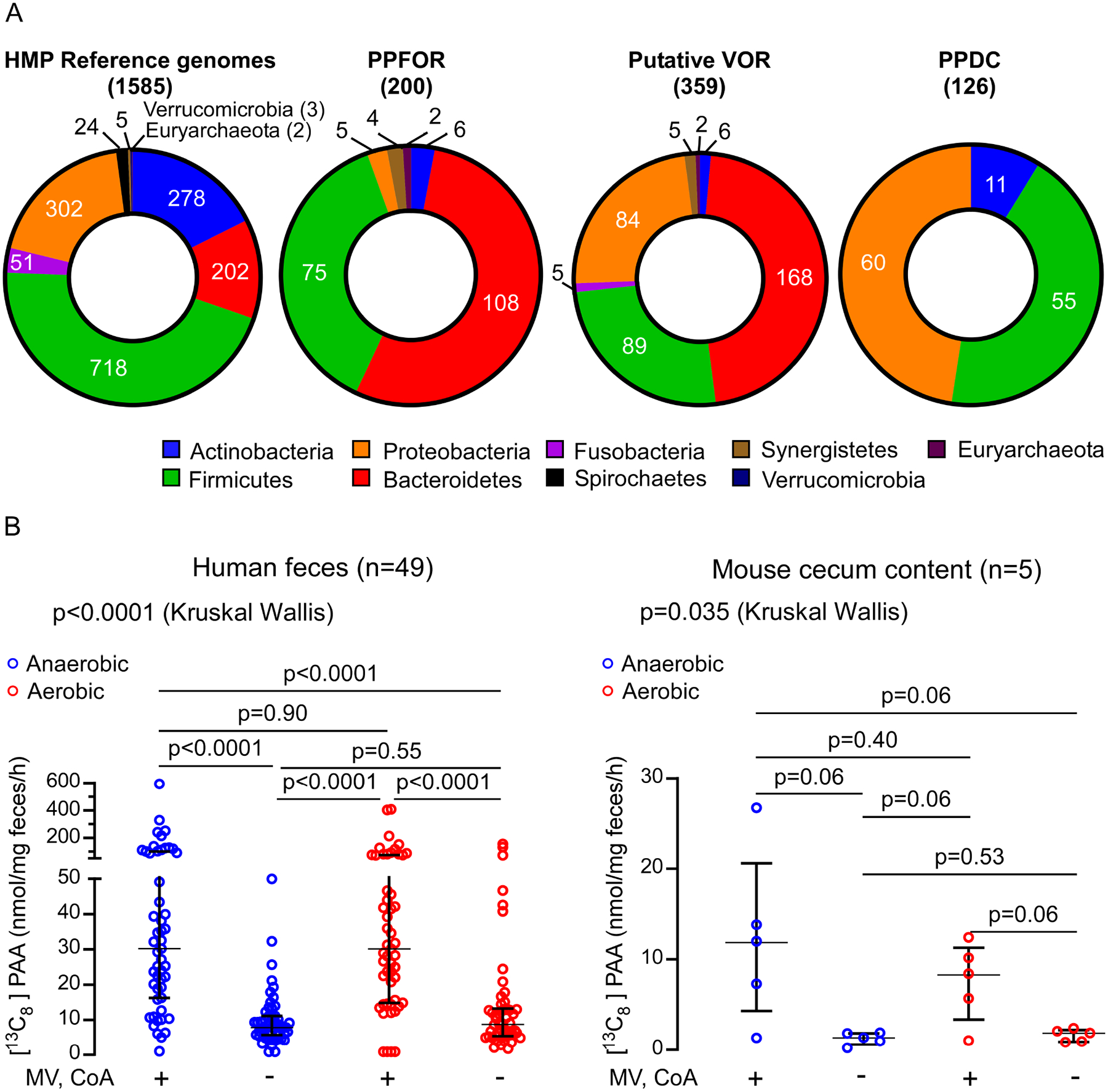
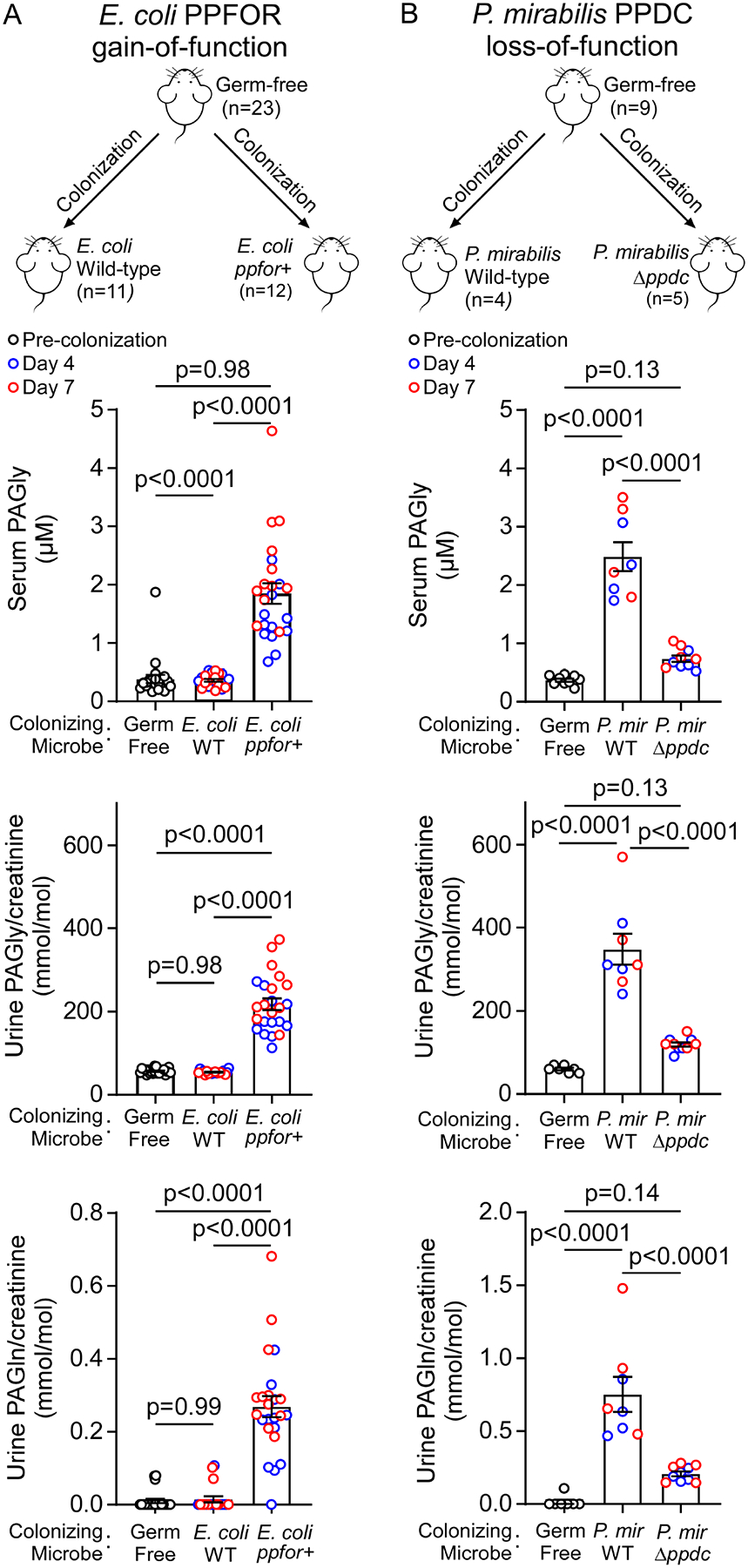
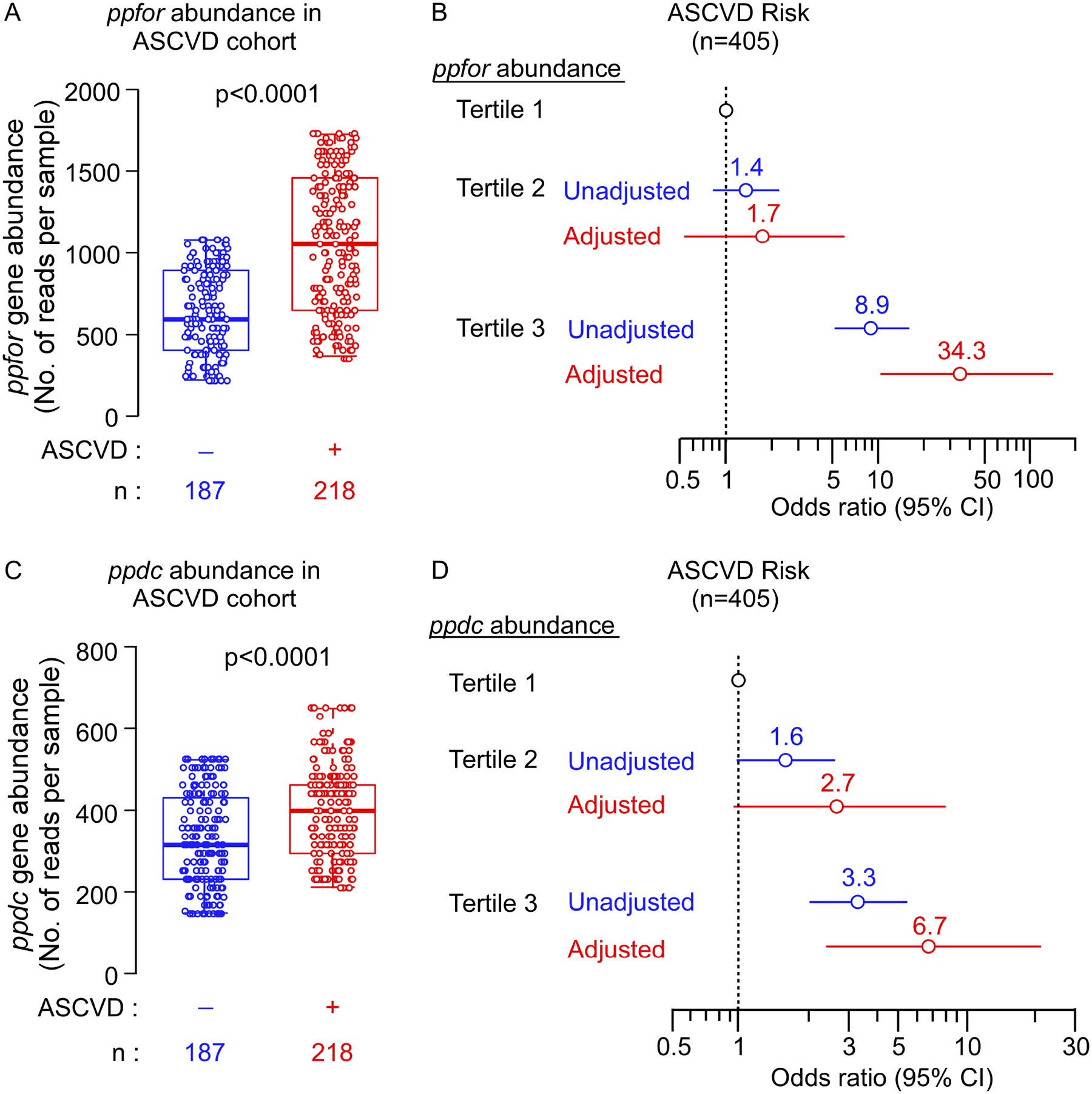
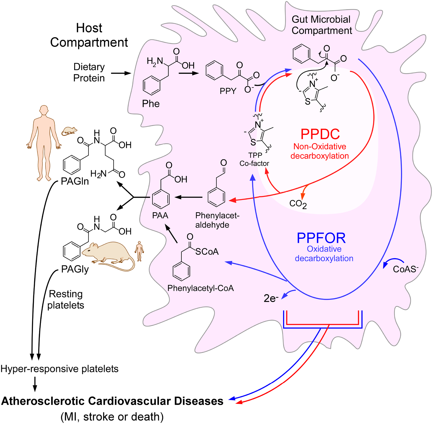
Recent studies show gut microbiota-dependent metabolism of dietary phenylalanine into phenylacetic acid (PAA) is critical in phenylacetylglutamine (PAGln) production, a metabolite linked to atherosclerotic cardiovascular disease (ASCVD). Accordingly, microbial enzymes involved in this transformation are of interest. Using genetic manipulation in selected microbes and monocolonization experiments in gnotobiotic mice, we identify two distinct gut microbial pathways for PAA formation; one is catalyzed by phenylpyruvate:ferredoxin oxidoreductase (PPFOR) and the other by phenylpyruvate decarboxylase (PPDC). PPFOR and PPDC play key roles in gut bacterial PAA production via oxidative and non-oxidative phenylpyruvate decarboxylation, respectively. Metagenomic analyses revealed a significantly higher abundance of both pathways in gut microbiomes of ASCVD patients compared with controls. The present studies show a role for these two divergent microbial catalytic strategies in the meta-organismal production of PAGln. Given the numerous links between PAGln and ASCVD, these findings will assist future efforts to therapeutically target PAGln formation in vivo.

Keywords: Atherosclerotic cardiovascular disease; gut microbes; phenylacetic acid; phenylacetylglutamine; phenylacetylglycine; phenylalanine; phenylpyruvate decarboxylase; phenylpyruvate:ferredoxin oxidoreductase.


Conflict of interest statement

Declaration of interests S.L.H., Z.W., and J.T.A. report being named as co-inventor on pending and issued patents held by the Cleveland Clinic relating to cardiovascular diagnostics and/or therapeutics and being eligible to receive royalty payments for inventions or discoveries related to cardiovascular diagnostics or therapeutics from Cleveland HeartLab, a wholly owned subsidiary of Quest Diagnostics, P&G, and Zehna Therapeutics. S.L.H. and J.T.A. report being paid consultants for Zehna Therapeutics. S.L.H. also reports having received research funds from P&G, Roche Diagnostics, and Zehna Therapeutics. The other authors have reported that they have no relationships relevant to the contents of this paper to disclose.

Figures

Figure 1.
Figure 1.. Phenylpyruvate:ferredoxin oxidoreductase (PPFOR) is the main contributor for phenylacetic acid (PAA) production in B. thetaiotaomicron.
(A) Scheme of the proposed VOR/PPFOR-mediated PAA generation pathway from phenylalanine. Fdxox, Oxidized ferredoxin; Fdxred, Reduced ferredoxin. (B) Domain structure of putative VOR of C. sporogenes, and B. thetaiotaomicron, putative PPFOR of B. thetaiotaomicron, and the identified VOR and PPFOR (IOR) of archaeon M. marburgensis. (C) Maximum likelihood phylogenetic tree of thiamine pyrophosphate (TPP) dependent 2-oxoacids:ferredoxin oxidoreductase (OFOR) family. Bootstrap values (500 replicates) greater than 90% are shown. Groups are assigned substrate specificity based upon those enzymes (bold) within the groups that have been biochemically characterized. The preferred substrate of each OFOR enzyme group is: POR, pyruvate; VOR, α-ketoisovalerate; OGOR, α-oxoglutarate; OOR, oxalate; PPFOR (IOR), phenylpyruvate. The locus tag of the α subunit of the enzyme is shown. The scale bar denotes the number of amino acid differences per site. * identified VOR and PPFOR of M. marburgensis. ** putative VOR and PPFOR of C. sporogenes and B. thetaiotaomicron. (D) Cell lysate of B. thetaiotaomicron oxidatively decarboxylate phenylpyruvic acid and forms phenylacetyl-CoA in the presence of coenzyme A (CoA) and the artificial electron acceptor, methyl viologen (MV). Phenylacetyl-CoA was quantified by LC-MS/MS. Data points represent the mean±SE from three independent replicates. (E) [13C8]-PAA generation of B. thetaiotaomicron Δtdk (Control), ΔtdkΔBT0430, ΔtdkΔBT0331, ΔtdkΔBT2836 and ΔtdkΔBT0430 ΔBT0331ΔBT2836 knockout mutants in BHI media supplemented with 100 μM [13C9,15N]-Phe at 37 °C under anaerobic condition. Supernatants were sampled after 24 hours of incubation, and [13C8]-PAA was quantified using LC-MS/MS. Data points represent the mean±SE from four independent replicates. Significance was determined using One-way ANOVA followed by Tukey’s multiple comparison test. (F) Enzymatic assay with cell lysate of E. coli BL21 DE3 overexpressing PPFOR. The PPFOR-overexpressing E.coli cell lysate was washed on 3 kDa MW cutoff filters followed by supplementation with CoA and/or TPP. MV reduction was monitored by measuring the absorption at 600 nm. Bar graphs represent the mean±SE from four independent replicates. Significance was determined using One-way ANOVA followed by Tukey’s multiple comparison test.
Figure 2.
Figure 2.. Inhibiting PPFOR/VOR-mediated oxidative phenylpyruvic acid decarboxylation is not sufficient to eliminate the gut microbial production of phenylacetic acid (PAA).
(A) Nitazoxanide (NTZ) inhibits B. thetaiotaomicron and C. sporogenes ΔfldH cell lysate mediated phenylpyruvic acid oxidative decarboxylation under anaerobic condition. Relative activity was measured by the reduction of methyl viologen (MV) monitored by measuring the absorption at 600 nm. Data points represent the mean±SE from three independent replicates. (B) NTZ inhibits B. thetaiotaomicron and C. sporogenes ΔfldH growth under anaerobic condition. (C) NTZ does not reduce P. mirabilis growth or PAA generation. P. mirabilis [13C8]-PAA production was measured after 24 hours of growth. Data points represent the mean±SE from three independent replicates. (D) Scheme for phenylacetylglycine (PAGly) and p-cresol sulfate (pCS) meta-organismal production from diet-derived phenylalalnine (Phe) and tyrosine (Tyr) through the initial microbial tranformation into PAA, and 4-hydroxy-phenylacetic acid (4-HPAA), respectively. PAA is then converted by host enzymes to PAGly. For pCS production, 4-HPAA is converted by other microbial enzymes into p-cresol followed by sulfonation by host enzymes to pCS. (E) NTZ does not significantly lower plasma and urine levels of PAA or PAGly, but does reduce pCS levels. Mice (n=5) were maintained on high protein diet (60% w/w egg white) only (Control, black) or on high protein diet supplemented with NTZ at 0.06% w/w (NTZ, red). Urine and plasma were collected at the indicated time points and analyzed by LC-MS/MS. Significance at each time point was determined using Wilcoxon rank sum test. Box and whisker plots are showing the median, lower and upper quartiles, and lower and upper extremes of data points.
Figure 3.
Figure 3.. Phenylpyruvate decarboxylase (PPDC) is responsible for phenylacetic acid (PAA) production in P. mirabilis.
(A) The conserved active site and thiamine pyrophosphate (TPP) binding motif of HMPREF0693_2975 of P. mirabilis and characterized 2-oxoacids decarboxylases. The enzymes included in the analysis are: PPDC of Azospirillum brasilense, PPDC of Saccharomyces cerevisiae, pyruvate decarboxylase isozyme 1 (PDC1) and isozyme 2 (PDC5) of S. cerevisiae, indolepyruvate decarboxylase (IPDC) of Enterobacter cloacae, and branched-chain keto acid decarboxylase (KDC) of Lactococcus lactis. (B, C) Quantification of PAA production and growth of P. mirabilis wild type and ΔHMPREF0693_2975 knockout mutant in M9 media with phenylalanine (Phe) as the sole nitrogen source under aerobic condition. OD600 and PAA in the supernatant were measured at the indicated time points. Data points represent the mean±SE from three independent replicates. (D) Enzymatic reaction of recombinant PPDC (HMPREF0693_2975). Scheme of PPDC reaction (top) and enzymatic assay with cell lysate of E. coli BL21 DE3 overexpressing PPDC (bottom). PPDC non-oxidatively degrades phenylpyruvic acid (PPY) to form phenylacetaldehyde. Bar graphs represent the mean±SE from three independent replicates. Phenylacetaldehyde was quantified by LC-MS after derivatization with 2,4-dinitrophenylhydrazine. Significance was determined using Student’s t-test. (E) PPDC is a thiamine-pyrophosphate (TPP) dependent enzyme. Bar graphs represent the mean±SE from three independent replicates. Significance was determined using Student’s t-test.
Figure 4.
Figure 4.. Phenylacetic acid (PAA) is produced by gut microbiota via oxidative and non-oxidative phenylpyruvic acid decarboxylation.
(A) Distribution of PPFOR, VOR and PPDC homologs in Human Microbiome Project (HMP) reference genomes. The pie charts show the total number of microbial genomes harboring the corresponding subject (PPFOR, VOR, or PPDC homologues) classified according to the phyla. The leftmost chart shows the total number of microbial genomes included in the analyses for each phylum. Analyses were performed using the COG functions; COG3961 (for PPDC), COG4231 and COG1014 (for PPFOR) and COG0674, COG1013, and COG1014 (for VOR). The gene cluster architecture of PPFOR and VOR homologs are inspected manually for each hit. See Supplementary Tables S3, for detailed gene locus tag, strain information and percent identity for each hit to the corresponding subject (PPFOR of B.thetaiotaomicron, VOR of C. sporogenes and PPDC of P. mirabilis). (B) PAA is produced both anaerobically and aerobically by human fecal microbiome (left, n=49) and mouse cecum microbiome (right, n=5). Human fecal and mouse cecal slurry supernatants were incubated with [13C9,15N]-Phe. [13C8]-PAA was quantified by LC-MS/MS. The reaction was done under anaerobic (blue) and aerobic (red) conditions. MV and CoA were included in the assay to facilitate PPFOR/VOR mediated oxidative decarboxylation. Each mouse cecum was done in three independent replicates and the averages of these replicates are plotted. Medians with interquartile ranges are shown. Significance was determined using Kruskal Wallis test.
Figure 5.
Figure 5.. Bacterial PFFOR and PPDC activity results in phenylacetylglycine (PAGly) and phenylacetylglutamine (PAGln) formation in circulation of monocolonized murine models.
(A) Gain of PPFOR function in E. coli enabled the production of PAGly and PAGln in circulation in monocolonized germ free mice. Male (M) and female (F) germ-free C57BL/6 mice were colonized with E. coli wild-type (n=11; F=6, M=5) or E. coli expressing BT0429/BT0430 genes (ppfor+) (n=12, F=7, M=5). (B) Loss of P. mirabilis PPDC function reduced PAGly and PAGln in circulation in monocolonized germ-free mice after colonization. Female germ-free C57BL/6 mice (n=9) were randomized, colonized with P.mirabilis wild-type (n=4) or P. mirabilis ΔHMPREF0693_2975ppdc) (n=5). Mice were maintained on sterile drinking water and chow diet. Serum and urine levels of PAGly were measured using LC-MS/MS before colonization (Germ-free; black open circles) then at day 4 (blue open circles), and day 7 (red open circles) post colonization. Data is shown as individual data points and mean±SE. No difference was seen between males and females, and therefore, the collective data is shown. Significance was determined using One-way ANOVA (p<0.0001 for seum and urine analyses in both A, and B) followed by Tukey’s multiple comparison test. For some mice, urine samples were only able to be collected at one time point (either 4 or 7 days).
Figure 6.
Figure 6.. The association of human fecal microbial abundances of ppdc gene, and ppfor gene and atherosclerotic cardiovascular disease (ASCVD).
The fecal metagenomics data from Jie et al study (N=405, Controls = 187, ASCVD patients = 218) was used to investigate the abundance of ppfor and ppdc gene homologues in control indviduals versus ASCVD patients. (A, C) Box–Whisker (5–95%) plots of ppfor (A) and ppdc (C) gene abundance in the gut metagenome of control indviduals versus ASCVD patients. P values were calculated using Wilcoxon-rank sum test. (B, D) Forest plots indicating the ASCVD prevelance according to the tertiles of ppfor (B) and ppdc (D) gene abundance. The multivariable logistic regression model for odds ratio in (B), and (D) included adjustments for age, sex, systolic blood pressure (SBP), HDL cholesterol, LDL cholesterol, triglycerides, and body mass index (BMI). The 5–95% confidence interval is indicated by line length.
Figure 7.
Figure 7.. Scheme of the two proposed gut bacterial phenylacetic acid (PAA) generation pathways.
Gut bacteria convert dietary phenylalanine (Phe) into phenylpyruvic acid (PPY). This initial step is then followed by conversion of PPY into PAA through two distinct thiamine pyrophosphate (TPP)-dependent microbial pathways; an oxidative decarboxylation pathway (blue) mediated by PPFOR, and non-oxidative decarboxylation pathway (red) mediated by PPDC enzyme with phenyacetyl-CoA and phenylacetaldehyde as intermediates, respectively. PAA is converted by host liver and kidney enzymes to either phenylacetylglycine (PAGly, dominant pathway in rodents) or phenylacetylglutamine (PAGln, dominant pathway in primates). PAGly and PAGln contribute to atherosclerotic cardiovascular disease (ASCVD) risk through enhancing platelet responsiveness and thrombosis.

Comment in

References

    1. Zmora N, Soffer E, and Elinav E (2019). Transforming medicine with the microbiome. Sci Transl Med 11. 10.1126/scitranslmed.aaw1815. - DOI - PubMed
    1. Ursell LK, Haiser HJ, Van Treuren W, Garg N, Reddivari L, Vanamala J, Dorrestein PC, Turnbaugh PJ, and Knight R (2014). The intestinal metabolome: an intersection between microbiota and host. Gastroenterology 146, 1470–1476. 10.1053/j.gastro.2014.03.001. - DOI - PMC - PubMed
    1. Donia MS, and Fischbach MA (2015). HUMAN MICROBIOTA. Small molecules from the human microbiota. Science 349, 1254766. 10.1126/science.1254766. - DOI - PMC - PubMed
    1. Witkowski M, Weeks TL, and Hazen SL (2020). Gut Microbiota and Cardiovascular Disease. Circ Res 127, 553–570. 10.1161/CIRCRESAHA.120.316242. - DOI - PMC - PubMed
    1. Brown JM, and Hazen SL (2017). Targeting of microbe-derived metabolites to improve human health: The next frontier for drug discovery. J Biol Chem 292, 8560–8568. 10.1074/jbc.R116.765388. - DOI - PMC - PubMed

LinkOut - more resources