. 2022 Dec 22;8(1):137.
doi: 10.1038/s41421-022-00502-2.

Structural insights into the covalent regulation of PAPP-A activity by proMBP and STC2

Affiliations

Structural insights into the covalent regulation of PAPP-A activity by proMBP and STC2

Qihang Zhong et al. Cell Discov. .

Abstract

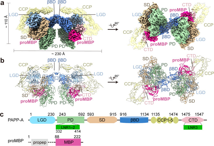
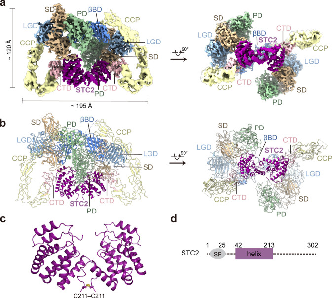
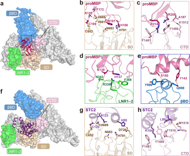
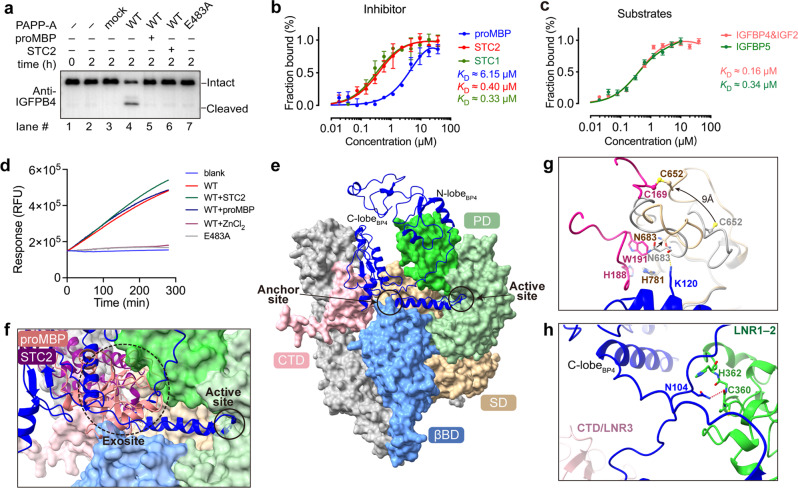
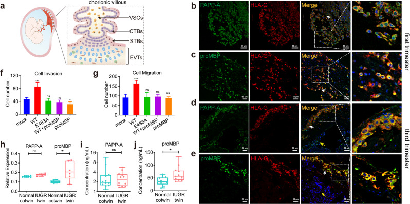
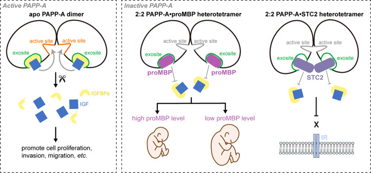
Originally discovered in the circulation of pregnant women as a protein secreted by placental trophoblasts, the metalloprotease pregnancy-associated plasma protein A (PAPP-A) is also widely expressed by many other tissues. It cleaves insulin-like growth factor-binding proteins (IGFBPs) to increase the bioavailability of IGFs and plays essential roles in multiple growth-promoting processes. While the vast majority of the circulatory PAPP-A in pregnancy is proteolytically inactive due to covalent inhibition by proform of eosinophil major basic protein (proMBP), the activity of PAPP-A can also be covalently inhibited by another less characterized modulator, stanniocalcin-2 (STC2). However, the structural basis of PAPP-A proteolysis and the mechanistic differences between these two modulators are poorly understood. Here we present two cryo-EM structures of endogenous purified PAPP-A in complex with either proMBP or STC2. Both modulators form 2:2 heterotetramer with PAPP-A and establish extensive interactions with multiple domains of PAPP-A that are distal to the catalytic cleft. This exosite-binding property results in a steric hindrance to prevent the binding and cleavage of IGFBPs, while the IGFBP linker region-derived peptides harboring the cleavage sites are no longer sensitive to the modulator treatment. Functional investigation into proMBP-mediated PAPP-A regulation in selective intrauterine growth restriction (sIUGR) pregnancy elucidates that PAPP-A and proMBP collaboratively regulate extravillous trophoblast invasion and the consequent fetal growth. Collectively, our work reveals a novel covalent exosite-competitive inhibition mechanism of PAPP-A and its regulatory effect on placental function.


Conflict of interest statement

The authors declare no competing interests.

Figures

Fig. 1
Fig. 1. Cryo-EM structure determination of the PAPP-A·proMBP complex.
a The overall EM density map of the PAPP-A·proMBP complex with a rotation of 90°. b The corresponding cartoon representation as in a. c Schematic domain organization for PAPP-A and proMBP proteins with domain boundaries defined in this study. LGD laminin-G like domain, PD proteolytic domain, LNR lin-12/Notch repeat, SD scarf domain, βBD β-barrel domain, CCP complement control protein, CTD C-terminal domain, propep pro-peptide, MBP matured form of major basic protein. The EM map and the structural figures were generated in either ChimeraX (www.cgl.ucsf.edu/chimeraX/) or PyMOL (www.pymol.org) with the same color scheme applied to all figures.
Fig. 2
Fig. 2. PAPP-A domain organization and the intramolecular interactions.
a Color-coded overall structure of PAPP-A. Detailed features are zoomed-in in be. b The LGD contains fifteen β-strands. The hydrogen bonds between the side chains of Arg71 and Arg98 of the LGD and the main-chain hydroxyl groups of CCP2 are colored red. Phe1257 inserts into a hydrophobic pocket of LGD. c The PD is superimposed to its homologs ulilysin (PDB code: 2J83) and mirolysin (PDB code: 6R7V). The Zn2+ ion at the active site and the Ca2+ ions are shown in blue and green spheres, respectively. d The SD composed of 17 β-strands is shown in rainbow color from N-terminus to C-terminus as running from blue to red, respectively. e The βBD contains two β-sheets composed of eight β-strands and three small helices.
Fig. 3
Fig. 3. The cryo-EM structure of the PAPP-A·STC2 complex.
a The overall color-coded EM density map of the PAPP-A·STC2 complex. b The corresponding cartoon representation. c The structure of the covalently linked STC2 dimer. d Schematic domain organization for STC2.
Fig. 4
Fig. 4. Structural properties of the binding interfaces between PAPP-A and proMBP or STC2.
a The representation of the four interfaces between PAPP-A and proMBP with PAPP-A shown in color-coded surface and proMBP in the cartoon. The boxes represent the close-up details shown in be. b The interface between proMBP and SD. Cys169 of proMBP and Cys652 of SD that form a disulfide bond are shown as ball-and-stick. The indole of Trp191 insets into a hydrophobic groove of the SD. Arg170 and Phe172 of proMBP form two hydrogen bonds with Val691 and His689 of SD, respectively (red dashed lines). c The interface between proMBP and CTD. Arg214–Phe1481 and Arg208–Tyr1486 form two cationic–π stacks (black dashed lines) and Ala187–Lys1512 form a hydrogen bond. d The interface between proMBP and LNR1–2. Side chains of His137 and Asn138 from proMBP and Arg338, Asn343, and Phe368 from LNR1–2 form several hydrogen bonds and hydrophobic stacks. e The interface between proMBP and βBD. Two perpendicular helices form several pairs of interactions that include Tyr143 of proMBP, Gln998 of βBD, Ser150 of proMBP, and Tyr996 of βBD. f Structural representation of the interactions between PAPP-A and STC2 (same view as in a). The close-up views are presented in g and h. g The interface between STC2 and SD (same view as in b). Cys120 of STC2 and Cys652 of PAPP-A form a disulfide bond. Two other hydrogen bonds are formed by Arg123 and His55 from STC2 and His689 and Asp726 from SD, respectively. h The interface between STC2 and CTD (same view as in c). The side chain of Lys104 from STC2 inserts into a groove formed by Phe1481, Tyr1486, and Phe1516, creating a strong interaction. The main chain of Met1518 of PAPP-A and Leu89 of STC2 form a hydrogen bond.
Fig. 5
Fig. 5. Proteolytic inhibition of PAPP-A activity requires the exosite binding of proMBP or STC2.
a Cleavage of the full-length IGFBP4 in the presence of IGF-2 was assessed using in vitro reaction. The bands of intact and cleaved IGFBP4 detected by western blot analysis are indicated. Reactions with empty vector (mock) and inactive PAPP-A (E483A) were used as negative controls. b Microscale thermophoresis (MST) analyses of proMBP, STC1, and STC2 bound to PAPP-A. The identical batch of PAPP-A protein was used. The signals were consonant with the bound fraction, generating a dissociation constant (KD) that was measured from three biologically independent repeats (n = 3). c MST analyses of substrates bound to PAPP-A. KD was measured from three biologically independent repeats (n = 3). d Fluorescence resonance energy transfer (FRET) analyses of PAPP-A cleavage on 4P1 under different inhibitory conditions. The absence of PAPP-A (blank), inactive PAPP-A (E483A), and the addition of ZnCl2 (WT + ZnCl2) were used as negative controls. e AlphaFold-predicted IGFBP4·PAPP-A complex model with IGFBP4 shown in cartoon (blue) and PAPP-A shown in surface (color-coded). f ProMBP or STC2 competes with the substrate for binding PAPP-A at the exosite. g Close-up view of proMBP and IGFBP4 binding on PAPP-A. SD is shown either in tan (in the PAPP-A·proMBP complex) or gray (in the predicted model). h In the predicted IGFBP4·PAPP-A structure, His362 of LNR1–2 inserts into a groove formed by a linker between the N-lobe and the anchor peptide, and Asn104 of IGFBP4 forms a hydrogen bond with the main chain of Cys360 of LNR1–2.
Fig. 6
Fig. 6. The modulation of PAPP-A by proMBP regulates the placental EVT function in sIUGR.
a Schematic illustration of a placental villous anchored to the maternal decidua during gestation. Atop the villous stroma cells (VSCs), the cytotrophoblasts (CTBs) undergo cell fusion to produce the outer multi-nuclear syncytiotrophoblasts (STBs), which then directly contact maternal blood. EVTs are located outside the villi and invade the maternal decidua. be Co-staining IF analysis of PAPP-A, proMBP, and HLA-G in first-trimester decidual tissue section (8th week) (b, c) and term placental tissue section (40th week) (d, e). HLA-G human leukocyte antigen G. White arrows, decidual face; yellow arrows, chorionic face. The inset dashed squares indicate a higher-magnification view. Scale bars, 50 μm. f, g Cellular invasion (f) and migration (g) assays of HTR8/SVneo cells under different conditions. Compared to the mock, *P < 0.05; ***P < 0.005; ns, non-significant. h The relative protein expressions of PAPP-A and proMBP in placentas from the IUGR-twin and Normal-cotwin in sIUGR. i, j Protein concentrations of PAPP-A (i) and proMBP (j) in umbilical cord blood from the IUGR-twin and Normal-cotwin in sIUGR.
Fig. 7
Fig. 7. A simplified model for the regulation of PAPP-A activity by proMBP or STC2.
Left, active PAPP-A dimer is able to bind the IGFBPIGF at the exosites and cleaves IGFBP at the active sites, releasing the bioactive IGF to promote cell proliferation, cell invasion, cell migration, etc. Right, PAPP-A is inactive when complexed with either proMBP or STC2 at the exosites. Higher proMBP concentration leads to compromised PAPP-A activity and restricts fetal growth. STC2 could also modulate PAPP-A activity and block the downstream IGF receptor (IR)-mediated signaling.

Similar articles

Cited by

References

    1. Junnila RK, et al. The GH/IGF-1 axis in ageing and longevity. Nat. Rev. Endocrinol. 2013;9:366–376. doi: 10.1038/nrendo.2013.67. - DOI - PMC - PubMed
    1. Pollak M. The insulin and insulin-like growth factor receptor family in neoplasia: an update. Nat. Rev. Cancer. 2012;12:159–169. doi: 10.1038/nrc3215. - DOI - PubMed
    1. Monget P, Oxvig C. PAPP-A and the IGF system. Ann. Endocrinol. (Paris) 2016;77:90–96. doi: 10.1016/j.ando.2016.04.015. - DOI - PubMed
    1. Overgaard MT, et al. Expression of recombinant human pregnancy-associated plasma protein-A and identification of the proform of eosinophil major basic protein as its physiological inhibitor. J. Biol. Chem. 2000;275:31128–31133. doi: 10.1074/jbc.M001384200. - DOI - PubMed
    1. Kloverpris S, et al. Stanniocalcin-1 potently inhibits the proteolytic activity of the metalloproteinase pregnancy-associated plasma protein-A. J. Biol. Chem. 2015;290:21915–21924. doi: 10.1074/jbc.M115.650143. - DOI - PMC - PubMed