Review
. 2023 Jul;209(4):641-662.
doi: 10.1007/s00359-022-01601-x. Epub 2022 Dec 23.

The sky compass network in the brain of the desert locust

Affiliations
Review

The sky compass network in the brain of the desert locust

Uwe Homberg et al. J Comp Physiol A Neuroethol Sens Neural Behav Physiol. 2023 Jul.

Abstract

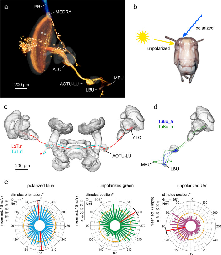
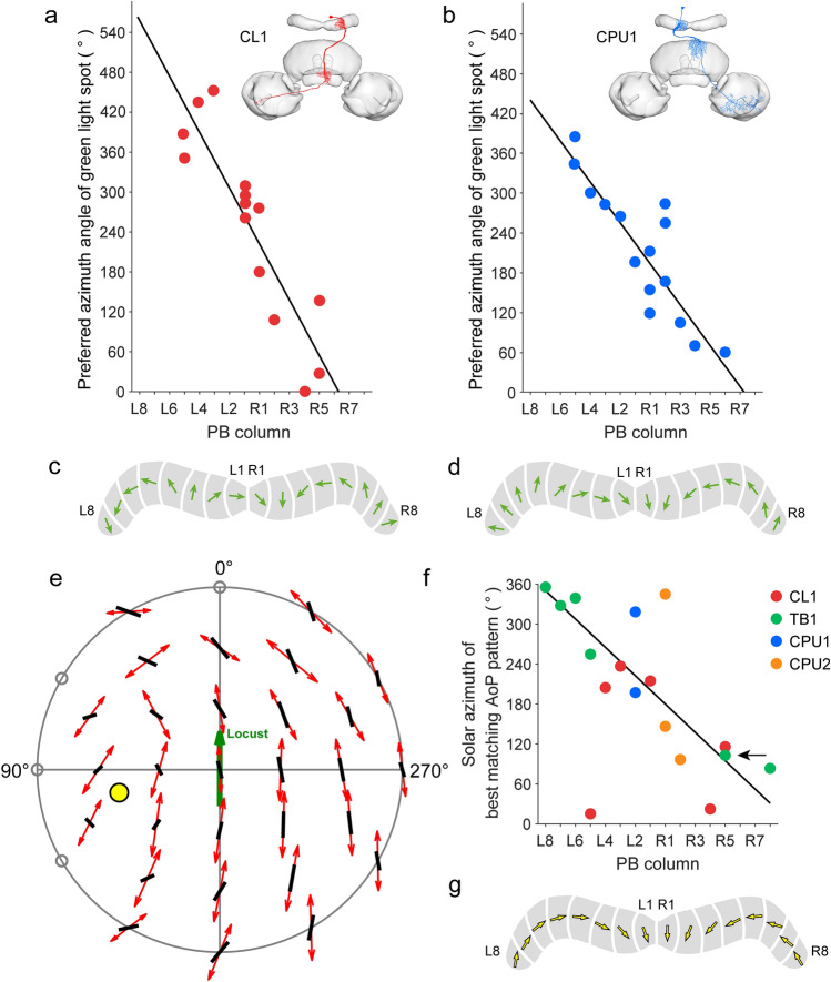
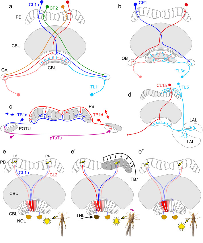
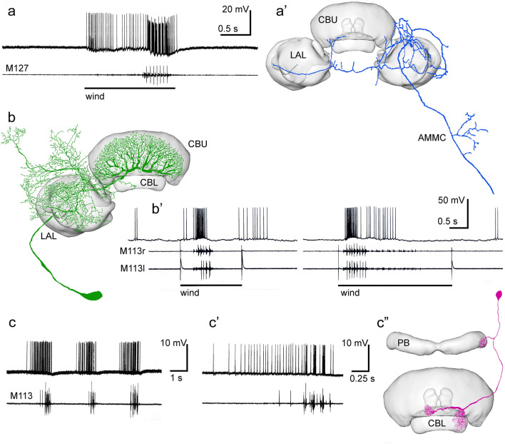
Many arthropods and vertebrates use celestial signals such as the position of the sun during the day or stars at night as compass cues for spatial orientation. The neural network underlying sky compass coding in the brain has been studied in great detail in the desert locust Schistocerca gregaria. These insects perform long-range migrations in Northern Africa and the Middle East following seasonal changes in rainfall. Highly specialized photoreceptors in a dorsal rim area of their compound eyes are sensitive to the polarization of the sky, generated by scattered sunlight. These signals are combined with direct information on the sun position in the optic lobe and anterior optic tubercle and converge from both eyes in a midline crossing brain structure, the central complex. Here, head direction coding is achieved by a compass-like arrangement of columns signaling solar azimuth through a 360° range of space by combining direct brightness cues from the sun with polarization cues matching the polarization pattern of the sky. Other directional cues derived from wind direction and internal self-rotation input are likely integrated. Signals are transmitted as coherent steering commands to descending neurons for directional control of locomotion and flight.

Keywords: Central complex; Desert locust; Intracellular recordings; Polarization vision; Sky compass coding.


Conflict of interest statement

The authors declare no competing interests.

Figures

Fig. 1
Fig. 1
a, b Compass signals of the daytime sky include the position of the sun, the chromatic gradient between short-wavelength light (UV) with uniform intensity across the sky (a) and long-wavelength light (green) with high intensity in the solar hemisphere and lower intensity in the anti-solar hemisphere of the sky (b), and the polarization pattern of the sky (a,b). Bar orientation indicates the angle of polarization (AoP). While direct sunlight is unpolarized, the degree of polarization, indicated by the thickness of the bars, increases with increasing angular distance from the sun. c Recession and invasion areas of the desert locust S. gregaria. a,b Modified from el Jundi et al. (2014), c modified from Cressman (2016) with permission from the publisher
Fig. 2
Fig. 2
Ommatidial subtypes in the compound eye of the desert locust. a Photoreceptor arrangement in type I and type II ommatidia of the main eye and in dorsal rim area (DRA) ommatidia. In main eye ommatidia, R2, R3, R5, R6, and R8 ommatidia contain blue and green opsin and R1 and R4, green opsin. R7 photoreceptors contain either UV opsin (type I ommatidia) or blue opsin (type II ommatidia). Both ommatidia types are stochastically distributed in the main retina with type I ommatidia constituting about 65% and type II ommatidia, 35% of all ommatidia. In DRA ommatidia, all photoreceptors contain blue opsin. Throughout the eye photoreceptor axons of R7 terminate in the medulla, all others in the lamina. b Spatial arrangement of ommatidia in the DRA. The T-shaped symbols indicate the orthogonal arrangement of microvilli within ommatidia as illustrated in the enlarged ommatidium. 1–8, photoreceptors R1-R8. c Visual field of the DRA along the transverse axis of the locust. Black bars indicate the relative sensitivity (rel. sens.) of pooled photoreceptors of the right DRA and gray bars, relative sensitivity of photoreceptors of the left DRA. d Dorsal view on the locust’s head illustrating different pigmentation of the main eye and DRA. Scale bar = 1 mm. a From Schmeling et al. (2014), b from Homberg (2004), c from Schmeling et al. (2015), d photograph by Erich Staudacher
Fig. 3
Fig. 3
Sky compass neurons of the optic lobe and the anterior optic tubercle. a Tracer injection into the lower unit of the anterior optic tubercle (AOTU-LU) reveals projections of TuBu neurons to the bulbs (LBU, lateral bulb, MBU, medial bulb) and transmedulla neurons in the optic lobe. Transmedulla neurons branch in the dorsal rim area of the medulla (MEDRA), where they overlap with photoreceptor terminals (PR) from the dorsal rim area (blue), additionally labeled by tracer injection into the dorsal rim area. The unbranched neurites of the transmedulla neurons run vertically through the medulla (ME) and, as a population, cover the entire anterior–posterior extent of the neuropil. b Transmedulla neurons receive polarization information through the dorsal rim area and unpolarized light information through the main part of the compound eye. c LoTu1 and TuTu1 neurons connect the AOTU-LU of both brain hemispheres. d Two types of TuBu neurons connect the AOTU-LU to the bulbs. e Circular plots of physiological responses of a TuTu1 neuron to dorsally presented polarized light (left), an unpolarized green light spot (simulated sun, middle) and an unpolarized ultraviolet light spot (right). Spiking activity is plotted in 10° bins, black T-bars indicate SD (if number of stimulus presentations (N) > 1), orange circles indicate background activity shown in impulses (imp) s−1. Φmax indicates the preferred angle of polarization or preferred azimuth for unpolarized light spot when the Rayleigh test indicated directedness. Φmax and its 95% confidence interval are indicated by a red line and a black arc, respectively. ALO, anterior lobe of the lobula complex. a After el Jundi et al. (2011), e after Pfeiffer and Homberg (2007)
Fig. 4
Fig. 4
Organization of the locust central complex. a 3D reconstruction of the locust brain with neuropils involved in sky polarization vision illustrated in color. b Schematic illustration of the central complex and prominent accessory neuropils. c Oblique view of a sagittal section through the central complex illustrating subunits and layers. d Schematic sagittal section illustrating layers of the upper division of the central body (CBU) and the subunits of the noduli. e Connectivity scheme of a system of 18 CL1a neurons connecting columns of the lower division of the central body (CBL) to columns of the protocerebral bridge (PB) and to the gall (GA). Axonal fibers from even-numbered columns of the PB (light blue, light red) take a slightly different course to the GA than axons from odd-numbered columns. f,g Major types of central-complex neurons involved in sky-compass coding. f Reconstruction of neurons embedded into semi-transparent neuropils; g proposed information flow through the CX compass network. Large red and blue arrows indicate input (IN) from TuBu- to TL neurons, brown arrow indicates output (OUT) from CPU neurons to other brain areas and descending pathways. AL, antennal lobe; ALI, anterior lip; AOTU-LU, lower unit of the anterior optic tubercle; CB, central body; CBL, lower division of the CB, consisting of layers 1–6; CBU, upper division of the CB with layers Ia, Ib, IIa, IIb, and III; CL1a, type 1a columnar neuron of the CBL; CPU1, type 1 columnar neuron of the PB and CBU; GA, gall; L0-L8, columns L0-L8 in the left hemisphere of the PB; LA, lamina, LADRA, dorsal rim area of the LA; LAL, lateral accessory lobe; LBU, lateral bulb; LOX, lobula complex; LX, lateral complex; MB, mushroom body; MBU, medial bulb; ME, medulla; MEDRA, dorsal rim area of the ME; NO, nodulus; NOL, lower unit of the nodulus, NOU, upper unit of the nodulus with layers I, II and III; OB, ovoid body; POTU, posterior optic tubercle; R1-R7, columns R1-R7 in the right hemisphere of the PB; TB1b, type 1b tangential neuron of the PB; TL2/3, type 2/3 tangential neuron of the CBL. Scale bar = 500 µm (a), 100 µm (c). c,d, Adapted from von Hadeln et al. (2020); e based on data from Heinze and Homberg (2008) and Hensgen et al. (2022)
Fig. 5
Fig. 5
Sky compass signaling at the input stage to the central complex. a TL2, TL3 and TL4 neurons provide layer-specific input to the lower division of the central body (CBL). b,c Stimulation of the locust from the zenithal direction with blue light passing through a rotating polarizer (b) and with an unpolarized green (or UV) light moved at an elevation of 45° around the head of the locust (c). d,e Spike trains from a TL2 neuron during 360° rotation of the polarizer (d) and 360° circular movement of the unpolarized green LED around the locust’s head (e). f-f’’ Circular plots from that neuron from 2 to 4 stimulations (N) with polarized blue light (f), an unpolarized green (simulated sun) (f’) and UV (f’’) light spot. Spiking activity is plotted in 10° bins, black T-bars indicate SD, orange circles indicate background activity shown in impulses (imp) s−1. Φmax, illustrated by a red line and a black arc (95% confidence interval) indicates preferred angle of polarization or preferred azimuth for unpolarized light spot when the Rayleigh test indicated directedness. Modified from Pegel et al. (2018). g-i’ Circular plots of neural activity of a TL3 neuron innervating the medial bulb to a rotating zenithal polarizer (g,h,i) and a green (simulated sun) light spot (g’,h’,i’) moved in a circular path around the head of the animal as illustrated in b and c. g-g’ Both eyes are uncovered, h–h’ the contralateral eye is covered with black paint, i-i’ paint on the contralateral eye removed and ipsilateral eye painted black. Preferred orientations (Φmax) of the neuron are indicated by red lines and confidence intervals (95%) by the black arcs. The neuron receives input to all stimuli only through the ipsilateral eye
Fig. 6
Fig. 6
Topographic representation of sky compass coding in the protocerebral bridge (PB). a,b Preferred azimuth angles for a green light spot (simulated sun) of CL1 neurons (a) and CPU1 neurons (b) plotted against the columns of innervation in the PB reveal linear clockwise shifts of preferred azimuths across the PB. Circular-linear regression shows a high correlation between the innervated column of the PB and the preferred azimuth for the green light spot (a: y =−42.5x + 562.9; b: y =−30.9x + 440.0). Insets show morphologies of the two cell types. c,d Schematic compass topographies in the PB for CL1 neurons (c) and CPU1 neurons (d) following the regression lines in a and b. e–g Matched-filter coding of polarization patterns in the sky. e Top view on the pattern of preferred angles of polarization (AoPs; red double arrows) of a TB1 neuron plotted against the best-matching sky polarization pattern (black bars). This pattern corresponds to a solar position (yellow dot) at 39° elevation and 103° azimuth. Open circles along the horizon indicate non-significant AoP responses. f Solar azimuth of best-matching pattern of preferred AoPs plotted against the columnar innervation domains in the PB for four types of central-complex neurons. Regression analysis shows a 320° topography of solar azimuths represented in the columns of the PB (y =−21.3x + 350.3). Arrow points at neuron recorded in e. g Schematic compass topography in the PB following the regression line in f. a-d Based on data from Pegel et al. (2019); e–g from Zittrell et al. (2020)
Fig. 7
Fig. 7
Recurrent circuits and lateral interactions in the compass network of the central complex. a,b Recurrent connections via the gall (GA) and ovoid body (OB). a The GA receives input from CL1 neurons, here illustrated by a CL1a cell, as well as CP2 columnar cells without ramifications in the central body. TL1 tangential neurons have dendrites in the GA and provide input to layers 1–5 of the CBL, thus providing a potential feedback from the PB and CBL back to the CBL. b A similar feedback may operate through the OBs. The OBs receive dense axonal input from CP1 columnar neurons of the PB and may contact TL3c tangential neurons innervating layers 6 and 2 of the CBL, shown in white. c Tangential neurons of the PB, type TB1 might interact with each other directly and via the posterior optic tubercles (POTU) to stabilize the compass topography across the PB. The image illustrates the input and output columns of a TB1a neuron in the left hemisphere and a TB1d neuron in the right hemisphere. Their tuning to polarization angle (double arrows, data from Pegel et al. 2018) is nearly orthogonal suggesting inhibitory interactions. Posterior tubercle neurons (pTuTu) connect the dorsal parts of the ipsilateral POTU to the ventral parts of the contralateral POTU. d TL5 tangential neurons of the CBL innervate one hemisphere of the PB and the lateral accessory lobe (LAL) and innervate all layers of the CBL, providing synchrony or feedback between these areas. e-e’’ CL2 columnar neurons of the CBL may be involved in shifting the activity in the PB during turns of the locust. e CL2 neurons receive input in the PB and lower unit of a nodulus and connect to single columns of the CBL shifted by one column relative to the projection matrix of CL1a neurons. In the CBL, CL1a neurons invade at least three columns, one central column and one column to the right and left (illustrated in shades of red). Repetitive circuits involving two CL1a and two CL2 neurons might serve to maintain tonic activity during straight-line locomotion. Yellow arrows in the respective PB columns, taken from Fig. 6g, indicate opposite tuning angles, suggesting that one of the connections between CL2 and CL1a, perhaps in the PB, is inhibitory. e’ During a right turn (magenta arrow) TNL and/or TB7 tangential neurons may be activated leading to increased activity in CL2 innervating column R4, followed by increased activity in CL1a innervating L5 in the PB, and reduced activity in CL1a innervating R4. As a consequence, columns further left in the CBL become activated shifting the activity profile in the PB to the left (e’’). Insets at the bottom of e-e’’ show the orientation of the locust relative to the sun (yellow dot). a,b Based on Hensgen et al. (2021a); c based on Beetz et al. (2015)
Fig. 8
Fig. 8
Changes in neural activity in CX neurons associated with flight. Intracellular recordings in tethered locusts combined with wire recordings from flight muscles. Legs and parts of the wings were clipped off as outlined in Homberg (1994). a,a’ Ascending interneuron projecting to the lateral accessory lobes (LAL). Frontal wind stimulation elicits spiking activity which turns into strong bursting synchronized with flight motor activity registered by electromyographic recoding from the first basalar muscle M127. The neuron has additional projections to the antennal mechanosensory and motor center (AMMC). b,b’ Tangential neuron of the upper division of the central body (CBU) with cell body near the vest (TUves1 neuron, also termed giant fan-shaped neuron). Wind-stimulus-elicited flight motor activity recorded from the right and left tergosternal muscle (M113r, M113l) is correlated with high spiking activity. c–c’’ Intracellular recording from a CL2 neuron. The animal showed spontaneous bouts of flight activity, which were preceded by high spiking activity. During the stationary flight, bursting activity of the neuron was correlated with flight motor activity. c’ Enlarged first burst of activity from c. CBL, lower division of the central body; PB, protocerebral bridge. a,a’ modified from Homberg (1994), b-c’’ modified from Müller (1997) with permission
Fig. 9
Fig. 9
Proposed connectivity of CPU1 and CPU2 neurons with descending pathways (DN) originating in the lateral accessory lobes (LALs). a CPU1 neurons innervating the left hemisphere of the protocerebral bridge (PB) are highly active when the sun (arrow) is on the right side of the animal. Asymmetric activity of CPU1 neurons leads to stronger activation of descending neurons from the left than from the right lateral accessory lobe resulting in a turn of the animal. b Symmetric activation of descending neurons by CPU2 neurons promotes forward locomotion along a particular menotactic course relative to the solar azimuth

References

    1. Baker PS, Gewecke M, Cooter RJ. Flight orientation of swarming Locusta migratoria. Physiol Entomol. 1984;9:247–252. doi: 10.1111/j.1365-3032.1984.tb00706.x. - DOI
    1. Bech M, Homberg U, Pfeiffer K. Receptive fields of locust brain neurons are matched to polarization patterns of the sky. Curr Biol. 2014;24:2124–2129. doi: 10.1016/j.cub.2014.07.045. - DOI - PubMed
    1. Beck M, Althaus V, Pegel U, Homberg U (2022) Neurons sensitive to non-celestial polarized light in the brain of the desert locust. J Comp Physiol A (in revision) - PMC - PubMed
    1. Beetz MJ, el Jundi B, Heinze S, Homberg U. Topographic organization and possible function of the posterior optic tubercles in the brain of the desert locust Schistocerca gregaria. J Comp Neurol. 2015;523:1589–1607. doi: 10.1002/cne.23736. - DOI - PubMed
    1. Beetz MJ, Pfeiffer K, Homberg U. Neurons in the brain of the desert locust Schistocerca gregaria sensitive to polarized light at low stimulus elevations. J Comp Physiol A. 2016;202:759–781. doi: 10.1007/s00359-016-1116-x. - DOI - PubMed

Publication types

LinkOut - more resources