. 2022 Dec 22;22(1):1345.
doi: 10.1186/s12885-022-10449-y.

Synthesis, characterization, and anticancer evaluation of 1,3-bistetrahydrofuran-2yl-5-FU as a potential agent for pancreatic cancer

Affiliations

Synthesis, characterization, and anticancer evaluation of 1,3-bistetrahydrofuran-2yl-5-FU as a potential agent for pancreatic cancer

Nkafu Bechem Ndemazie et al. BMC Cancer. .

Abstract

The failure of current chemotherapeutic agents for pancreatic cancer (PCa) makes it the most aggressive soft tissue tumor with a 5-year survival of slightly above 10% and is estimated to be the second leading cause of cancer death by 2030.

Objective: The main aim was to synthesize, characterize and evaluate the anticancer activity of 1,3-bistetrahydrofuran-2yl-5FU (MFU).

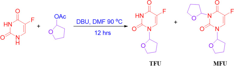
Methods: MFU was synthesized by using 5-fluorouracil (5-FU) and tetrahydrofuran acetate, and characterized by nuclear magnetic resonance (NMR), micro-elemental analysis, high-performance liquid chromatography (HPLC), and liquid chromatography with mass spectrophotometry (LC-MS). MFU and Gemcitabine hydrochloride (GemHCl) were tested for antiproliferative activity against MiaPaca-2 and Panc-1 cell lines.

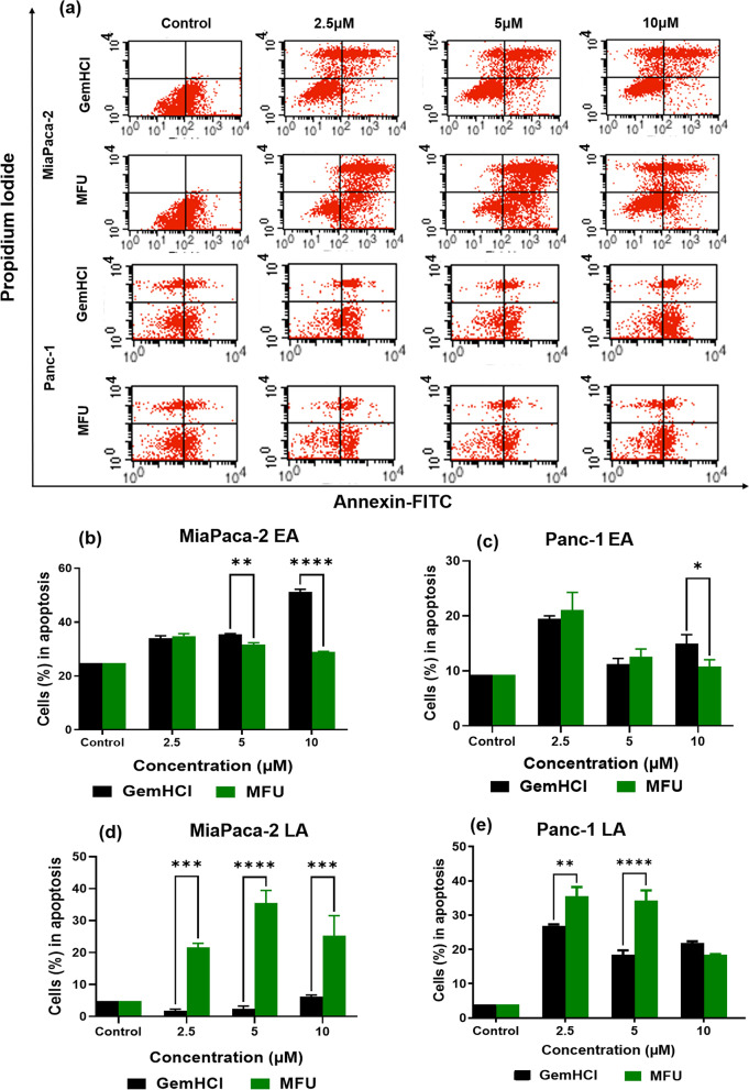
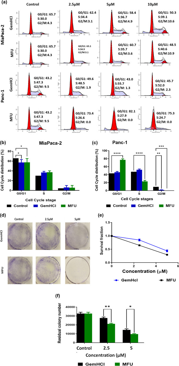
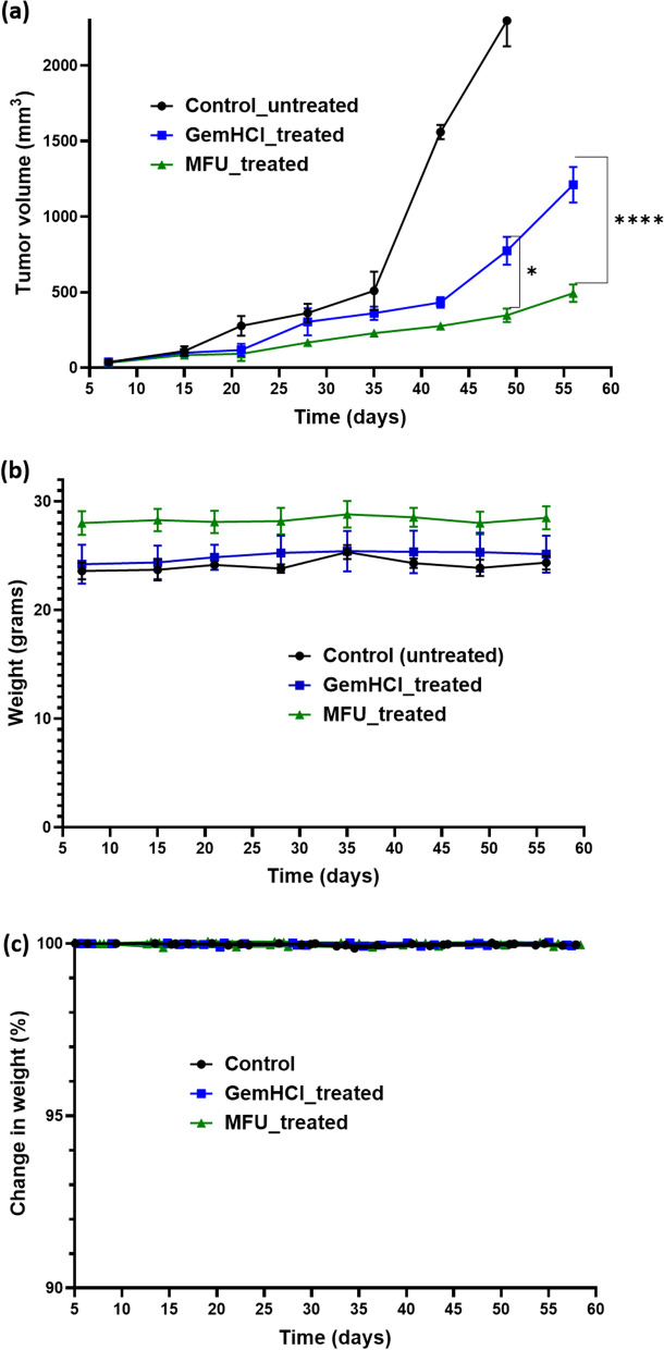
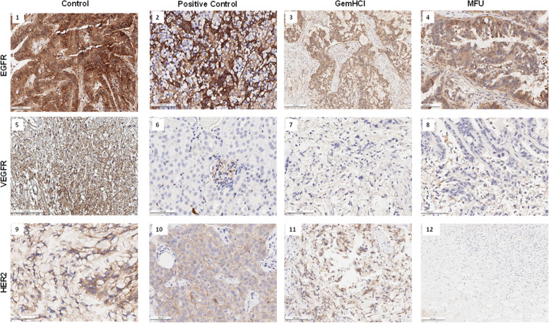
Results: The half-minimum inhibitory concentration (IC50) of MFU was twice lower than that of GemHCl when used in both cell lines. MiaPaca-2 cells (MFU-IC50 = 4.5 ± 1.2 μM vs. GemHCl-IC50 = 10.3 ± 1.1 μM); meanwhile similar trend was observed in Panc-1 cells (MFU-IC50 = 3.0 ± 1 μM vs. GemHCl-IC50 = 6.1 ± 1.03 μM). The MFU and GemHCl effects on 3D spheroids showed a similar trend (IC50-GemHCl = 14.3 ± 1.1 μM vs. IC50-MFU = 7.2 ± 1.1 μM) for MiaPaca-2 cells, and (IC50-GemHCl = 16.3 ± 1.1 μM vs. IC50-MFU = 9.2 ± 1.1 μM) for Panc-1 cells. MFU significantly inhibited clonogenic cell growth, and induced cell death via apoptosis. Cell cycle data showed mean PI for GemHCl (48.5-55.7) twice higher than MFU (24.7 to 27.9) for MiaPaca-2 cells, and similarly to Panc-1 cells. The in-vivo model showed intensely stained EGFR (stained brown) in all control, GemHCl and MFU-treated mice bearing subcutaneous PDX tumors, however, HER2 expression was less stained in MFU-treated tumors compared to GemHCl-treated tumors and controls. Mean tumor volume of MFU-treated mice (361 ± 33.5 mm3) was three-fold lower than GemHCl-treated mice (1074 ± 181.2 mm3) bearing pancreatic PDX tumors.

Conclusion: MFU was synthesized with high purity and may have potential anticancer activity against PCa.

Keywords: Apoptosis; Cytotoxicity; Immunohistochemistry; Modified fluorouracil; Pancreatic cancer.


Conflict of interest statement

The authors declare that they have no known competing financial interests or personal relationships that could have appeared to influence the work reported in this paper.

Figures

Fig. 1
Fig. 1
Synthesis of MFU. Synthesis and reaction conditions for the synthesis of MFU, which also yielded TFU (tegafur)
Fig. 2
Fig. 2
Cytotoxic activity of GemHCl and MFU on MiaPaCa-2 and Panc-1 cells after 48 h incubation and stability studies of MFU. (a) and (b) 2-D cultured cells showing GemHCl and MFU-treated MiaPaCa-2 and Panc-1 cells, respectively; (c) and (d) 3-D cultured cells) showing GemHCl and MFU-treated MiaPaCa-2 and Panc-1 cells, respectively. Fluorescent images, (e) and (f) showing 3-D organoids of MiaPaca-2 and Panc-1 cells treated with GemHCl and (g) and (h) showing 3-D organoids of MiaPaca-2 and Panc-1 cells treated with MFU. (i) Shows percent of MFU remaining after treatment of MiaPaca-2 and Panc-1 cells over 24 h; (j) showing percent of intact MFU remaining after exposure to human liver microsomes over 2 h
Fig. 3
Fig. 3
Flow cytometry images showing apoptosis studies at different stages following treatment with GemHCl and MFU using MiaPaca-2 and Panc-1 cells (a). (b) and (c) Miapaca-2 and Panc-1 cells showing early apoptosis, respectively; (d) and (e) MiaPaca-2 and Panc-1 cells at late apoptosis respectively
Fig. 4
Fig. 4
Cell cycle studies for MiaPaca-2 and Panc-1 cells following treatment with GemHCl and MFU and clonogenic studies using MiaPaca-2 cells. (a) Flow cytometry images of MaiPaca-2 and Panc-1 cells arrested at different phases of the cell cycle. (b) and (c) MiaPaca-2 and Panc-1 cells showing percentage of cells at different phases of the cell cycle, respectively. (d) photo images captured MiaPaca-2 cells after 2 weeks of growth following 48 h of incubation with GemHCl and MFU; (e) and (f) survival fraction and residual colony numbers respectively after 2 weeks growth following 48 h of incubation with GemHCl and MFU
Fig. 5
Fig. 5
Protein bands after treatment with MFU after treatment with GemHCl and MFU. For protein band expression, the blots were cut prior to hybridization with primary antibodies during the blotting. (a) Expression of apoptotic protein including Caspase-9, (b) quantitative protein bands expressed, (c) bands expressing cell cycle protein, (d) quantity of cell cycle proteins expressed
Fig. 6
Fig. 6
In-vivo efficacy of MFU in PDX mouse model and chronic toxicity. (a) Tumor growth curves of GemHCl and MFU treated mice bearing pancreatic PDX tumor and (b) body weight during treatment. (e) Percent change in the body weight of mice over the study period. Asterisks represents level of significance between control and treatment group (**p < 0.05, ***p < 0.001). All data represents mean ± SD, (n = 4/group)
Fig. 7
Fig. 7
Immunohistochemistry of tumors after treatment. Expression of EGFR, VEGFR and HER2 after treatment of PDX Pca mice bearing tumors with GemHCl and MFU with untreated control

References

    1. Bray F, et al. Global cancer statistics 2018: GLOBOCAN estimates of incidence and mortality worldwide for 36 cancers in 185 countries. CA Cancer J Clin. 2018;68:394–424. doi: 10.3322/caac.21492. - DOI - PubMed
    1. Ferlay J, et al. Global cancer observatory: cancer today. Lyon: International Agency for Research on Cancer; 2018.
    1. Binenbaum Y, Na’ara, S. & Gil, Z. Gemcitabine resistance in pancreatic ductal adenocarcinoma. Drug Resist Updat. 2015;23:55–68. doi: 10.1016/j.drup.2015.10.002. - DOI - PubMed
    1. Haeberle L, Esposito I. Pathology of pancreatic cancer. Transl Gastroenterol Hepatol. 2019;4. 10.21037/tgh.2019.06.02. - PMC - PubMed
    1. Mahajan UM, et al. Tumor-specific delivery of 5-fluorouracil-incorporated epidermal growth factor receptor-targeted Aptamers as an efficient treatment in pancreatic ductal adenocarcinoma models. Gastroenterology. 2021;161:996–1010.e1011. doi: 10.1053/j.gastro.2021.05.055. - DOI - PubMed