. 2022 Nov;28(6):609-615.
doi: 10.5152/dir.2022.211015.

Flow-diverter treatment for renal artery aneurysms: One-year follow-up of a multicentric preliminary experience

Affiliations

Flow-diverter treatment for renal artery aneurysms: One-year follow-up of a multicentric preliminary experience

Vittorio Semeraro et al. Diagn Interv Radiol. 2022 Nov.

Abstract

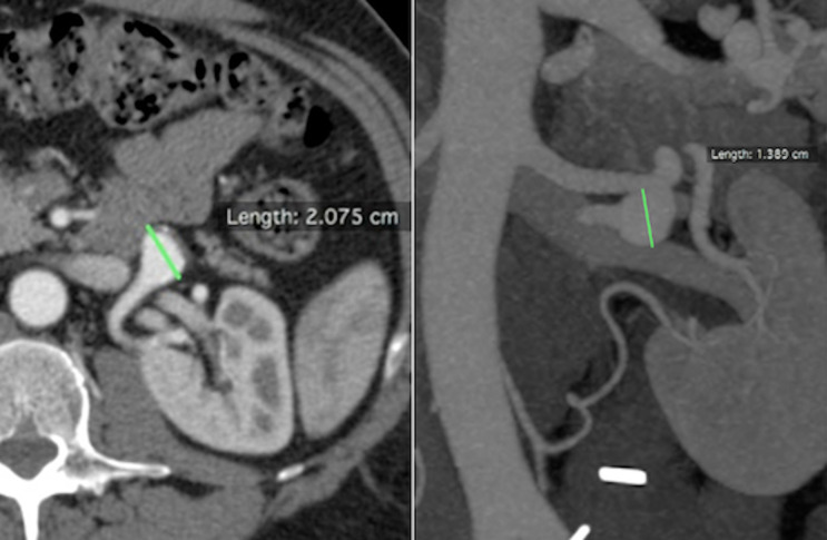
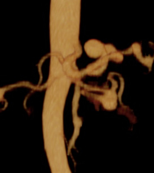
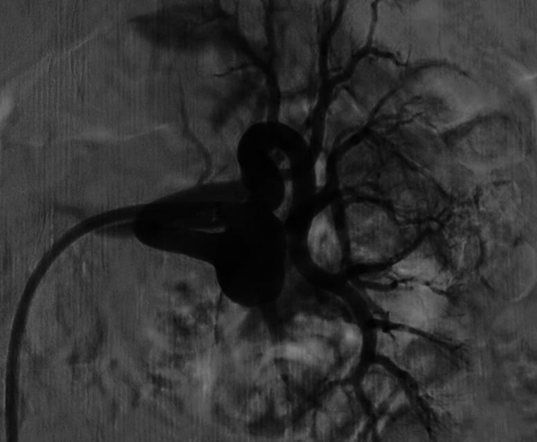
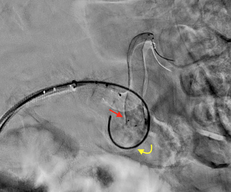
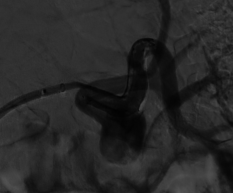
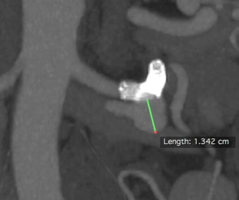
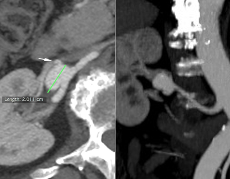
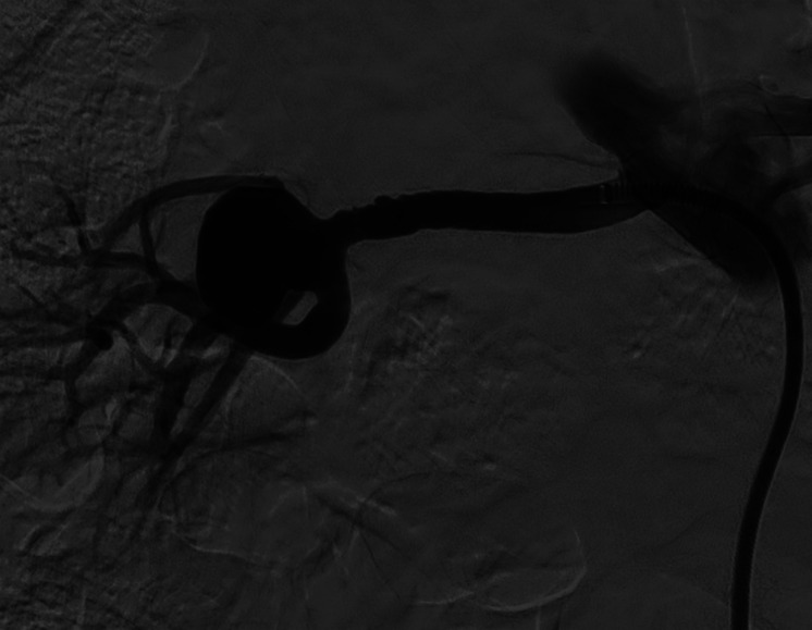
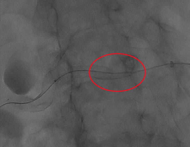
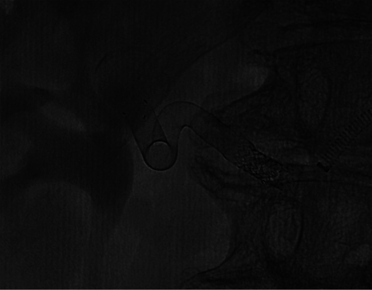
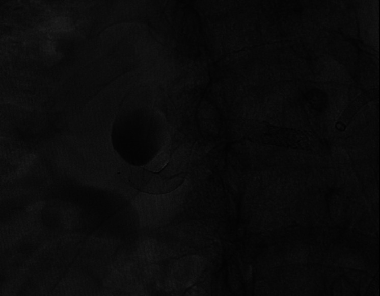
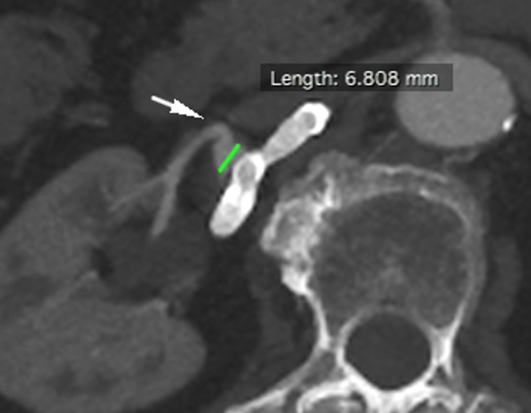
PURPOSE Renal artery aneurysms (RAAs) are rare in the general population, although the true incidence and natural history remain elusive. Conventional endovascular therapies such as coil embolization or covered stent graft may cause sidebranches occlusion, leading to organ infarction. Flow-diverters (FD) have been firstly designed to treat cerebrovascular aneurysms, but their use may be useful to treat complex RAAs presenting sidebraches arising from aneurysmal sac. To evaluate mid-term follow-up (FUP) safety and efficacy of FD during treatment of complex RAAs. METHODS Between November 2019 and April 2020, 7 RAAs were identified in 7 patients (4 men, 3 women; age range 55-82 years; median 67 years) and treated by FD. Procedural details, complications, morbidity and mortality, aneurysm occlusion and segmental artery patency were retrospectively reviewed. Twelve months computed tomography angiography (CTA) FUP was evaluated for all cases. RESULT Deployment of FD was successful in all cases. One intraprocedural technical complication was encountered with one FD felt down into aneurism sac which requiring additional telescopic stenting. One case at 3 months CTA FUP presented same complication, requiring same rescue technique. At 12 months CTA FUP 5 cases of size shrinkage and 2 cases of stable size were documented. No rescue surgery or major intraprocedural or mid-term FUP complication was seen. CONCLUSION Complex RAAs with two or more sidebranches can be safely treated by FD. FD efficacy for RAA needs a further validation at long term FUP by additional large prospective studies.


Figures

Figure 1.
Figure 1.
Patient number 2. Axial and coronal CTA. CTA, computed tomography angiography.
Figure 2.
Figure 2.
Patient number 2. 3D CTA. 3D, three-dimensional.
Figure 3.
Figure 3.
Patient number 2. Selective left renal artery DSA demonstrated a hilar bifurcation RAA. DSA, digital subtraction angiography; RAA, renal artery aneurysm.
Figure 4.
Figure 4.
Patient number 2. During treatment, proximal FD prolapsed into the aneurysm sac, just after deployment. The red straight arrow indicates the prolapsed stent, while the yellow curved arrow refers to the hydrophilic 0.035″ guidewire used for FD renavigation. FD, flow diverter.
Figure 5.
Figure 5.
Patient number 2. The malpositioning was rescued by an additional telescopic FD.
Figure 6.
Figure 6.
Patient number 2. One-year CTA FUP documented stent patency and aneurysm dimension stability. FUP, follow-up.
Figure 7.
Figure 7.
Patient number 3. Axial and coronal CTA. The white arrow shows a rising branch artery.
Figure 8.
Figure 8.
Patient number 3. Selective right renal artery DSA showed a hilar trifurcation RAA.
Figure 9.
Figure 9.
Patient number 3. During FD placement, a stent fishmouth was observed proximally (red circle).
Figure 10.
Figure 10.
Patient number 3. A stent fishmouth was subsequently treated by balloon expandable stent delivery (RX Herculink Renal Stent System).
Figure 11.
Figure 11.
Patient number 3. Final DSA showed aneurysmal contrast stasis
Figure 12.
Figure 12.
Patient number 3. One-year CTA FUP demonstrated parental artery patency, no renal ischemia, aneurysmal size reduction, with small residual close to a rising branch artery (white arrow).

References

    1. Tham G, Ekelund L, Herrlin K, Lindstedt EL, Olin T, Bergentz SE. Renal artery aneurysms. Natural history and prognosis. Ann Surg. 1983;197(3):348 352. 10.1097/00000658-198303000-00016) - DOI - PMC - PubMed
    1. Elaassar O, Auriol J, Marquez R, Tall P, Rousseau H, Joffre F. Endovascular techniques for the treatment of renal artery aneurysms. Cardiovasc Intervent Radiol. 2011;34(5):926 935. 10.1007/s00270-011-0127-9) - DOI - PubMed
    1. Henke PK, Cardneau JD, Welling TH.et al. Renal artery aneurysms: a 35-year clinical experience with 252 aneurysms in 168 patients. Ann Surg. 2001;234(4):454 62; discussion 462. 10.1097/00000658-200110000-00005) - DOI - PMC - PubMed
    1. Coleman DM, Stanley JC. Renal artery aneurysms. J Vasc Surg. 2015;62(3):779 785. 10.1016/j.jvs.2015.05.034) - DOI - PubMed
    1. Etezadi V, Gandhi RT, Benenati JF.et al. Endovascular treatment of visceral and renal artery aneurysms. J Vasc Interv Radiol. 2011;22(9):1246 1253. 10.1016/j.jvir.2011.05.012) - DOI - PubMed