

The Osteogenic Potential of Falciform Ligament-Derived Stromal Cells-A Comparative Analysis between Two Osteogenic Induction Programs
- PMID: 36551016
- PMCID: PMC9774535
- DOI: 10.3390/bioengineering9120810
The Osteogenic Potential of Falciform Ligament-Derived Stromal Cells-A Comparative Analysis between Two Osteogenic Induction Programs
Abstract
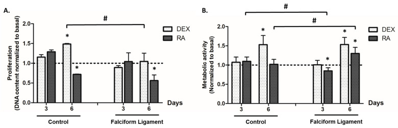
Mesenchymal stromal cells (MSCs) have gained special relevance in bone tissue regenerative applications. MSCs have been isolated from different depots, with adipose tissue being acknowledged as one of the most convenient sources, given the wide availability, high cellular yield, and obtainability. Recently, the falciform ligament (FL) has been regarded as a potential depot for adipose tissue-derived stromal cells (FL-ADSCs) isolation. Nonetheless, the osteogenic capability of FL-ADSCs has not been previously characterized. Thus, the present study aimed the detailed characterization of FL-ADSCs' functionality upon osteogenic induction through a classic (dexamethasone-based-DEX) or an innovative strategy with retinoic acid (RA) in a comparative approach with ADSCs from a control visceral region. Cultures were characterized for cell proliferation, metabolic activity, cellular morphology, fluorescent cytoskeletal and mitochondrial organization, and osteogenic activity-gene expression analysis and cytochemical staining. FL-derived populations expressed significantly higher levels of osteogenic genes and cytochemical markers, particularly with DEX induction, as compared to control ADSCs that were more responsive to RA. FL-ADSCs were identified as a potential source for bone regenerative applications, given the heightened osteogenic functionality. Furthermore, data highlighted the importance of the selection of the most adequate osteogenic-inducing program concerning the specificities of the basal cell population.
Keywords: adipose tissue; falciform ligament; mesenchymal stromal cells; osteogenesis; retinoic acid.
Conflict of interest statement
The authors declare no conflict of interest.
Figures


Similar articles
-
Influence of the Anatomical Site on Adipose Tissue-Derived Stromal Cells' Biological Profile and Osteogenic Potential in Companion Animals.Vet Sci. 2023 Nov 24;10(12):673. doi: 10.3390/vetsci10120673. Vet Sci. 2023. PMID: 38133224 Free PMC article. Review.
-
Retinoic acid induces the osteogenic differentiation of cat adipose tissue-derived stromal cells from distinct anatomical sites.J Anat. 2023 Feb;242(2):277-288. doi: 10.1111/joa.13758. Epub 2022 Sep 2. J Anat. 2023. PMID: 36056547 Free PMC article.
-
Are Adipose-Derived Stem Cells From Liver Falciform Ligaments Another Possible Source of Mesenchymal Stem Cells?Cell Transplant. 2017 May 9;26(5):855-866. doi: 10.3727/096368916X693833. Epub 2016 Nov 24. Cell Transplant. 2017. PMID: 27938473 Free PMC article.
-
Gluteal and abdominal subcutaneous adipose tissue depots as stroma cell source: gluteal cells display increased adipogenic and osteogenic differentiation potentials.Exp Dermatol. 2014 Jun;23(6):395-400. doi: 10.1111/exd.12406. Exp Dermatol. 2014. PMID: 24689514
-
Bone marrow-derived stem/stromal cells and adipose tissue-derived stem/stromal cells: Their comparative efficacies and synergistic effects.J Biomed Mater Res A. 2017 Sep;105(9):2640-2648. doi: 10.1002/jbm.a.36089. Epub 2017 May 17. J Biomed Mater Res A. 2017. PMID: 28419760 Review.
Cited by
-
Influence of the Anatomical Site on Adipose Tissue-Derived Stromal Cells' Biological Profile and Osteogenic Potential in Companion Animals.Vet Sci. 2023 Nov 24;10(12):673. doi: 10.3390/vetsci10120673. Vet Sci. 2023. PMID: 38133224 Free PMC article. Review.
References
-
- Requicha J.F., Viegas C.A., Albuquerque C.M., Azevedo J.M., Reis R.L., Gomes M.E. Effect of Anatomical Origin and Cell Passage Number on the Stemness and Osteogenic Differentiation Potential of Canine Adipose-Derived Stem Cells. Stem Cell Rev. Rep. 2012;8:1211–1222. doi: 10.1007/s12015-012-9397-0. - DOI - PubMed
-
- Voga M., Kovač V., Majdic G. Comparison of Canine and Feline Adipose-Derived Mesenchymal Stem Cells/Medicinal Signaling Cells with Regard to Cell Surface Marker Expression, Viability, Proliferation, and Differentiation Potential. Front. Vet. Sci. 2021;7:610240. doi: 10.3389/fvets.2020.610240. - DOI - PMC - PubMed
-
- Szydlarska J., Weiss C., Marycz K. The Effect of Methyl-β-cyclodextrin on Apoptosis, Proliferative Activity, and Oxidative Stress in Adipose-Derived Mesenchymal Stromal Cells of Horses Suffering from Metabolic Syndrome (EMS) Molecules. 2018;23:287. doi: 10.3390/molecules23020287. - DOI - PMC - PubMed
Grants and funding
LinkOut - more resources
Full Text Sources
