Review
. 2022 Nov 29;12(12):1094.
doi: 10.3390/bios12121094.

Paper-Based Biosensors for the Detection of Nucleic Acids from Pathogens

Affiliations
Review

Paper-Based Biosensors for the Detection of Nucleic Acids from Pathogens

Jiangshan Wang et al. Biosensors (Basel). .

Abstract

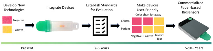
Paper-based biosensors are microfluidic analytical devices used for the detection of biochemical substances. The unique properties of paper-based biosensors, including low cost, portability, disposability, and ease of use, make them an excellent tool for point-of-care testing. Among all analyte detection methods, nucleic acid-based pathogen detection offers versatility due to the ease of nucleic acid synthesis. In a point-of-care testing context, the combination of nucleic acid detection and a paper-based platform allows for accurate detection. This review offers an overview of contemporary paper-based biosensors for detecting nucleic acids from pathogens. The methods and limitations of implementing an integrated portable paper-based platform are discussed. The review concludes with potential directions for future research in the development of paper-based biosensors.

Keywords: paper-based biosensors; pathogens; point-of-care testing.


Conflict of interest statement

M.S.V. has interests in Krishi, Inc., which is a startup company developing paper-based molecular assays. Krishi, Inc. did not fund this work.

Figures

Figure 2
Figure 2
Structure-recognition biosensors. (A) Schematic of the 3D Cu-based metal–organic frameworks (MOF) interacting with two different fluorophore-labeled DNA probes, which can be used as effective sensing platforms for simultaneous detection of Dengue and Zika virus RNA sequences. The MOF can interact with two fluorophore-labeled DNA probes and quench the fluorescence. In the presence of Dengue virus and Zika virus RNA sequences, the DNA probes form a double-stranded DNA–RNA structure that results in fluorescence recovery. Adapted from [31] with copyright permissions from the publisher. Copyright © 2022, Elsevier. (B) Schematic design for the C60-rRNA detector–reporter complex, which enables bacterium detection at a specific excitation frequency. The C60-rRNA detector–reporter complex can enter the microbial cell, and when the rRNA detector hybridizes with the bacterial rRNA, the fluorophore-labeled complementary DNA is released and emits a fluorescence signal. Adapted from [32]. Copyright © The Author(s) 2017. (C) Schematic of hybrid capture fluorescence immunoassay (HC-FIA). The amplification-free nucleic acid immunoassay employs DNA probes that are designed to bind to the conserved regions of the SARS-CoV-2 genome and a fluorescent-nanoparticle-labeled monoclonal antibody that binds to double-stranded DNA–RNA hybrids. Adapted from [37] with copyright permissions from the publisher. Copyright © 2022, Springer Nature. (D) Schematic of non-enzymatic isothermal strand displacement and amplification (NISDA) assay for rapid detection of SARS-CoV-2 RNA. The viral RNA/DNA displaces the short DNA template of the initiator. The released DNA template initiates the cascade unfolding of two DNA molecular beacon structures (M1 and M2), resulting in a substantial increase in the fluorescence intensity of M1. Adapted from [46]. Copyright © The Author(s) 2021.
Figure 1
Figure 1
Schematic of the three essential components of paper-based biosensors for detecting nucleic acids from pathogens.
Figure 3
Figure 3
The basic schematic principles of isothermal amplification methods. (A) The principle of rolling circle amplification (RCA). (B) The principle of recombinase polymerase amplification (RPA). (C) The principle of loop-mediated isothermal amplification (LAMP). Created with BioRender.com.
Figure 4
Figure 4
The basic schematic principle of CRISPR/Cas-based detection. DNA or RNA are amplified by isothermal amplification. Binding of the crRNA to the complementary target sequence activates the Cas enzyme and triggers collateral cleavage of quenched fluorescent reporters. Thereby, Cas13a (used in SHERLOCK) or Cas12a (used in DETECTR) indicate the presence of RNA or DNA target sequences, respectively. Created with BioRender.com.
Figure 5
Figure 5
Summary of recent paper-based biosensors. (A) Paper-based microfluidic device that enables multiplex loop-mediated isothermal amplification (LAMP)-based detection of malaria in blood. Following the LAMP reactions in the buffer chamber, the test samples were then transferred to the lateral flow strip by pressing manually on the buffer chambers. This device can run up to three LAMP reactions at a time with a positive control to account for environmental factors and handling. Adapted from [193]. Copyright © The Author(s) 2019. (B) All-in-one nucleic acid testing paper chip for simultaneous detection of Zika, Dengue, and Chikungunya. The device consolidated the complete nucleic acid testing process, including sampling, extraction, amplification, and detection, onto a single paper chip. Adapted from [185] with copyright permissions from the publisher. Copyright © 2022, Elsevier. (C) Detection of HIV diluted in whole blood on microfluidic rapid and autonomous analytical device (microrad) with pre-dried reagents. Whole blood is deposited into the microRAAD’s sample inlet. The membrane separates HIV from blood cells, and it then migrates by capillary force to the amplification zone. Followed by amplification, the solution is released to lateral flow immunoassay (LFIA) for detection. Adapted from [197]. Copyright © The Author(s) 2019. (D) Equipment and consumables needed for running DNA endonuclease-targeted CRISPR trans reporter (DETECTR). The assay performs reverse-transcription LAMP (RT-LAMP) for extracted SARS-CoV-2 RNA, followed by Cas12 detection. Afterwards, a lateral flow strip is added to the reaction tube and the result is visualized after approximately 2 min. Adapted from [104]. Copyright © 2022, Springer Nature. (E) Schematic layout of the paper device for detecting SARS-CoV-2 in saliva. The paper-based device utilizes RT-LAMP to detect SARS-CoV-2 in whole saliva without RNA extraction. Adapted from [83]. Copyright © Elsevier 2022. (F) Paper-based all-in-one origami microdevice for nucleic acid amplification testing for rapid colorimetric identification of live cells for point-of-care testing. This origami paper device was successfully applied to determine the viability of foodborne pathogens by implementing a propidium monoazide treatment. Adapted from [170]. Copyright © 2022 American Chemical Society.
Figure 6
Figure 6
An overview of the process, challenges, and future prospects of developing commercial-grade paper-based biosensors. The review paper concluded that sufficient technologies were developed for paper-based biosensors, however, this is merely the first step in the development from the lab to the field. For the future success of paper-based biosensors and to facilitate point-of-care applications, certain aspects, such as device integration, establishing a general standard for device evaluation, and improving the user-friendliness of the device, should be addressed in the near future. To illustrate the step-by-step accomplishment, we have used data from our lab as examples of integrated devices and potential commercial prototypes. The integrated device is a cartridge that contains paper-based strips in the middle. The device under ‘commercialized paper-based biosensors’ is a heating unit that takes the cartridge and provides an output signal. These devices were built in collaboration between Purdue, Raytheon BBN, Cortex Design Inc., and Portascience Inc. (now DCNDx).

Similar articles

Cited by

References

    1. Abayasekara L.M., Perera J., Chandrasekharan V., Gnanam V.S., Udunuwara N.A., Liyanage D.S., Bulathsinhala N.E., Adikary S., Aluthmuhandiram J.V.S., Thanaseelan C.S., et al. Detection of Bacterial Pathogens from Clinical Specimens Using Conventional Microbial Culture and 16S Metagenomics: A Comparative Study. BMC Infect. Dis. 2017;17:631. doi: 10.1186/s12879-017-2727-8. - DOI - PMC - PubMed
    1. Kok J., Blyth C.C., Foo H., Patterson J., Taylor J., McPhie K., Ratnamohan V.M., Iredell J.R., Dwyer D.E. Comparison of a Rapid Antigen Test with Nucleic Acid Testing during Cocirculation of Pandemic Influenza A/H1N1 2009 and Seasonal Influenza A/H3N2. J. Clin. Microbiol. 2010;48:290–291. doi: 10.1128/JCM.01465-09. - DOI - PMC - PubMed
    1. Craw P., Balachandran W. Isothermal Nucleic Acid Amplification Technologies for Point-of-Care Diagnostics: A Critical Review. Lab Chip. 2012;12:2469–2486. doi: 10.1039/c2lc40100b. - DOI - PubMed
    1. Morozumi M., Chiba N., Igarashi Y., Mitsuhashi N., Wajima T., Iwata S., Ubukata K. Direct Identification of Streptococcus Agalactiae and Capsular Type by Real-Time PCR in Vaginal Swabs from Pregnant Women. J. Infect. Chemother. 2015;21:34–38. doi: 10.1016/j.jiac.2014.08.024. - DOI - PubMed
    1. Park J.S., Cho D.-H., Yang J.H., Kim M.Y., Shin S.M., Kim E.-C., Park S.S., Seong M.-W. Usefulness of a Rapid Real-Time PCR Assay in Prenatal Screening for Group B Streptococcus Colonization. Ann. Lab. Med. 2012;33:39–44. doi: 10.3343/alm.2013.33.1.39. - DOI - PMC - PubMed

LinkOut - more resources