

Development and Application of an SPR Nanobiosensor Based on AuNPs for the Detection of SARS-CoV-2 on Food Surfaces
- PMID: 36551068
- PMCID: PMC9776341
- DOI: 10.3390/bios12121101
Development and Application of an SPR Nanobiosensor Based on AuNPs for the Detection of SARS-CoV-2 on Food Surfaces
Abstract
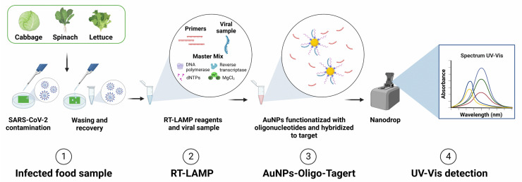
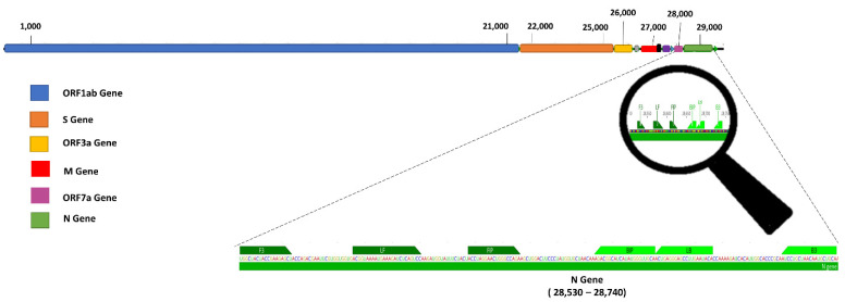
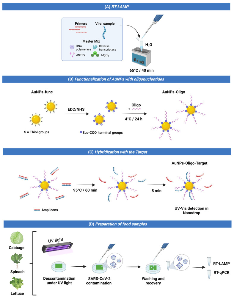
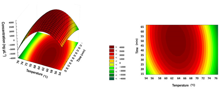
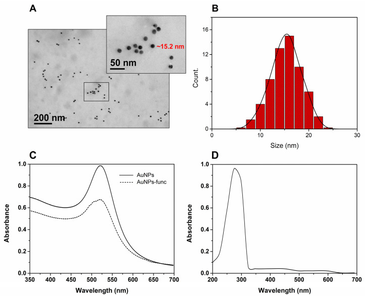
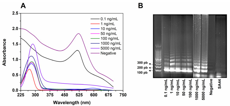
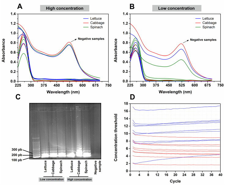
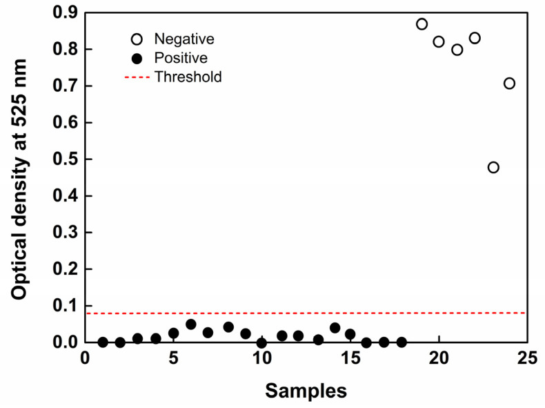
A new transmission route of SARS-CoV-2 through food was recently considered by the World Health Organization (WHO), and, given the pandemic scenario, the search for fast, sensitive, and low-cost methods is necessary. Biosensors have become a viable alternative for large-scale testing because they overcome the limitations of standard techniques. Herein, we investigated the ability of gold spherical nanoparticles (AuNPs) functionalized with oligonucleotides to detect SARS-CoV-2 and demonstrated their potential to be used as plasmonic nanobiosensors. The loop-mediated isothermal amplification (LAMP) technique was used to amplify the viral genetic material from the raw virus-containing solution without any preparation. The detection of virus presence or absence was performed by ultraviolet-visible (UV-Vis) absorption spectroscopy, by monitoring the absorption band of the surface plasmonic resonance (SPR) of the AuNPs. The displacement of the peak by 525 nm from the functionalized AuNPs indicated the absence of the virus (particular region of gold). On the other hand, the region ~300 nm indicated the presence of the virus when RNA bound to the functionalized AuNPs. The nanobiosensor system was designed to detect a region of the N gene in a dynamic concentration range from 0.1 to 50 × 103 ng·mL-1 with a limit of detection (LOD) of 1 ng·mL-1 (2.7 × 10³ copy per µL), indicating excellent sensitivity. The nanobiosensor was applied to detect the SARS-CoV-2 virus on the surfaces of vegetables and showed 100% accuracy compared to the standard quantitative reverse transcription polymerase chain reaction (RT-qPCR) technique. Therefore, the nanobiosensor is sensitive, selective, and simple, providing a viable alternative for the rapid detection of SARS-CoV-2 in ready-to-eat vegetables.
Keywords: COVID-19; LAMP; SPR; UV–Vis; optical biosensor.
Conflict of interest statement
The authors declare no conflict of interest.
Figures
Similar articles
-
Ultra-sensitive colorimetric detection of SARS-CoV-2 by novel gold nanoparticle (AuNP)-assisted loop-mediated isothermal amplification (LAMP) and freezing methods.Mikrochim Acta. 2024 May 24;191(6):339. doi: 10.1007/s00604-024-06422-0. Mikrochim Acta. 2024. PMID: 38789855
-
Rapid and Visual Detection of SARS-CoV-2 Using Multiplex Reverse Transcription Loop-Mediated Isothermal Amplification Linked With Gold Nanoparticle-Based Lateral Flow Biosensor.Front Cell Infect Microbiol. 2021 Jul 14;11:581239. doi: 10.3389/fcimb.2021.581239. eCollection 2021. Front Cell Infect Microbiol. 2021. PMID: 34336708 Free PMC article.
-
Development and Clinical Application of a Rapid and Sensitive Loop-Mediated Isothermal Amplification Test for SARS-CoV-2 Infection.mSphere. 2020 Aug 26;5(4):e00808-20. doi: 10.1128/mSphere.00808-20. mSphere. 2020. PMID: 32848011 Free PMC article.
-
Recent advances in methods for the diagnosis of Corona Virus Disease 2019.J Clin Lab Anal. 2022 Jan;36(1):e24178. doi: 10.1002/jcla.24178. Epub 2021 Dec 17. J Clin Lab Anal. 2022. PMID: 34921443 Free PMC article. Review.
-
Biosensors as Nano-Analytical Tools for COVID-19 Detection.Sensors (Basel). 2021 Nov 24;21(23):7823. doi: 10.3390/s21237823. Sensors (Basel). 2021. PMID: 34883826 Free PMC article. Review.
Cited by
-
Ultra-sensitive colorimetric detection of SARS-CoV-2 by novel gold nanoparticle (AuNP)-assisted loop-mediated isothermal amplification (LAMP) and freezing methods.Mikrochim Acta. 2024 May 24;191(6):339. doi: 10.1007/s00604-024-06422-0. Mikrochim Acta. 2024. PMID: 38789855
-
COVID severity test (CoST sensor)-An electrochemical immunosensing approach to stratify disease severity.Bioeng Transl Med. 2023 Jul 11;8(5):e10566. doi: 10.1002/btm2.10566. eCollection 2023 Sep. Bioeng Transl Med. 2023. PMID: 37693054 Free PMC article.
-
Application of Nanoparticles in Human Nutrition: A Review.Nutrients. 2024 Feb 25;16(5):636. doi: 10.3390/nu16050636. Nutrients. 2024. PMID: 38474764 Free PMC article. Review.
-
Emerging Applications of Nanobiosensors in Pathogen Detection in Water and Food.Biosensors (Basel). 2023 Oct 11;13(10):922. doi: 10.3390/bios13100922. Biosensors (Basel). 2023. PMID: 37887115 Free PMC article. Review.
-
Biosensors for Monitoring of Biologically Relevant Molecules.Biosensors (Basel). 2023 Jul 17;13(7):738. doi: 10.3390/bios13070738. Biosensors (Basel). 2023. PMID: 37504136 Free PMC article.
References
-
- WHO WHO Coronavirus (COVID-19) Dashboard 2022. [(accessed on 30 September 2022)]. Available online: https://covid19.who.int/
-
- Erkoc P., Ulucan-Karnak F. Nanotechnology-Based Antimicrobial and Antiviral Surface Coating Strategies. Prosthesis. 2021;3:25–52. doi: 10.3390/prosthesis3010005. - DOI
MeSH terms
Substances
Grants and funding
- E-26/200.691/2021/Fundação Carlos Chagas Filho de Amparo à Pesquisa do Estado do Rio de Janeiro
- E-26/202.000/2021/Fundação Carlos Chagas Filho de Amparo à Pesquisa do Estado do Rio de Janeiro
- E-26/201.813/2022/Fundação Carlos Chagas Filho de Amparo à Pesquisa do Estado do Rio de Janeiro
- ; E-26/204.254/2021/Fundação Carlos Chagas Filho de Amparo à Pesquisa do Estado do Rio de Janeiro
- 313119/2020-1/National Council for Scientific and Technological Development
- 152275/2022-3/National Council for Scientific and Technological Development
- [88887.518753/2020-00/Coordenação de Aperfeicoamento de Pessoal de Nível Superior
- FinanceCode001/Coordenação de Aperfeicoamento de Pessoal de Nível Superior
- 164569/2020-0/National Council for Scientific and Technological Development
- 151200/2020-0/National Council for Scientific and Technological Development
- 423952/2018-8/National Council for Scientific and Technological Development
LinkOut - more resources
Full Text Sources
Medical
Miscellaneous
