Review
. 2022 Nov 26;12(12):1761.
doi: 10.3390/biom12121761.

Regulation Mechanisms of Meiotic Recombination Revealed from the Analysis of a Fission Yeast Recombination Hotspot ade6-M26

Affiliations
Review

Regulation Mechanisms of Meiotic Recombination Revealed from the Analysis of a Fission Yeast Recombination Hotspot ade6-M26

Kouji Hirota. Biomolecules. .

Abstract

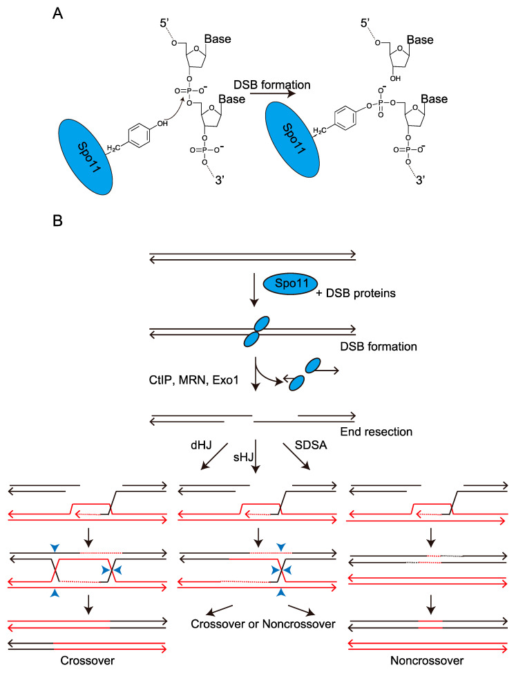
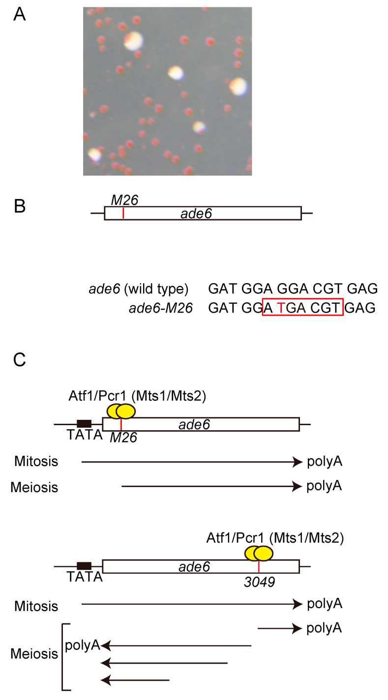
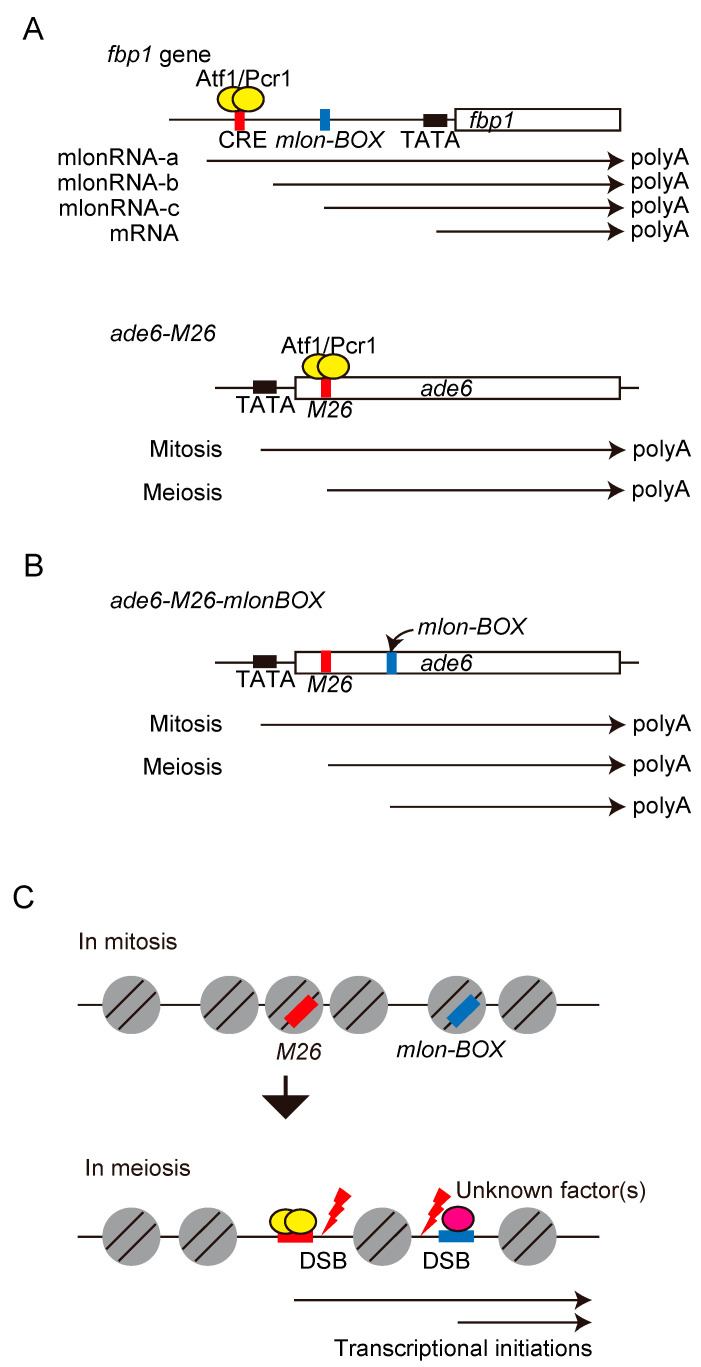
Meiotic recombination is a pivotal event that ensures faithful chromosome segregation and creates genetic diversity in gametes. Meiotic recombination is initiated by programmed double-strand breaks (DSBs), which are catalyzed by the conserved Spo11 protein. Spo11 is an enzyme with structural similarity to topoisomerase II and induces DSBs through the nucleophilic attack of the phosphodiester bond by the hydroxy group of its tyrosine (Tyr) catalytic residue. DSBs caused by Spo11 are repaired by homologous recombination using homologous chromosomes as donors, resulting in crossovers/chiasmata, which ensure physical contact between homologous chromosomes. Thus, the site of meiotic recombination is determined by the site of the induced DSB on the chromosome. Meiotic recombination is not uniformly induced, and sites showing high recombination rates are referred to as recombination hotspots. In fission yeast, ade6-M26, a nonsense point mutation of ade6 is a well-characterized meiotic recombination hotspot caused by the heptanucleotide sequence 5'-ATGACGT-3' at the M26 mutation point. In this review, we summarize the meiotic recombination mechanisms revealed by the analysis of the fission ade6-M26 gene as a model system.

Keywords: Rec12 (fission yeast Spo11 homolog); S. pombe; chromatin; double strand break (DSB); meiotic recombination; transcription factor.


Conflict of interest statement

The authors declare no conflict of interest.

Figures

Figure 1
Figure 1
Meiotic double-strand break (DSB) formation and homologous recombination induced by the programed DSBs. (A) Mechanism of the DSB formation catalyzed by Spo11 protein. The tyrosine (Tyr) side chain on the Spo11 protein carries out nucleophilic attack on the DNA phosphodiester backbone. This transesterase reaction cuts the DNA backbone and covalently links the Spo11 protein to the DNA end via a tyrosyl phosphodiester linkage. Spo11 protein is represented by a blue circle. (B) Schematic representation of meiotic recombination. Spo11 protein is represented by a blue circle. Homologous donor DNA is indicated by the red line. Only one of the two sister chromatids from each homolog has been shown.
Figure 2
Figure 2
ade6-M26 meiotic recombination hotspot created by single nucleotide substitution. (A) Image of pink colonies on a medium containing limited adenine concentration. (B) Schematic representation of ade6-M26 allele. The white box shows ade6 gene. The red vertical line shows the M26 mutation point. The sequences around M26 mutation are indicated. The red letter indicates the single nucleotide substitution in the ade6-M26 allele. The red square shows cyclic adenosine monophosphate (cAMP)-responsive element (CRE)-like heptanucleotide sequence. (C) Schematic representation of ade6-M26 and ade6-3049 alleles. White boxes show ade6 gene. Red vertical lines show each mutation. Yellow circles show Atf1 and Pcr1 (Mst1 and Mts2) transcription factors. Arrows indicate transcripts. In mitosis, ade6-M26 and ade6-3049 strains express ade6 gene from TATA box, while the transcriptional initiation site was shifted to Atf1/Pcr1 binding sites in meiosis.
Figure 3
Figure 3
A model of meiotic recombination at ade6-M26 by histone acetylation-mediated chromatin remodeling. (A) During meiosis, the state of the chromatin at the M26 mutation site changes to open configuration. This chromatin alteration is dependent on Atf1/Pcr1 and CRE-like heptanucleotide sequence, 5′-ATGACGT-3′. The binding of Atf1 to M26 site is enhanced in meiosis. (B) Histones around M26 are acetylated by Gcn5 histone acetyl transferase and probably other histone acetyl transferases, which are recruited to the M26 site via Atf1/Pcr1. Acetylated histones are recognized by ATP-dependent chromatin remodelers including Snf22. DSB machinery is recruited to the open chromatin region created around M26, thereby activating meiotic recombination.
Figure 4
Figure 4
Schematic representation showing the position of DSBs within ade6 gene. The red box indicates the position of the CRE-like heptanucleotide sequence generated due to M26 mutation. DSBs were detected on both sides near the M26 mutation site surrounded by closely spaced M26 sites, whereas the M26 site itself lies in a region free of visible breaks.
Figure 5
Figure 5
The conserved role of promoter-lncRNAs, mlonRNAs identified in the upstream region of fbp1 during the regulation of chromatin. (A) Schematic representation of mlonRNA transcriptions from the fbp1 and ade6 loci. In the fbp1 locus, during glucose rich condition, the longest mlonRNA (mlonRNA-a) is weakly expressed. Initially, during glucose starvation (10–20 min of glucose starvation), mlonRNA-b and -c are progressively expressed. At 60–180 min of glucose starvation, fbp1-mRNA is massively induced. mlon-BOX (mlonRNA-c initiation element), is located approximately 100 bp upstream from mlonRNA-c transcription start site. Notably, mlon-BOX is around 200 bp downstream from the Atf1 binding site (CRE). Red, blue, and black boxes show CRE, mlon-BOX, and TATA-BOX, respectively. In the ade6 locus, during mitotic cell phase, mRNA is expressed from TATA-BOX. During meiosis, the transcription initiation site is shifted to the CRE site. (B) Schematic representation of ade6 gene locus. Position of TATA-box, M26, and the insertion point of mlon-BOX are indicated by black, red and blue boxes, respectively. The ade6 mRNA and transcripts induced from the M26 site or mlon-BOX are indicated by arrows. (C) Chromatin configuration around M26 site and mlon-BOX insertion point. Transcriptional activations occur form M26 site and mlon-BOX insertion point in meiosis. Then, chromatin configuration changes to open state and DSBs are induced at the M26 site and mlon-BOX insertion point. The DSB sites are shown.

Similar articles

Cited by

References

    1. Láscarez-Lagunas L., Martinez-Garcia M., Colaiácovo M. SnapShot: Meiosis—Prophase I. Cell. 2020;181:1442–1442.e1. doi: 10.1016/j.cell.2020.04.038. - DOI - PubMed
    1. Zickler D., Kleckner N. Meiotic chromosomes: Integrating structure and function. Annu. Rev. Genet. 1999;33:603–754. doi: 10.1146/annurev.genet.33.1.603. - DOI - PubMed
    1. Keeney S., Neale M.J. Initiation of meiotic recombination by formation of DNA double-strand breaks: Mechanism and regulation. Biochem. Soc. Trans. 2006;34:523–525. doi: 10.1042/BST0340523. - DOI - PubMed
    1. Kim S., Peterson S.E., Jasin M., Keeney S. Mechanisms of germ line genome instability. Semin. Cell Dev. Biol. 2016;54:177–187. doi: 10.1016/j.semcdb.2016.02.019. - DOI - PubMed
    1. Lam I., Keeney S. Mechanism and regulation of meiotic recombination initiation. Cold Spring Harb. Perspect. Biol. 2014;7:a016634. doi: 10.1101/cshperspect.a016634. - DOI - PMC - PubMed

Publication types

MeSH terms

Substances

LinkOut - more resources