

Galanin Receptors (GALR1, GALR2, and GALR3) Immunoexpression in Enteric Plexuses of Colorectal Cancer Patients: Correlation with the Clinico-Pathological Parameters
- PMID: 36551197
- PMCID: PMC9775555
- DOI: 10.3390/biom12121769
Galanin Receptors (GALR1, GALR2, and GALR3) Immunoexpression in Enteric Plexuses of Colorectal Cancer Patients: Correlation with the Clinico-Pathological Parameters
Abstract
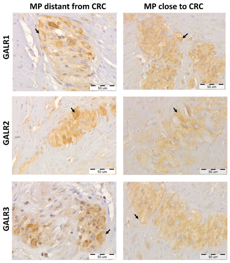
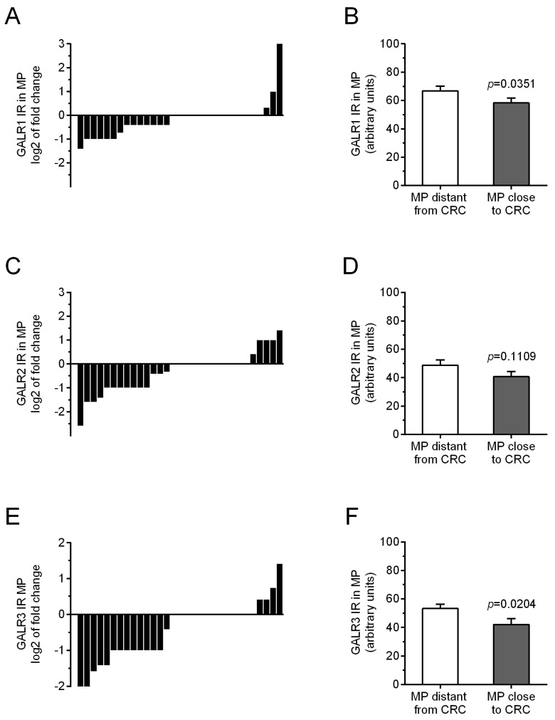
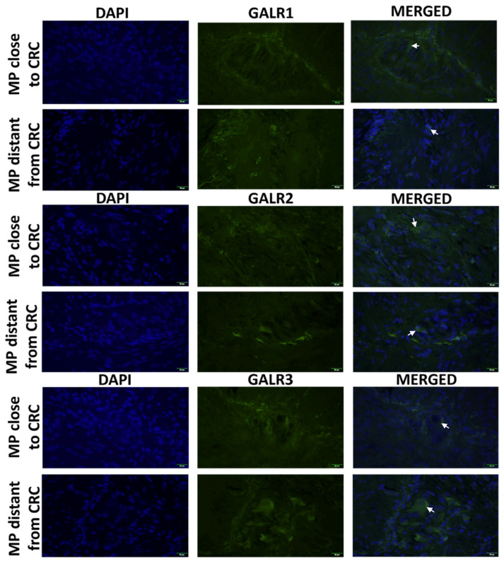
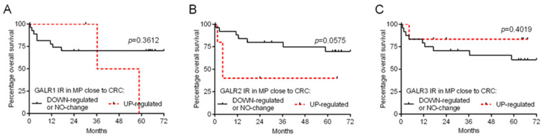
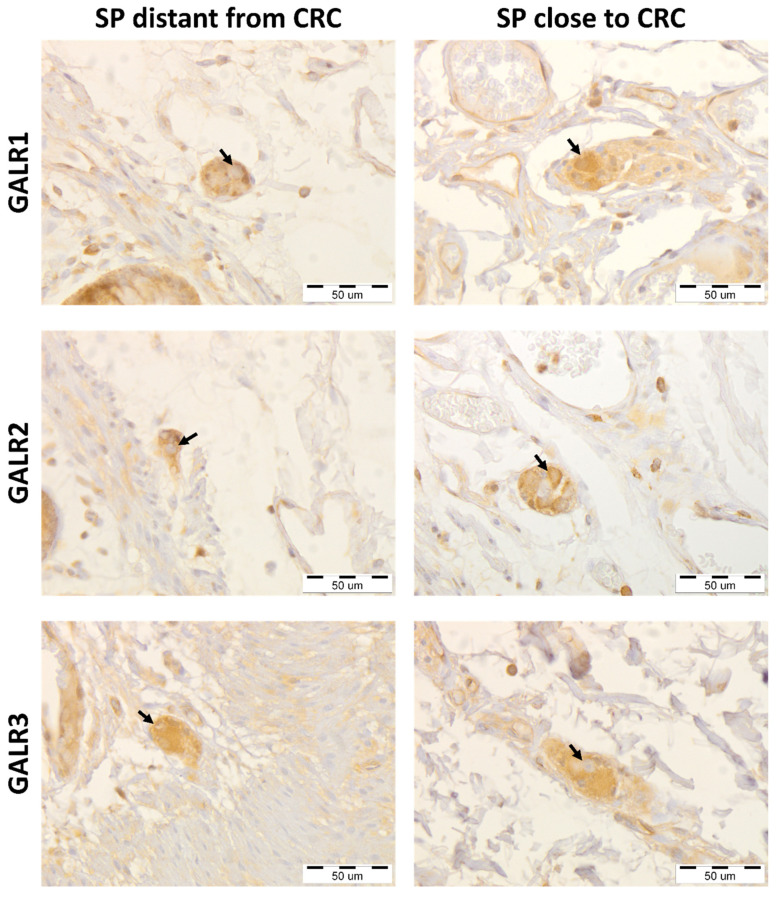
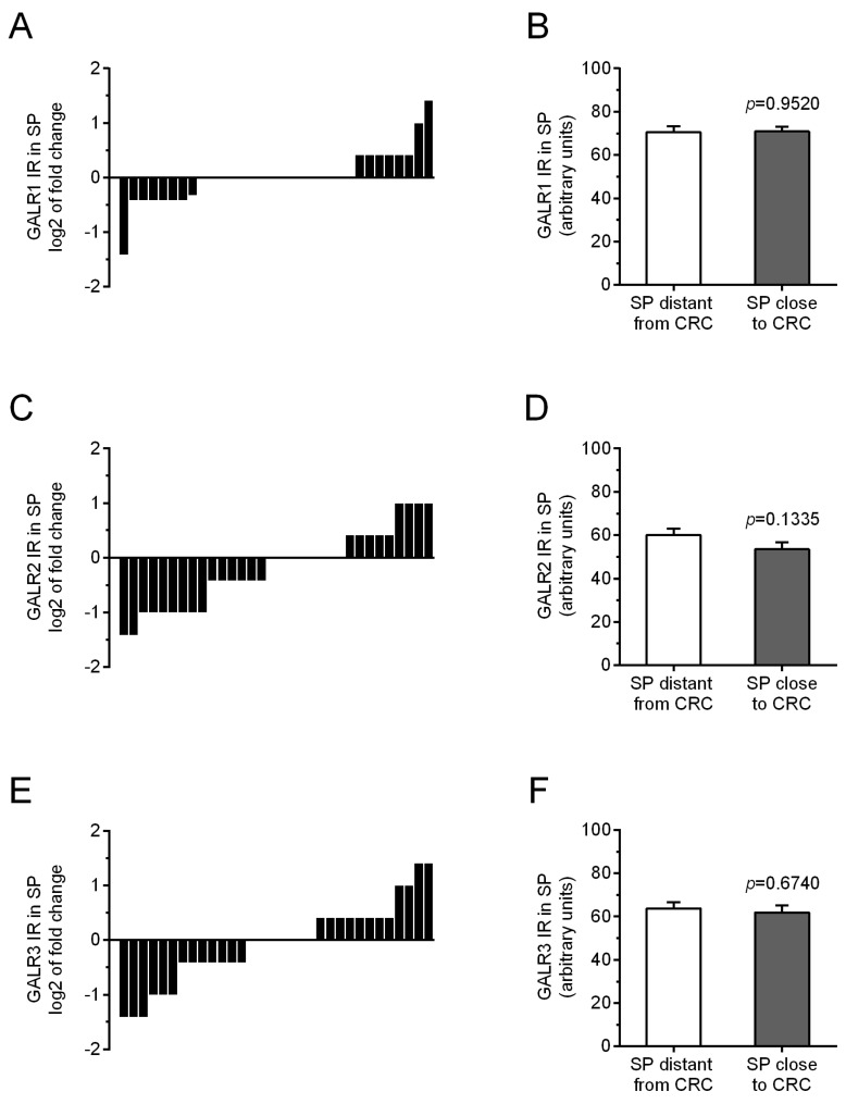
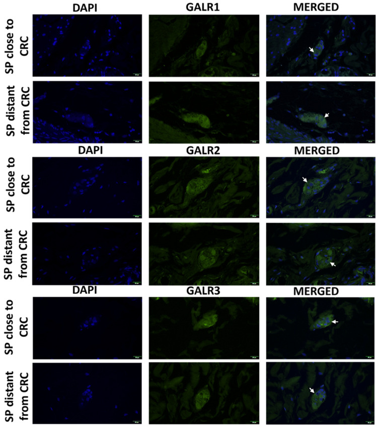
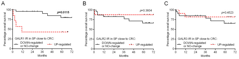
Galanin (GAL) is an important neurotransmitter released by the enteric nervous system (ENS) neurons located in the muscularis externa and submucosa enteric plexuses that acts by binding to GAL receptors 1, 2 and 3 (GALR1, 2 and 3). In our previous studies, the GAL immunoexpression was compared in colorectal cancer (CRC) tissue and the adjacent parts of the large intestine wall including myenteric and submucosal plexuses. Recently we have also found that expression levels of GALR1 and GALR3 proteins are elevated in CRC tissue as compared with their expression in epithelial cells of unchanged mucosa. Moreover, higher GALR3 immunoreactivity in CRC cells correlated with better prognosis of CRC patients. To understand the distribution of GALRs in enteric plexuses distal and close to CRC invasion, in the present study we decided to evaluate GALRs expression within the myenteric and submucosal plexuses located proximally and distally to the cancer invasion and correlated the GALRs expression levels with the clinico-pathological data of CRC patients. The immunohistochemical and immunofluorescent methods showed only slightly decreased immunoexpression of GALR1 and GALR3 in myenteric plexuses close to cancer but did not reveal any correlation in the immunoexpression of all three GAL receptors in myenteric plexuses and tumour progression. No significant changes were found between the expression levels of GALRs in submucosal plexuses distal and close to the tumour. However, elevated GALR1 expression in submucosal plexuses in vicinity of CRC correlated with poor prognosis, higher tumour grading and shorter overall survival. When myenteric plexuses undergo morphological and functional alterations characteristic for atrophy, GALRs maintain or only slightly decrease their expression status. In contrast, the correlation between high expression of GALR1 in the submucosal plexuses and overall survival of CRC patients suggest that GAL and GALRs can act as a components of local neuro-paracrine pro-proliferative pathways accelerating the invasion and metastasis of cancer cell. The obtained results suggest an important role of GALR1 in submucosal plexuses function during the progression of CRC and imply that GALR1 expression in submucosal plexuses of ENS could be an important predictive factor for CRC progression.
Keywords: colorectal cancer; enteric plexuses; galanin receptors; immunohistochemistry; prognosis.
Conflict of interest statement
The authors declare no conflict of interest.
Figures
Similar articles
-
Galanin Receptors (GalR1, GalR2, and GalR3) Expression in Colorectal Cancer Tissue and Correlations to the Overall Survival and Poor Prognosis of CRC Patients.Int J Mol Sci. 2022 Mar 29;23(7):3735. doi: 10.3390/ijms23073735. Int J Mol Sci. 2022. PMID: 35409094 Free PMC article.
-
Myenteric plexuses atrophy in the vicinity of colorectal cancer tissue is not caused by apoptosis or necrosis.Folia Histochem Cytobiol. 2016;54(2):99-107. doi: 10.5603/FHC.a2016.0012. Epub 2016 Jul 21. Folia Histochem Cytobiol. 2016. PMID: 27439439
-
The role of galanin in the progression and prognosis of colorectal cancer: the unfinished story.Eur J Histochem. 2024 Mar 6;68(1):3990. doi: 10.4081/ejh.2024.3990. Eur J Histochem. 2024. PMID: 38568200 Free PMC article.
-
Colorectal Cancer Invasion and Atrophy of the Enteric Nervous System: Potential Feedback and Impact on Cancer Progression.Int J Mol Sci. 2020 May 11;21(9):3391. doi: 10.3390/ijms21093391. Int J Mol Sci. 2020. PMID: 32403316 Free PMC article. Review.
-
Galanin receptors in the rat gastrointestinal tract.Neuropeptides. 2005 Jun;39(3):349-52. doi: 10.1016/j.npep.2004.12.023. Neuropeptides. 2005. PMID: 16044511 Free PMC article. Review.
Cited by
-
Increased galanin-galanin receptor 1 signaling, inflammation, and insulin resistance are associated with affective symptoms and chronic fatigue syndrome due to long COVID.PLoS One. 2025 Mar 6;20(3):e0316373. doi: 10.1371/journal.pone.0316373. eCollection 2025. PLoS One. 2025. PMID: 40048451 Free PMC article.
-
The immunoreactivity of GLI1 and VEGFA is a potential prognostic factor in kidney renal clear cell carcinoma.BMC Cancer. 2023 Nov 14;23(1):1110. doi: 10.1186/s12885-023-11622-7. BMC Cancer. 2023. PMID: 37964226 Free PMC article.
-
Galanin System in the Human Bile Duct and Perihilar Cholangiocarcinoma.Cells. 2023 Jun 21;12(13):1678. doi: 10.3390/cells12131678. Cells. 2023. PMID: 37443714 Free PMC article.
-
The Use of Biologics for Targeting GPCRs in Metastatic Cancers.BioTech (Basel). 2025 Jan 30;14(1):7. doi: 10.3390/biotech14010007. BioTech (Basel). 2025. PMID: 39982274 Free PMC article. Review.
-
Advancements in 3D In Vitro Models for Colorectal Cancer.Adv Sci (Weinh). 2024 Aug;11(32):e2405084. doi: 10.1002/advs.202405084. Epub 2024 Jul 4. Adv Sci (Weinh). 2024. PMID: 38962943 Free PMC article. Review.
References
-
- Kiezun J., Godlewski J., Krazinski B.E., Kozielec Z., Kmiec Z. Galanin Receptors (GalR1, GalR2, and GalR3) Expression in Colorectal Cancer Tissue and Correlations to the Overall Survival and Poor Prognosis of CRC Patients. Int. J. Mol. Sci. 2022;23:3735. doi: 10.3390/ijms23073735. - DOI - PMC - PubMed
Publication types
MeSH terms
Substances
Grants and funding
LinkOut - more resources
Full Text Sources
Medical
