

Gabapentin Disrupts Binding of Perlecan to the α2δ1 Voltage Sensitive Calcium Channel Subunit and Impairs Skeletal Mechanosensation
- PMID: 36551284
- PMCID: PMC9776037
- DOI: 10.3390/biom12121857
Gabapentin Disrupts Binding of Perlecan to the α2δ1 Voltage Sensitive Calcium Channel Subunit and Impairs Skeletal Mechanosensation
Abstract
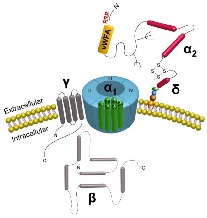
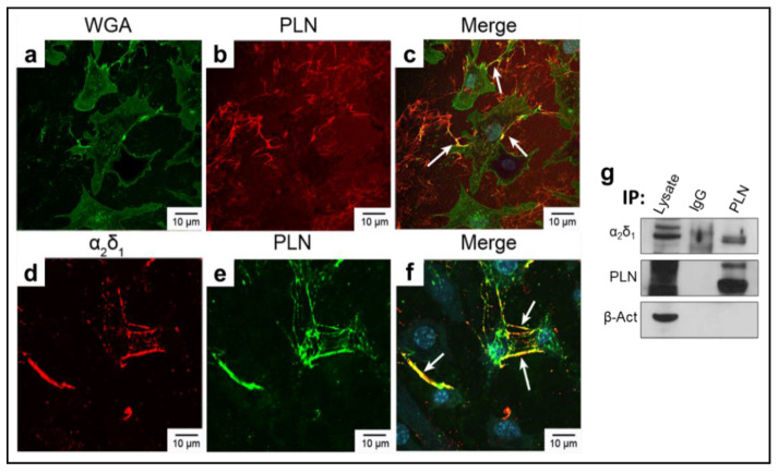
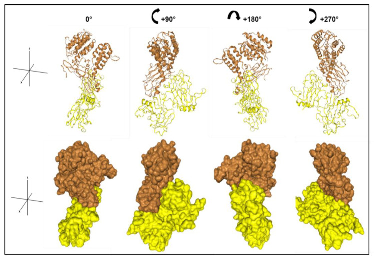
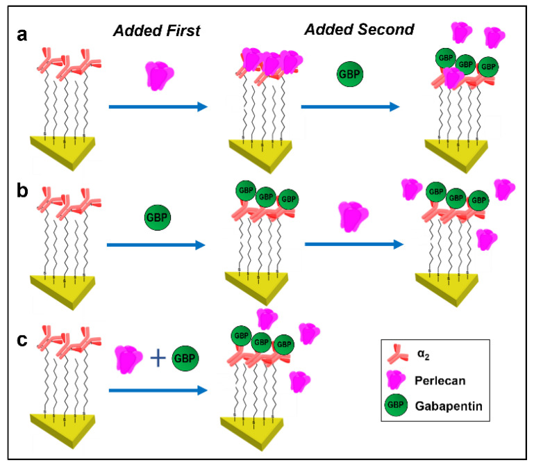
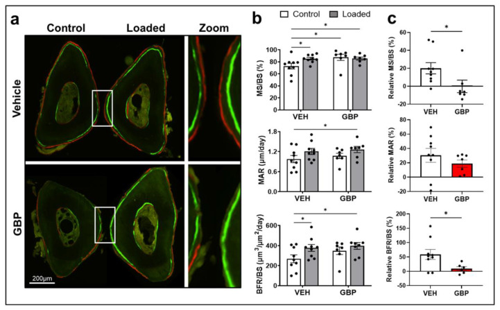
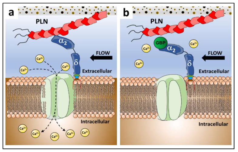
Our understanding of how osteocytes, the principal mechanosensors within bone, sense and perceive force remains unclear. Previous work identified "tethering elements" (TEs) spanning the pericellular space of osteocytes and transmitting mechanical information into biochemical signals. While we identified the heparan sulfate proteoglycan perlecan (PLN) as a component of these TEs, PLN must attach to the cell surface to induce biochemical responses. As voltage-sensitive calcium channels (VSCCs) are critical for bone mechanotransduction, we hypothesized that PLN binds the extracellular α2δ1 subunit of VSCCs to couple the bone matrix to the osteocyte membrane. Here, we showed co-localization of PLN and α2δ1 along osteocyte dendritic processes. Additionally, we quantified the molecular interactions between α2δ1 and PLN domains and demonstrated for the first time that α2δ1 strongly associates with PLN via its domain III. Furthermore, α2δ1 is the binding site for the commonly used pain drug, gabapentin (GBP), which is associated with adverse skeletal effects when used chronically. We found that GBP disrupts PLN::α2δ1 binding in vitro, and GBP treatment in vivo results in impaired bone mechanosensation. Our work identified a novel mechanosensory complex within osteocytes composed of PLN and α2δ1, necessary for bone force transmission and sensitive to the drug GBP.
Keywords: bone; gabapentin; mechanosensation; osteocytes; perlecan; voltage-sensitive calcium channels; α2δ1.
Conflict of interest statement
K.E.W. receives royalties for licensing FGF23 to Kyowa Hakko Kirin Co., Ltd.; had previous funding from Akebia, and current funding from Calico Labs. K.E.W. also owns equity interest in FGF Therapeutics. The other authors have nothing to declare.
Figures
Similar articles
-
Effects of Gabapentin and Pregabalin on Calcium Homeostasis: Implications for Physical Rehabilitation of Musculoskeletal Tissues.Curr Osteoporos Rep. 2022 Dec;20(6):365-378. doi: 10.1007/s11914-022-00750-x. Epub 2022 Sep 23. Curr Osteoporos Rep. 2022. PMID: 36149592 Free PMC article. Review.
-
Deletion of the auxiliary α2δ1 voltage sensitive calcium channel subunit in osteocytes and late-stage osteoblasts impairs femur strength and load-induced bone formation in male mice.J Bone Miner Res. 2024 Apr 19;39(3):298-314. doi: 10.1093/jbmr/zjae010. J Bone Miner Res. 2024. PMID: 38477790
-
Loss of the auxiliary α2δ1 voltage-sensitive calcium channel subunit impairs bone formation and anabolic responses to mechanical loading.JBMR Plus. 2024 Jan 10;8(2):ziad008. doi: 10.1093/jbmrpl/ziad008. eCollection 2024 Feb. JBMR Plus. 2024. PMID: 38505532 Free PMC article.
-
Single molecule force measurements of perlecan/HSPG2: A key component of the osteocyte pericellular matrix.Matrix Biol. 2016 Mar;50:27-38. doi: 10.1016/j.matbio.2015.11.001. Epub 2015 Nov 4. Matrix Biol. 2016. PMID: 26546708 Free PMC article.
-
Perlecan in Pericellular Mechanosensory Cell-Matrix Communication, Extracellular Matrix Stabilisation and Mechanoregulation of Load-Bearing Connective Tissues.Int J Mol Sci. 2021 Mar 8;22(5):2716. doi: 10.3390/ijms22052716. Int J Mol Sci. 2021. PMID: 33800241 Free PMC article. Review.
Cited by
-
Effects of Gabapentin and Pregabalin on Calcium Homeostasis: Implications for Physical Rehabilitation of Musculoskeletal Tissues.Curr Osteoporos Rep. 2022 Dec;20(6):365-378. doi: 10.1007/s11914-022-00750-x. Epub 2022 Sep 23. Curr Osteoporos Rep. 2022. PMID: 36149592 Free PMC article. Review.
-
Examining Mechanisms for Voltage-Sensitive Calcium Channel-Mediated Secretion Events in Bone Cells.Calcif Tissue Int. 2023 Jul;113(1):126-142. doi: 10.1007/s00223-023-01097-w. Epub 2023 Jun 1. Calcif Tissue Int. 2023. PMID: 37261463 Free PMC article. Review.
-
Influence of the calcium voltage-gated channel auxiliary subunit (CACNA2D1) absence on intraocular pressure in mice.Exp Eye Res. 2024 Apr;241:109835. doi: 10.1016/j.exer.2024.109835. Epub 2024 Feb 17. Exp Eye Res. 2024. PMID: 38373629 Free PMC article.
-
Proteoglycans in Mechanobiology of Tissues and Organs: Normal Functions and Mechanopathology.Proteoglycan Res. 2024 Apr-Jun;2(2):e21. doi: 10.1002/pgr2.21. Epub 2024 May 20. Proteoglycan Res. 2024. PMID: 39584146 Free PMC article.
-
Proteoglycans of basement membranes: Crucial controllers of angiogenesis, neurogenesis, and autophagy.Proteoglycan Res. 2024 Jul-Sep;2(3):e22. doi: 10.1002/pgr2.22. Epub 2024 Jun 29. Proteoglycan Res. 2024. PMID: 39184370 Free PMC article.
References
-
- You J., Yellowley C.E., Donahue H.J., Zhang Y., Chen Q., Jacobs C.R. Substrate deformation levels associated with routine physical activity are less stimulatory to bone cells relative to loading-induced oscillatory fluid flow. J. Biomech. Eng. Trans. ASME. 2000;122:387–393. doi: 10.1115/1.1287161. - DOI - PubMed
Publication types
MeSH terms
Substances
Grants and funding
LinkOut - more resources
Full Text Sources
Research Materials
