Review
. 2022 Dec 13;12(12):1862.
doi: 10.3390/biom12121862.

Alterations of Cytoskeleton Networks in Cell Fate Determination and Cancer Development

Affiliations
Review

Alterations of Cytoskeleton Networks in Cell Fate Determination and Cancer Development

Evan Ja-Yang Wang et al. Biomolecules. .

Abstract

Cytoskeleton proteins have been long recognized as structural proteins that provide the necessary mechanical architecture for cell development and tissue homeostasis. With the completion of the cancer genome project, scientists were surprised to learn that huge numbers of mutated genes are annotated as cytoskeletal or associated proteins. Although most of these mutations are considered as passenger mutations during cancer development and evolution, some genes show high mutation rates that can even determine clinical outcomes. In addition, (phospho)proteomics study confirms that many cytoskeleton-associated proteins, e.g., β-catenin, PIK3CA, and MB21D2, are important signaling mediators, further suggesting their biofunctional roles in cancer development. With emerging evidence to indicate the involvement of mechanotransduction in stemness formation and cell differentiation, mutations in these key cytoskeleton components may change the physical/mechanical properties of the cells and determine the cell fate during cancer development. In particular, tumor microenvironment remodeling triggered by such alterations has been known to play important roles in autophagy, metabolism, cancer dormancy, and immune evasion. In this review paper, we will highlight the current understanding of how aberrant cytoskeleton networks affect cancer behaviors and cellular functions through mechanotransduction.

Keywords: autophagy; cancer; cytoskeleton; dormancy; immune evasion; mechanotransduction; metabolism reprogramming; tumor microenvironment.


Conflict of interest statement

The authors declare no conflict of interest.

Figures

Figure 1
Figure 1
Cell softening and cancer stemness triggered by altered cytoskeleton networks. Cells with wild-type cytoskeleton networks maintain a mechanical structure in a symmetrical organization. When cells are at low cell density, Yap1 can be translocated into the nucleus and trigger cell proliferation. Once cells reach a high cell density with complete cell–cell contacts, the Hippo signaling is activated, and then subsequently suppresses Yap1 nuclear translocation and cell proliferation. Mutations in genes for cytoskeletal and its associated proteins disturb cytoskeleton networks, resulting in the failure of cell–cell contact formation. Thus, the cells cannot sense the contact inhibition signaling and keep proliferating continuously. Without mechanical support, cells tend to become softer and grow in multiple layers. Long-term effects of Yap1-mediated carcinogenesis will trigger cancer stemness and aggressiveness.
Figure 2
Figure 2
Functional impacts of cytokeratin fusion variants on wild-type (WT) K14 networks. K6-K14 cytokeratin fusion variants (tagged with GFP) and WT K14 (tagged with mCherry) were cotransfected into Cal-27 oral cancer cells. Permeable DAPI was utilized to stain the nucleus (blue). WT K6 and WT K14 tagged with GFP were utilized as reference controls. The representative images were taken 36 h after transfection. The interaction between each fusion variant and WT K14 was analyzed by fluorescence resonance energy transfer (FRET) assay. These data were reproduced from the original paper [51].
Figure 3
Figure 3
Modulation of the tumor microenvironment (TME) is associated with alterations of cytoskeleton networks. Cells with altered cytoskeleton network show softer mechanical properties and higher cancer stemness, which is associated with frequent membrane protrusion and extracellular vesicle (EV) release. When oxygen and nutrient gradients are present, the TME within the 3D lesions can be influenced by many factors and cells. The main remodeling factors, either released by direct secretion or embedded EVs, modulate cellular functions and behaviors of neighboring cancer cells, tumor-associated macrophages (TAMs), and cancer-associated fibroblasts (CAFs). In addition, altered cytoskeleton networks in cancer cells trigger DNA damage, leading to cGAS-STING activation and associated inflammation responses in cancer lesions.
Figure 4
Figure 4
Involvement of mechanotransduction in metabolism reprogramming and cancer cell evolution/clonal selection. Alterations in ECM stiffness influence the glucose metabolism in cancer cells through mechanotransduction. Activation of the YAP/TAZ and integrin-FAK-P13K-AKT pathways can enhance glucose uptake and glycolysis via endocytosis, glucose transportation by GLUT, gluconeogenesis, activation of glycolytic enzymes or glycogen synthase, and upregulation of the pentose phosphate pathway. Due to the Warburg effect in cancer cells, active glycolysis can generate ROS and free radicals, leading to oxidative stress, which in turn can change cellular metabolism into a cycle of overnutrition. On the other hand, ROS can cause DNA breakage and genome instability, which serves as a driving force for uncontrolled cell proliferation, and tumorigenesis. Oxidative stress also leads to autophagy and cancer dormancy, where autophagy aids the survival of dormant cancer cells and allows cancer cells to undergo clonal evolution.
Figure 5
Figure 5
Key roles of PFKFB3 expression in ECM stiffness-induced autophagy through changes in glucose metabolism. (Left panel) In dormant cancer cells, autophagy is activated through several ways: (i) trans-farnesylthiosalicylic acid inhibits the RAS pathway, leading to the downregulation of HIF-1α and subsequent PFKFB3 expression; (ii) upregulation of MTG16, a transcriptional repressor functioning as a tumor suppressor, reducing PFKFB3 expression. PFKFB3 downregulation inhibits tumor growth, cell proliferation, and tumorigenesis, and promotes cell death. In addition, the activity of F2,6BP can be also inhibited, leading to downregulation of PFK-1 and glycolysis. (Right panel) During cancer development or dormancy reactivation, autophagy is not prevalent through several means that can upregulate PFKFB3 expression: (i) mechanosensing through cell protrusion; (ii) hypoxia-induced HIF activation through AMPK pathway; (iii) Her2 overexpression; (iv) inactivation of p53 and PTEN; (v) progestin- and estradiol-triggered nuclear receptor activation. PFKFB3 upregulation can further promote pEMT and cancer metastasis via TGF-β signaling. Cell proliferation and tumorigenesis can be also enhanced. Glycolysis activity can be recovered by activating F2,6BP and PFK-1.
Figure 6
Figure 6
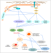
Cellular plasticity of tumor cells is governed by partial EMT (pEMT). Several factors are involved in the regulation of MET-pEMT-EMT transition and TME remodeling during tumor progression, including hypoxia, anticancer treatment, metabolic changes, and TGF-β secreted by cancer stem cells. These factors guide tumor cells in going into cellular transformation from an epithelial program to a partial epithelial-to-mesenchymal transition (pEMT) program. Cancer cells with the pEMT phenotype provide the necessary cell–cell contact for collective cell migration toward a secondary site. On the other hand, extracellular vesicles secreted by cancer stem cells recruit tumor-associated macrophages (TAMs), which can further assist cellular plasticity during metastasis by secreting a variety of cytokines and chemokines. Examples include angiogenesis and cluster cell migration promoted by VEGF and FGF2; TME remodeling for a protumor microenvironment by MMPs, TGF-β, and certain chemokines; cell proliferation and survival by TNF, IL-1, IL-6, polyamines, and NO; cancer progression and metastasis by TNF, IL-1, IL-6, MMPs; adaptive immunity by IL-10, TGF-β, PG, ROI, and certain chemokines; and cancer dormancy.

Similar articles

Cited by

References

    1. Majeski H.E., Yang J. The 2016 John J. Abel Award Lecture: Targeting the Mechanical Microenvironment in Cancer. Mol. Pharmacol. 2016;90:744–754. doi: 10.1124/mol.116.106765. - DOI - PMC - PubMed
    1. Przybyla L., Muncie J.M., Weaver V.M. Mechanical Control of Epithelial-to-Mesenchymal Transitions in Development and Cancer. Annu. Rev. Cell Dev. Biol. 2016;32:527–554. doi: 10.1146/annurev-cellbio-111315-125150. - DOI - PubMed
    1. Ciasca G., Papi M., Minelli E., Palmieri V., De Spirito M. Changes in cellular mechanical properties during onset or progression of colorectal cancer. World J. Gastroenterol. 2016;22:7203–7214. doi: 10.3748/wjg.v22.i32.7203. - DOI - PMC - PubMed
    1. Fletcher D.A., Mullins R.D. Cell mechanics and the cytoskeleton. Nature. 2010;463:485–492. doi: 10.1038/nature08908. - DOI - PMC - PubMed
    1. Chien S. Mechanotransduction and endothelial cell homeostasis: The wisdom of the cell. Am. J. Physiol. Heart Circ. Physiol. 2007;292:H1209–H1224. doi: 10.1152/ajpheart.01047.2006. - DOI - PubMed

Publication types

Substances