. 2022 Dec 13;12(12):1865.
doi: 10.3390/biom12121865.

Eosinophils Infiltration in Esophageal Muscularis Propria Induces Achalasia-like Esophageal Motility Disorder in Mice

Affiliations

Eosinophils Infiltration in Esophageal Muscularis Propria Induces Achalasia-like Esophageal Motility Disorder in Mice

Wei Zhao et al. Biomolecules. .

Abstract

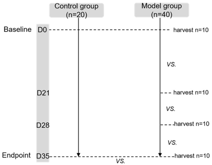
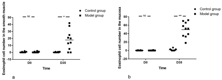
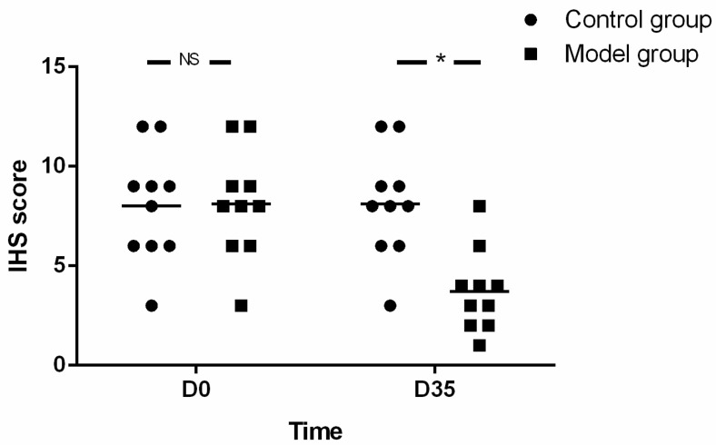
Eosinophil infiltration in esophageal muscularis propria is common in achalasia (AC). This study aims to evaluate the effect of eosinophil infiltration in muscularis propria of the esophagus on esophageal motility in mice. A mouse model with eosinophil infiltration in the esophageal muscle layer was established by long term Ovalbumin (OVA) exposure. The histopathology features of esophageal muscularis propria as well as parameters of esophageal motility, such as lower esophageal sphincter pressure (LESP) and esophageal emptying, were compared between model and control group. In addition, the histopathology and motility of esophagus at each time point in the model group were compared. The esophageal motor function severely deteriorated in the model group, mimicking the abnormal esophageal motility of AC, with more eosinophils and fewer SOX-10-IR cells in esophageal muscularis propria in the model group, compared with control. With the prolongation of OVA treatment, esophageal motility disorder was aggravated, accompanied by increased eosinophils in the the muscle layer of esophagus and decreased SOX-10-IR cells in the model group. In addition, the eosinophil count was negatively correlated with SOX-10-IR cells. Long-term exposure to OVA assisted by alum may induce eosinophil infiltration in esophageal muscularis propria, reduced SOX-10-IR cells and abnormal esophageal motility, which simulates the functional and histopathological features of some AC patients. This suggests that eosinophil infiltration in esophageal muscularis propria may play a role in the pathogenesis of a subgroup of AC.

Keywords: achalasia; eosinophil; mice.


Figures

Figure 1
Figure 1
Flow chart of the study. In the model group, the LESP, esophageal emptying and histological results at every viewpoint (D21, D28, D35) were compared with baseline (D0). At the endpoint of the study (D35), the low esophageal sphincter (LES) pressure LESP, esophageal emptying and histology of mice in the control group and model group were compared. In each viewpoint, the LESP was first programmed, and followed by the esophageal emptying test. Finally, 10 mice were selected randomly and sacrificed for esophageal histology at every time point in model group, and at D0 and D35 respectively in the control group. LESP: lower esophageal sphincter pressure.
Figure 2
Figure 2
Eosinophil infiltration induction protocol. In the model group, at D1 and D14, mice were sensitized by intraperitoneal injection of 50 μg OVA and 1 mg alum in PBS (50 μg/1.0 mg/0.5 mL), and further challenged with 150 μg OVA (50 μL) intra-nasally, under the condition of anesthesia, 3 times per week for 3 weeks. Mice in the control group were sensitized and challenged with the same volume of PBS solution as the model group. OVA: ovalbumin.
Figure 3
Figure 3
Esophageal emptying test in mouse. (A) Contour of the esophagus with 0.1 mL iohexol as contrast agent. The release position of the X-ray opaque marker was at the level of sternum angle shown with black arrow. (B) The movement of the X-ray opaque marker through esophagus. White arrow refers to the marker.
Figure 4
Figure 4
Body weight of mice. With the prolongation of OVA exposure time, body weight of mice in the model group was lower than in control group at the end of the experiment. Unpaired t-tests was used in comparison between groups and among different viewpoints in the model group. LESP: lower esophageal sphincter pressure. * p < 0.05, NS—not significant.
Figure 5
Figure 5
The LESP during baseline and different time points, esophageal emptying test and esophageal radiography. (a) Although there was no difference between model group and control group at baseline, the LESP was higher at D28 and D35. (b) During OVA treatment, the esophageal body transit time of mice in model group was longer than that of control group at D21, D28 and D35, respectively. (c) Although there was no difference between groups at baseline, the cardiac passing time of model group was longer than that of control group at the end of the experiment. (d) At every time point, there was no significant difference in the esophageal width between groups. The number of mice in control group was 20, 10, 10 and 10, for each time point (D0-D35). In contrast, the number of mice in model group was 40, 30, 20 and10 for each time point, respectively. Unpaired t-tests were used in comparison between groups and among different viewpoints in model group. LESP: lower esophageal sphincter pressure. * p < 0.05, NS—not significant.
Figure 6
Figure 6
Esophageal histopathology of mice. (a) Eosinophil cell number in the smooth muscle and (b) Eosinophil cell number in the mucosa. There were almost no eosinophils in the esophagus at baseline in both groups by HE staining. The eosinophils in model group outnumbered that in control group with the prolongation of OVA treatment, in both mucosa and smooth muscle layer of esophagus. The number of mice in both groups was 10 at each time point. Mann-Whitney U-Test was used in comparison between groups and among different viewpoints in model group. * p < 0.05, ** p < 0.001, NS—not significant.
Figure 7
Figure 7
Immuno-histochemical staining of the esophageal tissue of mice in control and model group at D35. (a,b) HE staining of the esophagus, which shows eosinophils in model group (b) outnumbered that in control group (a) in both mucosa and muscularis propria. * Indicates mucosa, ▲ indicates muscularis propria, black arrows indicate eosinophils. (ch) Immuno-histochemical staining showed more MBP, ECP and EDN positive cells in the esophageal smooth muscle of mice in model group (d,f,h) compared with controls (c,e,g). Black arrows indicate MBP, ECP and EDN positive cells, respectively. (ij) Immuno-histochemical staining for the localization of the primary antibody to SOX-10, which indicates ganglion cells in esophageal tissue of mice in model group (j) and control group (i). Black arrows indicate SOX-10 positive cells. Magnification ×400.
Figure 8
Figure 8
The expression of SOX-10. The expression of SOX-10 in model group was significantly reduced compared with that of control group at the end stage of the experiment. The number of mice in both groups was 10 at each time point. Unpaired two-tailed t-Test was used in comparison between groups and among different viewpoints in model group. IHS: immune-histochemical score. * p < 0.05, NS—not significant.
Figure 9
Figure 9
The correlation between eosinophil count and the SOX-10-IR Cells. The number of eosinophils in the smooth muscle layer of esophagus was negatively correlated with SOX-10-IR Cells. n = 40, r = −0.5739, r2 = 0.3294, p = 0.0001, 95% CI (−0.7512, −0.3195). IHS: immuno-histochemical score. Spearman rank correlation was used.

Similar articles

References

    1. Boeckxstaens G.E., Zaninotto G., Richter J.E. Achalasia. Lancet. 2014;383:83–93. doi: 10.1016/S0140-6736(13)60651-0. - DOI - PubMed
    1. Mundre P., Black C.J., Mohammed N., Ford A.C. Efficacy of surgical or endoscopic treatment of idiopathic achalasia: A systematic review and network meta-analysis. Lancet Gastroenterol. Hepatol. 2021;6:30–38. doi: 10.1016/S2468-1253(20)30296-X. - DOI - PubMed
    1. Chen Z., Bessell J.R., Chew A., Watson D.I. Laparoscopic cardiomyotomy for achalasia: Clinical outcomes beyond 5 years. J. Gastrointest. Surg. 2010;14:594–600. doi: 10.1007/s11605-010-1158-2. - DOI - PubMed
    1. Kilic A., Owens S.R., Pennathur A., Luketich J.D., Landreneau R.J., Schuchert M.J. An increased proportion of inflammatory cells express tumor necrosis factor alpha in idiopathic achalasia of the esophagus. Dis. Esophagus. 2009;22:382–385. doi: 10.1111/j.1442-2050.2008.00922.x. - DOI - PubMed
    1. Liu Z.-Q., Chen W.-F., Wang Y., Xu X.-Y., Zeng Y.-G., Dillon D.L., Cheng J., Xu M.-D., Zhong Y.-S., Zhang Y.-Q., et al. Mast cell infiltration associated with loss of interstitial cells of Cajal and neuronal degeneration in achalasia. Neurogastroenterol. Motil. 2019;31:e13565. doi: 10.1111/nmo.13565. - DOI - PubMed

Publication types

MeSH terms