. 2022 Dec 18;12(12):1898.
doi: 10.3390/biom12121898.

Genetic Ablation of Ankrd1 Mitigates Cardiac Damage during Experimental Autoimmune Myocarditis in Mice

Affiliations

Genetic Ablation of Ankrd1 Mitigates Cardiac Damage during Experimental Autoimmune Myocarditis in Mice

Ieva Rinkūnaitė et al. Biomolecules. .

Abstract

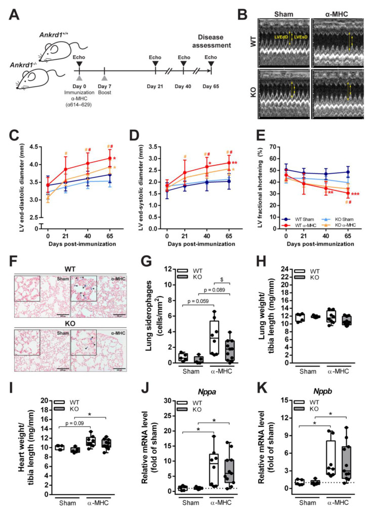
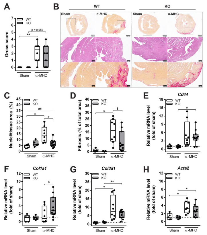
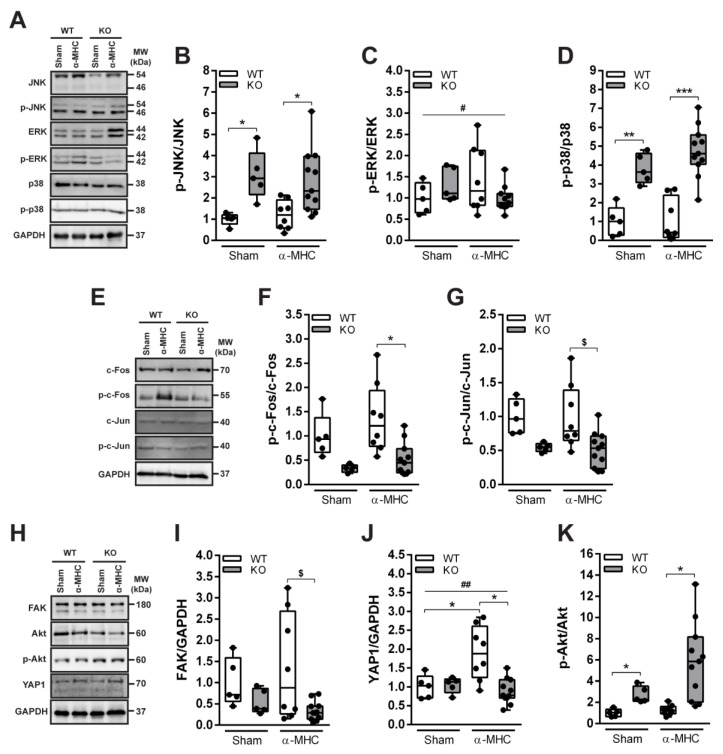
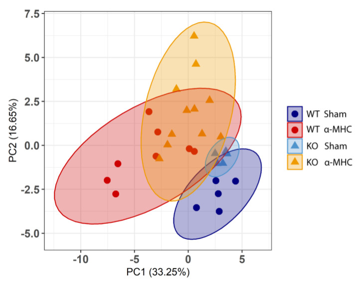
Myocarditis (MC) is an inflammatory disease of the myocardium that can cause sudden death in the acute phase, and dilated cardiomyopathy (DCM) with chronic heart failure as its major long-term outcome. However, the molecular mechanisms beyond the acute MC phase remain poorly understood. The ankyrin repeat domain 1 (ANKRD1) is a functionally pleiotropic stress/stretch-inducible protein, which can modulate cardiac stress response during various forms of pathological stimuli; however, its involvement in post-MC cardiac remodeling leading to DCM is not known. To address this, we induced experimental autoimmune myocarditis (EAM) in ANKRD1-deficient mice, and evaluated post-MC consequences at the DCM stage mice hearts. We demonstrated that ANKRD1 does not significantly modulate heart failure; nevertheless, the genetic ablation of Ankrd1 blunted the cardiac damage/remodeling and preserved heart function during post-MC DCM.

Keywords: ANKRD1; dilated cardiomyopathy; heart failure; inflammation; myocardial remodeling; myocarditis.


Conflict of interest statement

The authors declare no conflict of interest.

Figures

Figure 1
Figure 1
Development of DCM and HF in response to EAM induction. (A) Experimental design scheme. (B) Representative images of M-mode echocardiograms at the short parasternal axis of sham- and α-MHC-immunized WT and Ankrd1 KO mice hearts 65 days pi. Yellow arrows indicate LVEdD—left ventricular end-diastolic dimension and LVEsD—left ventricular end-systolic dimension. (C–E) Echocardiographic parameters of mice hearts during the 65-day time course. LV—left ventricular. Data represented as mean ± 95% CI. *—marks statistically significant differences from respective sham, #—differences from day 0, the color of the mark corresponds to the colors of the groups. *,# (p < 0.05), **,## (p < 0.01), *** (p < 0.001)—marks statistically significant differences, by robust ANOVA followed by Lincon post hoc. (F) Representative Prussian blue staining of lung sections (scale bar – 200 µm) showing HF cells—siderophages (blue, denoted with black arrows) in the lung parenchyma of sham- and α-MHC-immunized mice and (G) quantitative analysis of their distribution in the lung tissue. (H) Lung weight to tibia length ratios. (I) Heart weight to tibia length ratios. (J,K) Quantitative RT-PCR analysis of Nppa and Nppb transcripts levels, data were normalized to corresponding 18S rRNA levels and expressed as the fold change versus the corresponding sham control; the dotted line represents sham levels. WT-Sham (n = 5), WT-α-MHC (n = 8), KO-Sham (n = 5) and KO-α-MHC (n = 11). Data represented as box-plots depicting median ± IQR, black dots denote individual animals. * (p < 0.05), ** (p < 0.01), *** (p < 0.001)—marks statistically significant differences, by robust ANOVA followed by Lincon post hoc. $ (p < 0.05)—marks statistically significant differences between α-MHC-immunized groups, by ANCOVA (LVEdD was used as a covariate) followed by EMMs post hoc.
Figure 2
Figure 2
Histological and biochemical characterization of cardiac remodeling induced by EAM. (A) The gross score of the dissected mice hearts 65 days after EAM induction. ** (p < 0.01) marks statistically significant differences, by the Kruskal–Wallis test, and differences between groups were determined by Dunn’s post hoc test. (B) Representative sections of whole mouse hearts 65 days after EAM induction (upper panel, 1.5× magnification) stained with Sirius red for collagen (red) (scale bar—1000 µm), sections stained with H&E (middle panel, 6× magnification) (scale bar—200 µm) and Sirius red (lower panel, 6× magnification) (scale bar—200 µm). (C) Quantitative analysis of infiltrates found in the heart. (D) Quantification of heart fibrosis. The mRNA expression of (E) Cd44, (F) Col1a1, (G) Col3a1, and (H) Acta2, data were normalized to corresponding 18S rRNA levels and presented as the fold change versus the corresponding sham control; the dotted line marks sham levels. WT-Sham (n = 5), WT-α-MHC (n = 8), KO-Sham (n = 5) and KO-α-MHC (n = 11). Data represented as median ± IQR. Black dots denote individual animals. * (p < 0.05), ** (p < 0.01) marks statistically significant differences and ## (p < 0.01) marks statistically significant differences in interaction between genotype and immunization, by robust ANOVA followed by Lincon post hoc. $ (p < 0.05) marks statistically significant differences between α-MHC-immunized groups, by ANCOVA (Gross score was used as a covariate) followed by EMMs post hoc.
Figure 3
Figure 3
Western blot analysis of the MAPK/AP1 and mechanosensing-related proteins (FAK, Akt, and YAP1) levels in mice 65 days pi (post-MC DCM stage). (A) Representative immunoblots and (BD) quantification analysis of MAPK’s JNK, ERK, and p38 proteins’ activation. (E) Representative Western blot and (F,G) quantitative densitometric analysis of AP1 components c-Fos and c-Jun proteins phosphorylation. (H) Representative immunoblots and (IK) quantification of FAK, Akt and YAP1 proteins. WT-Sham (n = 5), WT-α-MHC (n = 8), KO-Sham (n = 5) and KO-α-MHC (n = 11). GAPDH served as a loading control. Data are represented as the median ± IQR, black dots denote individual animals. * (p < 0.05), ** (p < 0.01), *** (p < 0.001) marks statistically significant differences, # (p < 0.05), ## (p < 0.01) marks statistically significant differences in interaction between genotype and immunization, by robust ANOVA followed by Lincon post hoc. $ (p < 0.05) marks statistically significant differences between α-MHC-immunized groups, by ANCOVA (Gross score was used as a covariate) followed by EMMs post hoc.
Figure 4
Figure 4
Principal component analysis (PCA) of overall experimental data. Plot shows distribution of WT-Sham (n = 5) (blue, dot), WT-α-MHC (n = 8) (red, dot), KO-Sham (n = 5) (light blue, triangle) and KO-α-MHC (n = 11) (yellow, triangle) across the first two principal components (32 features per PCA were used). The explained variance per component (PC1, PC2) (%) is indicated in the panel. The 95% confidence ellipses are represented by the corresponding color of the animal group.
Figure 5
Figure 5
Summarized interactions between heart damage and the levels of proteins found in mice during post-MC DCM (65 days pi). Linear regressions of (A) p-JNK/JNK, (C) p-p38/p38, (E) p-c-Fos/c-Fos, (G) FAK/GAPDH, (I) YAP1/GAPDH, (K) p-Akt/Akt on Nuclei/tissue area (cellular infiltration). Linear regressions of (B) p-JNK/JNK, (D) p-p38/p38, (F) p-c-Fos/c-Fos, (H) FAK/GAPDH, (J) YAP1/GAPDH, (L) p-Akt/Akt on Fibrosis. The WT-α-MHC (white dots) and Ankrd1 KO- α-MHC (gray dots) groups were combined. The regression line is represented within a 95% confidence interval. The coefficients of determination (R2) from linear regression analysis and p-values are shown on the graphs. * (p < 0.05), ** (p < 0.01), *** (p < 0.001) marks statistically significant correlations.

Similar articles

Cited by

References

    1. GBD 2017 Disease and Injury Incidence and Prevalence Collaborators Global, regional, and national incidence, prevalence, and years lived with disability for 354 diseases and injuries for 195 countries and territories, 1990–2017: A systematic analysis for the Global Burden of Disease Study. Lancet. 2018;392:1789–1858. doi: 10.1016/S0140-6736(18)32279-7. - DOI - PMC - PubMed
    1. Ammirati E., Frigerio M., Adler E.D., Basso C., Birnie D.H., Brambatti M., Friedrich M.G., Klingel K., Lehtonen J., Moslehi J.J., et al. Management of Acute Myocarditis and Chronic Inflammatory Cardiomyopathy. Circ. Heart Fail. 2020;13:e007405. doi: 10.1161/CIRCHEARTFAILURE.120.007405. - DOI - PMC - PubMed
    1. D’Ambrosio A. The fate of acute myocarditis between spontaneous improvement and evolution to dilated cardiomyopathy: A review. Heart. 2001;85:499–504. doi: 10.1136/heart.85.5.499. - DOI - PMC - PubMed
    1. Tschöpe C., Cooper L., Torre-Amione G., Van Linthout S. Management of Myocarditis-Related Cardiomyopathy in Adults. Circ. Res. 2019;124:1568–1583. doi: 10.1161/CIRCRESAHA.118.313578. - DOI - PubMed
    1. Brüstle K., Heidecker B. Checkpoint inhibitor induced cardiotoxicity: Managing the drawbacks of our newest agents against cancer. Oncotarget. 2017;8:106165–106166. doi: 10.18632/oncotarget.22579. - DOI - PMC - PubMed

Publication types

LinkOut - more resources