

Cytotoxicity and Antibacterial Efficacy of AgCu and AgFe NanoAlloys: A Comparative Study
- PMID: 36551394
- PMCID: PMC9774506
- DOI: 10.3390/antibiotics11121737
Cytotoxicity and Antibacterial Efficacy of AgCu and AgFe NanoAlloys: A Comparative Study
Abstract
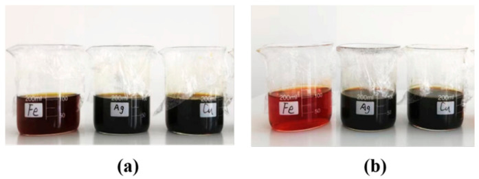
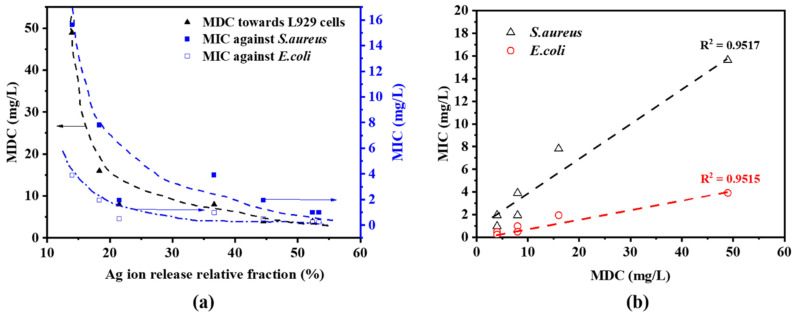
Although Ag nanoparticles (NPs) have been widely applied in daily life and in biomedical and industrial fields, there is a demand for Ag-based bimetallic nanoalloys (NAs), such as AgCu and AgFe, due to their enhanced antibacterial efficacy and reduced Ag consumption. In this work, we present a comparison study on the antibacterial efficacy and cytotoxicity rates of Ag NPs and AgCu and AgFe NAs to L929 mouse fibroblast cells using the CCK-8 technique based on the relative cell viability. The concept of the minimum death concentration (MDC) is introduced to estimate the cytotoxicity to the cells. It is found that the minimum inhibitory concentrations (MICs) of the NPs against E. coli and S. aureus decrease with the addition of both Cu and Fe. There is a strong correlation between the MDC and MIC, implying that the mechanisms of both antibacterial efficacy and cytotoxicity are similar. The enhanced antibacterial efficacy to bacteria and cytotoxicity toward the cell are attributed to Ag+ release. The following order is found for both the MIC and MDC: AgFe < AgCu < Ag NPs. However, there is no cytotoxicity to the L929 cells for AgFe and AgCu NAs at their MIC Ag concentrations against S. aureus.
Keywords: AgCu; AgFe; L929 cells; antibacterial efficacy; cytotoxicity; nanoalloy.
Conflict of interest statement
The authors declare no conflict of interest.
Figures


References
-
- CDC . Antibiotic Resistance Threats in the United States. U.S. Departmnet of Health and Human Services; Washington DC, USA: 2019.
-
- Marambio-Jones C., Hoek E. A review of the antibacterial effects of silver nanomaterials and potential implications for human health and the environment. J. Nanopart. Res. 2010;12:1531–1551. doi: 10.1007/s11051-010-9900-y. - DOI
-
- Marassi V., Di Cristo L., Smith S.G., Ortelli S., Blosi M., Costa A.L., Reschiglian P., Volkov Y., Prina-Mello A. Silver nanoparticles as a medical device in healthcare settings: A five-step approach for candidate screening of coating agents. R. Soc. Open Sci. 2018;5:171113. doi: 10.1098/rsos.171113. - DOI - PMC - PubMed
LinkOut - more resources
Full Text Sources
Molecular Biology Databases
