Review
. 2022 Dec 15;11(12):1822.
doi: 10.3390/antibiotics11121822.

Strategies to Mitigate and Treat Orthopaedic Device-Associated Infections

Affiliations
Review

Strategies to Mitigate and Treat Orthopaedic Device-Associated Infections

Darragh G Kennedy et al. Antibiotics (Basel). .

Abstract

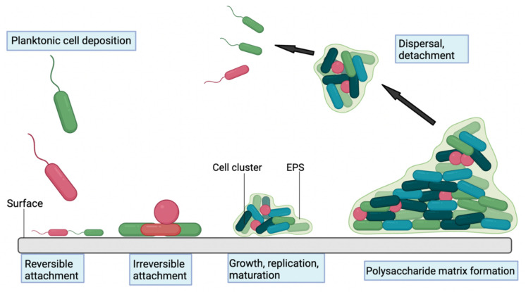
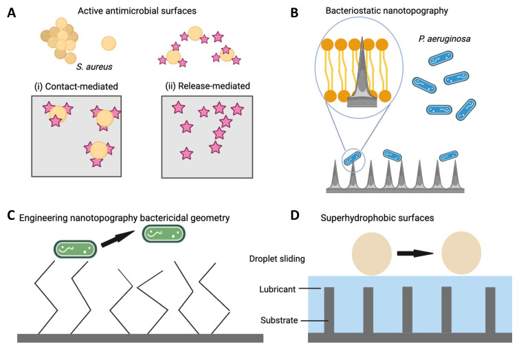
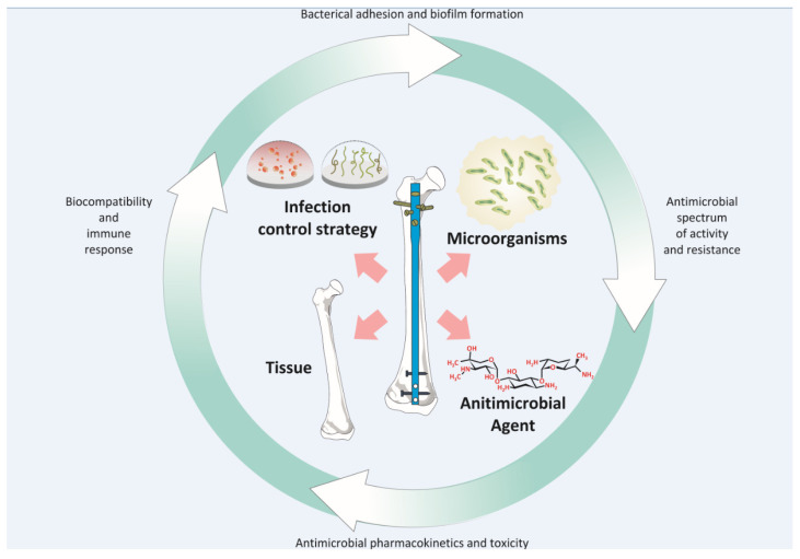
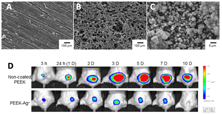
Orthopaedic device implants play a crucial role in restoring functionality to patients suffering from debilitating musculoskeletal diseases or to those who have experienced traumatic injury. However, the surgical implantation of these devices carries a risk of infection, which represents a significant burden for patients and healthcare providers. This review delineates the pathogenesis of orthopaedic implant infections and the challenges that arise due to biofilm formation and the implications for treatment. It focuses on research advancements in the development of next-generation orthopaedic medical devices to mitigate against implant-related infections. Key considerations impacting the development of devices, which must often perform multiple biological and mechanical roles, are delineated. We review technologies designed to exert spatial and temporal control over antimicrobial presentation and the use of antimicrobial surfaces with intrinsic antibacterial activity. A range of measures to control bio-interfacial interactions including approaches that modify implant surface chemistry or topography to reduce the capacity of bacteria to colonise the surface, form biofilms and cause infections at the device interface and surrounding tissues are also reviewed.

Keywords: antimicrobial; biofilm; bioinspired; drug delivery; implant coating; infection; medical device; nanotechnology; orthopaedic implants; polymer.


Conflict of interest statement

The authors declare no conflict of interest. The funders had no role in the design of the study; in the collection, analyses, or interpretation of data; in the writing of the manuscript, or in the decision to publish the results.

Figures

Figure 1
Figure 1
Schematic representation of the main steps in biofilm formation on a surface. Biofilm formation is a cyclic process that begins with surface contact by single planktonic cells. Cells go through reversible attachment whereby bacteria attach to a surface via their flagella or cell pole, to irreversible attachment when flagella reversal rates decrease, and biofilm matrix component production occurs. Biofilm maturation follows, and dispersion occurs due to extracellular polymeric substance (EPS) degradation. Created with BioRender.com.
Figure 2
Figure 2
Schematic illustration of various antimicrobial and drug-free strategies currently under investigation to combat device-related infection. (A) Active antimicrobial surfaces utilise antimicrobial agents that have been coated on the surface or encapsulated within coatings to kill or impede bacteria growth. They exert their effect when (i) bacteria contact the antimicrobial surface or (ii) when released. Drug-free strategies include engineering (B) bactericidal nanotopographies and (C) device surfaces to minimise bacterial adhesion including polymeric brushes that impede microbial attachment owing to steric hindrance, or (D) the use of superhydrophobic surfaces to repel microbes, such as SLIPS (slippery liquid-infused porous surfaces). Created with BioRender.com.
Figure 3
Figure 3
Important considerations impacting the design and clinical performance of orthopaedic devices incorporating antimicrobial agents to control device-related infection.
Figure 4
Figure 4
Example of an ionic silver antimicrobial coating immobilized onto hydroxyapatite covering a polyether–ether–ketone (PEEK) implant coating. The surface of PEEK (A) was immersed in 98% concentrated sulfuric acid (H2SO4) for 10 min (B), after which a porous configuration was observed on the surface by SEM. (C) Hydroxyapatite aggregates were homogeneously coated on the PEEK coated with immobilized Ag+ ions (PEEK-Ag+). (D). Sequential analysis of the bacterial bioluminescence in the mouse soft tissue infection model, where a non-coated PEEK of PEEK-Ag+ plate was placed into the superficial gluteus muscle of mouse, followed by inoculation with a bioluminescent strain of S. aureus. The bacterial photon density was measured at 3, 12 and 24 h, and then each day until 10 days after the operation). Modified and reproduced from [190] under CC BY 4.0 http://creativecommons.org/licenses/by/4.0/ (accessed on 27 July 2022).
Figure 5
Figure 5
SEM images of titania nanotubes (TNT) grown on Ti wire using the anodization technique. (A) The top surface showing cracks, (B) the entire structure showing TNT on Ti wire with dimensions, (C) the cross-section showing array of TNTs and (D) the hollow nanotubes. (E) Overall release and (F) burst release of gentamicin (corresponding to the first 6 h of fast diffusion of drug) from TNT-Ti wire. Modified and reproduced from [216] CC BY 2.0 http://creativecommons.org/licenses/by/2.0 (accessed on 18 October 2022).
Figure 6
Figure 6
Architectural representation of various bactericidal or repellent surface engineering strategies outlined in Section 7. (A) Nanotubular Ti after anodization, adapted with permission from [220]. (B) Multilevel (micro- and nano-) roughened aluminium alloys adapted from [221] under CC BY 4.0 http://creativecommons.org/licenses/by/4.0/ (accessed on accessed on 18 October 2022). (C) Reduced graphene oxide (rGO) nanosheets on silicon wafer adapted with permission from [222]. (D) Hyperbranched poly-L-lysine coating on Ti implant adapted with permission from [223].
Figure 7
Figure 7
SEM of P. aeruginosa response to the (A) D900 uncoated flat control (top, middle rows). Flagella were observed on the control samples (arrows) (bottom row in column A). (B) P. aeruginosa response to the 90 s pPEA-2 hr fibronectin (FN), bone morphogenetic protein-2 (BMP2)-coated D900 surface (top, middle rows, column B). Flagella were absent in bacterial cells observed on 90 s pPEA-2 hr FN/BMP2-coated D900, with ruptured bacterial cells also observed on the nanowires (arrowheads). In the control uncoated samples, large areas of confluent biofilms were observed in contrast to small, more diffuse bacterial accumulation on the nanowire-coated samples. Modified and reproduced with permission from [234].
Figure 8
Figure 8
(A) Schematic of the fabrication process of LOIS. A stainless-steel bare substrate is chemically etched using hydrofluoric acid (HF) and passivated with nitric acid (HNO3) to slow corrosion. The surface is then modified with a self-assembled monolayer (SAM) to increase the chemical affinity between the surface and the subsequently added slippery perfluorocarbon-based lubricant. (B) Fluorescence microscopy images of each material (bare, etched, superhydrophobic (SHP), lubricated orthopaedic implant surface (LOIS) incubated in P. aeruginosa and MRSA suspension for 12 and 72 h. (C) Quantification of adherent CFUs of P. aeruginosa and methicillin-resistant Staphylococcus aureus (MRSA) on each group of surfaces. (D) Quantitative analysis of the callus formation outside cortical bone with (1) micro-CT and (2) osteoclast activity based on TRAP activity. (E) X-ray images of fractured bone of bare negative (without being exposed to bacterial suspension) surface and LOIS 6 weeks post-implantation. Statistical significance, ns (not significant), * p < 0.05; ** p < 0.01, *** p < 0.001, and **** p < 0.0001. Modified and reproduced from [259] under CC BY-NC 4.0 https://creativecommons.org/licenses/by-nc/4.0/ (accessed on 27 July 2022).

Similar articles

Cited by

References

    1. Goodman S.B., Yao Z., Keeney M., Yang F. The future of biologic coatings for orthopaedic implants. Biomaterials. 2013;34:3174–3183. doi: 10.1016/j.biomaterials.2013.01.074. - DOI - PMC - PubMed
    1. McMillan D.J., Lutton C., Rosenzweig N., Sriprakash K.S., Goss B., Stemberger M., Schuetz M.A., Steck R. Prevention of staphylococcus aures biofilm formation on metallic surgical implants via controlled release of gentamicin. J. Biomed. Sci. Eng. 2011;4:535–542. doi: 10.4236/jbise.2011.48069. - DOI
    1. Evans N.T., Torstrick F.B., Lee C.S.D., Dupont K.M., Safranski D.L., Chang W.A., Macedo A.E., Lin A.S.P., Boothby J.M., Whittingslow D.C., et al. High-strength, surface-porous polyether-ether-ketone for load-bearing orthopedic implants. Acta Biomater. 2015;13:159–167. doi: 10.1016/j.actbio.2014.11.030. - DOI - PMC - PubMed
    1. O’ Sullivan C., Kennedy G., O’ Neill L., Crean A.M., Ryan K.B. Biomedical Applications of Inorganic Materials. The Royal Society of Chemistry; Cambridge, UK: 2022. Chapter 5 Inorganic Biomaterials to Support the Formation and Repair of Bone Tissue; pp. 242–304.
    1. Stigter M., Bezemer J., de Groot K., Layrolle P. Incorporation of different antibiotics into carbonated hydroxyapatite coatings on titanium implants, release and antibiotic efficacy. J. Control. Release. 2004;99:127–137. doi: 10.1016/j.jconrel.2004.06.011. - DOI - PubMed

LinkOut - more resources