. 2022 Dec 12;14(24):6132.
doi: 10.3390/cancers14246132.

A Novel Therapeutic Target for Small-Cell Lung Cancer: Tumor-Associated Repair-like Schwann Cells

Affiliations

A Novel Therapeutic Target for Small-Cell Lung Cancer: Tumor-Associated Repair-like Schwann Cells

Shuhui Cao et al. Cancers (Basel). .

Abstract

Small-cell lung cancer (SCLC), representing 15-20% of all lung cancers, is an aggressive malignancy with a distinct natural history, poor prognosis, and limited treatment options. We have previously identified Schwann cells (SCs), the main glial cells of the peripheral nervous system, in tumor tissues and demonstrated that they may support tumor spreading and metastasis formation in the in vitro and in vivo models. However, the role of SCs in the progression of SCLC has not been investigated. To clarify this issue, the cell proliferation assay, the annexin V apoptosis assay, and the transwell migration and invasion assay were conducted to elucidate the roles in SCLC of tumor-associated SCs (TA-SCs) in the proliferation, apoptosis, migration, and invasion of SCLC cells in vitro, compared to control group. In addition, the animal models to assess SC action's effects on SCLC in vivo were also developed. The result confirmed that TA-SCs have a well-established and significant role in facilitating SCLC cell cancer migration and invasion of SCLC in vitro, and we also observed that SC promotes tumor growth of SCLC in vivo and that TA-SCs exhibited an advantage and show a repair-like phenotype, which allowed defining them as tumor-associated repair SCs (TAR-SCs). Potential molecular mechanisms of pro-tumorigenic activity of TAR-SCs were investigated by the screening of differentially expressed genes and constructing networks of messenger-, micro-, and long- non-coding RNA (mRNA-miRNA-lncRNA) using DMS114 cells, a human SCLC, stimulated with media from DMS114-activated SCs, non-stimulated SCs, and appropriate controls. This study improves our understanding of how SCs, especially tumor-activated SCs, may promote SCLC progression. Our results highlight a new functional phenotype of SCs in cancer and bring new insights into the characterization of the nervous system-tumor crosstalk.

Keywords: Schwann cells; gene expression; miRNA; small-cell lung cancer; tumor progression.


Conflict of interest statement

The authors declare no conflict of interest.

Figures

Figure 1
Figure 1
SCs increase the migration (A) and invasion (B) of small-cell lung cancer cells. DMS114, DMS53, and H1048 cells were cultured in Transwell inserts (8-μm pore size) without (migration assay) or with a Matrigel layer (invasion assay). The bottom chambers contained medium (medium) as a control, SC-conditioned medium (SC-con med), or conditioned medium from SCs pretreated with DMS114, DMS53, or H1048 cells (SC/DMS114-con, SC/DMS53-con medium, or SC/H1048-con medium). The tumor cells were allowed to transmigrate for 20 h in the migration assay and 40 h in the invasion assay at 37 °C. Cells that penetrated the underside surfaces of the membranes were fixed, stained, and counted on a grid in 10 HPF. Stained membranes from the representative experiment are shown. The results are shown as a relative invasion cell number—transmigrating to control cell ratio. Each assay was carried out in duplicates and all experiments were repeated at least three times. Scale bar is 100 um. **, p < 0.01, ***, p < 0.001, ****, p < 0.0001. SC/DMS114-con, SC/DMS53-con, SC/H1048-con medium, or SC-con vs. medium (one-way ANOVA).
Figure 2
Figure 2
The SC effects on the SCLC cell proliferation and viability. (A) SCs may increase the proliferation of the SCLC cells in vitro. DMS114, DMS53, and H1048 cells were cultured for 24 h in a complete RPMI-1640 medium containing 10% (v/v) SC medium (medium), 10% (v/v) SC-conditioned medium (SC−con med), and 10% (v/v) conditioned medium from SCs pretreated with DMS114, DMS53, and H1048 cells (SC/DMS114-con med, SC/DMS53-con med, or SC/H1048-con med). Cell proliferation was determined with the cell counting-8 assay. The results are expressed as the mean ± SEM from three independent experiments. **, p < 0.01; SC/DMS53-con, or SC/H1048-con vs. medium. (B) SCs do not alter the apoptosis of the SCLC cells in vitro. DMS114, DMS53, and H1048 cells were cultured for 48 h in a complete RPMI medium containing 10% (v/v) SC medium (medium), 10% (v/v) SC-conditioned medium (SC-con med), and 10% (v/v) conditioned medium from SCs pretreated with DMS114, DMS53, or H1048 cells (SC/DMS114-con med, SC/DMS53-con med, or SC/H1048-con med). Then, cells were stained with PI and FITC-conjugated Annexin V, and analyzed by flow cytometry. Q1: necrotic, AV−/PI+; Q2: late-apoptotic, AV+/PI+; Q3: live, AV−/PI−; Q4: early-apoptotic, AV+/PI−. The results are expressed as the mean ± SEM from three independent experiments.
Figure 3
Figure 3
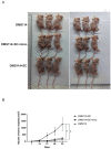
SCs promote tumorigenicity of DMS114 cells in vivo. Nude mice (n = 6/group) were injected with DMS114 cells (DMS114), DMS114 cells plus SCs (DMS114+SC), and DMS114 cells plus minocycline-pretreated SCs (DMS114+SC+mino). Tumor growth was assessed for five weeks. Representative pictures of three nude mice in each group are shown (A). Tumor growth curves of all six mice in each group are shown (B). The results are expressed as the mean ± SEM. *, p < 0.05, DMS114+SC vs. DMS114+SC+mino group; ^, and p < 0.05, DMS114+SC vs. DMS114 group.
Figure 4
Figure 4
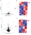
Expression profiles of distinct mRNAs in SC-treated SCLC cells. Volcano plot (A) and heat map (B) of the differentially expressed mRNAs in SCLC cells treated with the tumor-activated SC-conditioned medium (SC114MP-1 and SC114MP-2) and control medium (con-1 and con-2). Volcano plot (C) and heat map (D) of the differentially expressed mRNAs in SCLC cells treated with the tumor-activated SC-conditioned medium (SC114MP-1 and SC114MP-2) and SC-conditioned medium (SCSMP-1 and SCSMP-2). Volcano plot (E) and heat map (F) of the differentially expressed mRNAs in SCLC cells treated with the SC-conditioned medium (SCSMP-1 and SCSMP-2) and control medium (con-1 and con-2).
Figure 4
Figure 4
Expression profiles of distinct mRNAs in SC-treated SCLC cells. Volcano plot (A) and heat map (B) of the differentially expressed mRNAs in SCLC cells treated with the tumor-activated SC-conditioned medium (SC114MP-1 and SC114MP-2) and control medium (con-1 and con-2). Volcano plot (C) and heat map (D) of the differentially expressed mRNAs in SCLC cells treated with the tumor-activated SC-conditioned medium (SC114MP-1 and SC114MP-2) and SC-conditioned medium (SCSMP-1 and SCSMP-2). Volcano plot (E) and heat map (F) of the differentially expressed mRNAs in SCLC cells treated with the SC-conditioned medium (SCSMP-1 and SCSMP-2) and control medium (con-1 and con-2).
Figure 5
Figure 5
Expression profiles of distinct lncRNAs in SC-treated SCLC cells. Volcano plot (A) and heat map (B) of the differentially expressed lncRNAs in SCLC cells treated with the tumor-activated SC-conditioned medium (SC114MP-1 and SC114MP-2) and control medium (con-1 and con-2). Volcano plot (C) and heat map (D) of the differentially expressed lncRNAs in SCLC cells treated with the tumor-activated SC-conditioned medium (SC114MP-1 and SC114MP-2) and SC-conditioned medium (SCSMP-1 and SCSMP-2). Volcano plot (E) and heat map (F) of the differentially expressed lncRNAs in SCLC cells treated with the SC-conditioned medium (SCSMP-1 and SCSMP-2) and control medium (con-1 and con-2).
Figure 5
Figure 5
Expression profiles of distinct lncRNAs in SC-treated SCLC cells. Volcano plot (A) and heat map (B) of the differentially expressed lncRNAs in SCLC cells treated with the tumor-activated SC-conditioned medium (SC114MP-1 and SC114MP-2) and control medium (con-1 and con-2). Volcano plot (C) and heat map (D) of the differentially expressed lncRNAs in SCLC cells treated with the tumor-activated SC-conditioned medium (SC114MP-1 and SC114MP-2) and SC-conditioned medium (SCSMP-1 and SCSMP-2). Volcano plot (E) and heat map (F) of the differentially expressed lncRNAs in SCLC cells treated with the SC-conditioned medium (SCSMP-1 and SCSMP-2) and control medium (con-1 and con-2).
Figure 6
Figure 6
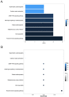
Gene ontology (GO) enrichment annotations of pathological progression of differentially expressed mRNAs in the SC-treated SCLC cells was categorized into three groups: biological process (BP), cellular component (CC) and molecular function (MF). (A,B): Enrichment result of GO of the differentially expressed mRNAs in treated SCLC cells comparing the SC/DMS114-conditioned group (SC114MP-1 and SC114MP-2) and control group (con-1 and con-2). (C,D): Enrichment result of GO of the differentially expressed mRNAs in treated SCLC cells comparing the SC/DMS114-conditioned group (SC114MP-1 and SC114MP-2) and SC-conditioned group (SCSMP-1 and SCSMP-2). (E,F): Enrichment result of GO of the differentially expressed mRNAs in treated SCLC cells comparing the SC-conditioned group (SCSMP-1 and SCSMP-2) and control group (con-1 and con-2).
Figure 6
Figure 6
Gene ontology (GO) enrichment annotations of pathological progression of differentially expressed mRNAs in the SC-treated SCLC cells was categorized into three groups: biological process (BP), cellular component (CC) and molecular function (MF). (A,B): Enrichment result of GO of the differentially expressed mRNAs in treated SCLC cells comparing the SC/DMS114-conditioned group (SC114MP-1 and SC114MP-2) and control group (con-1 and con-2). (C,D): Enrichment result of GO of the differentially expressed mRNAs in treated SCLC cells comparing the SC/DMS114-conditioned group (SC114MP-1 and SC114MP-2) and SC-conditioned group (SCSMP-1 and SCSMP-2). (E,F): Enrichment result of GO of the differentially expressed mRNAs in treated SCLC cells comparing the SC-conditioned group (SCSMP-1 and SCSMP-2) and control group (con-1 and con-2).
Figure 7
Figure 7
The Kyoto Encyclopedia of Genes and Genomes (KEGG) pathway enrichment analysis of the differentially expressed mRNAs in treated SCLC cells compared to the SC/DMS114-conditioned group, SC-conditioned group, and control group. (A,B): Enrichment result of the KEGG analysis of the differentially expressed mRNAs in SCLC cells treated by the tumor-activated SC-conditioned medium (SC114MP-1 and SC114MP-2) and SC-conditioned medium (SCSMP-1 and SCSMP-2). (C,D): Enrichment result of the KEGG analysis of the differentially expressed mRNAs in SCLC cells treated by the SC-conditioned group (SCSMP-1 and SCSMP-2) and control medium (con-1 and con-2).
Figure 7
Figure 7
The Kyoto Encyclopedia of Genes and Genomes (KEGG) pathway enrichment analysis of the differentially expressed mRNAs in treated SCLC cells compared to the SC/DMS114-conditioned group, SC-conditioned group, and control group. (A,B): Enrichment result of the KEGG analysis of the differentially expressed mRNAs in SCLC cells treated by the tumor-activated SC-conditioned medium (SC114MP-1 and SC114MP-2) and SC-conditioned medium (SCSMP-1 and SCSMP-2). (C,D): Enrichment result of the KEGG analysis of the differentially expressed mRNAs in SCLC cells treated by the SC-conditioned group (SCSMP-1 and SCSMP-2) and control medium (con-1 and con-2).
Figure 8
Figure 8
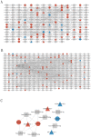
mRNA-miRNA-lncRNA and ceRNA networks in small-cell lung cancer, conditioned by SCs. According to the targeting relationship between miRNA-mRNA and miRNA-lncRNA, and the expression trend of lncRNA and mRNA, mRNA-miRNA-lncRNA and ceRNA networks between the SC/DMS114-conditioned group and control group (A), the SC-conditioned group and control group (B), and the SC/DMS114-conditioned group and SC-conditioned group (C) were conducted. The gray rectangle represents miRNA, the circle represents mRNA, the triangle represents lncRNA, the red color represents the gene up-regulation in the corresponding group comparison, and the blue color represents the down-regulation.

Similar articles

Cited by

References

    1. Siegel R.L., Miller K.D., Jemal A. Cancer Statistics, 2017. CA Cancer J. Clin. 2017;67:7–30. doi: 10.3322/caac.21387. - DOI - PubMed
    1. Torre L.A., Siegel R.L., Jemal A. Lung Cancer Statistics. Adv. Exp. Med. Biol. 2016;893:1–19. doi: 10.1007/978-3-319-24223-1_1. - DOI - PubMed
    1. Hanahan D., Weinberg R.A. Hallmarks of cancer: The next generation. Cell. 2011;144:646–674. doi: 10.1016/j.cell.2011.02.013. - DOI - PubMed
    1. Dyachuk V., Furlan A., Shahidi M.K., Giovenco M., Kaukua N., Konstantinidou C., Pachnis V., Memic F., Marklund U., Müller T., et al. Parasympathetic neurons originate from nerve-associated peripheral glial progenitors. Science. 2014;345:82–87. doi: 10.1126/science.1253281. - DOI - PubMed
    1. Baraldi J.H., Martyn G.V., Shurin G.V., Shurin M.R. Tumor Innervation: History, Methodologies, and Significance. Cancers. 2022;14:1979. doi: 10.3390/cancers14081979. - DOI - PMC - PubMed

LinkOut - more resources