

Prostate-Specific Membrane Antigen Targeted Pet/CT Imaging in Patients with Colon, Gastric and Pancreatic Cancer
- PMID: 36551695
- PMCID: PMC9777210
- DOI: 10.3390/cancers14246209
Prostate-Specific Membrane Antigen Targeted Pet/CT Imaging in Patients with Colon, Gastric and Pancreatic Cancer
Abstract
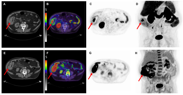
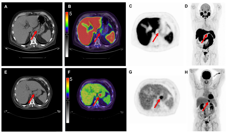
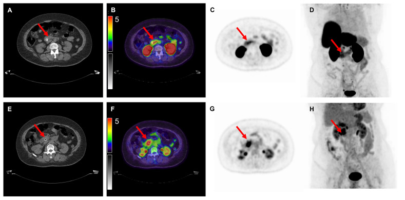
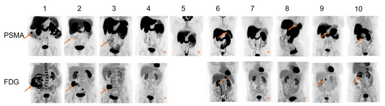
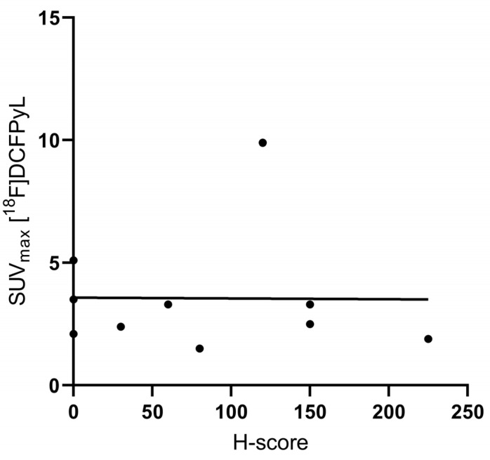
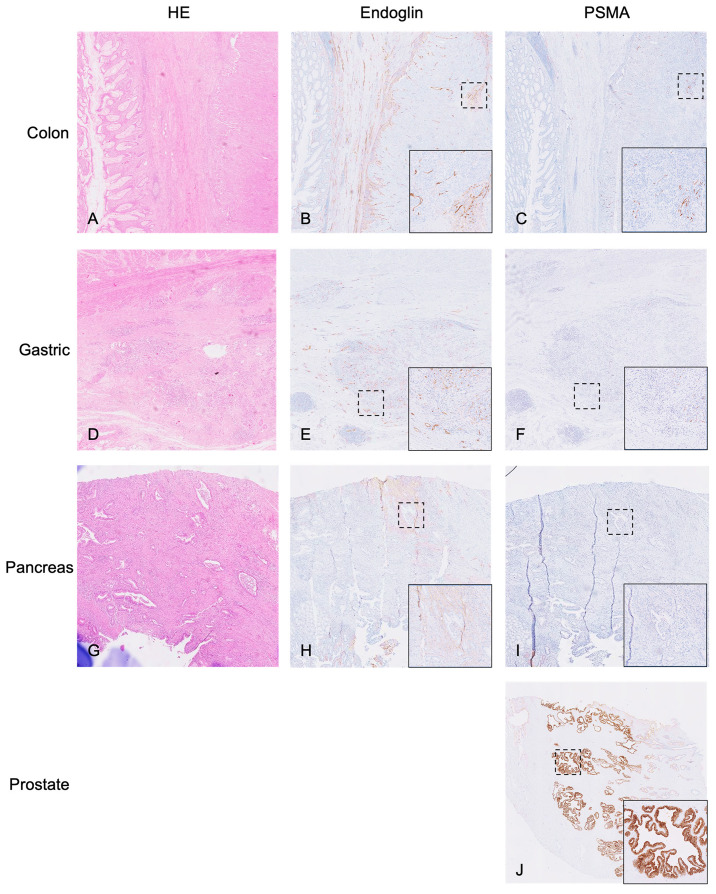
Current imaging modalities frequently misjudge disease stage in colorectal, gastric and pancreatic cancer. As treatment decisions are dependent on disease stage, incorrect staging has serious consequences. Previous preclinical research and case reports indicate that prostate-specific membrane antigen (PSMA)-targeted PET/CT imaging might provide a solution to some of these challenges. This prospective clinical study aims to assess the feasibility of [18F]DCFPyL PET/CT imaging to target and visualize primary colon, gastric and pancreatic cancer. In this prospective clinical trial, patients with colon, gastric and pancreatic cancer were included and underwent both [18F]DCFPyL and [18F]FDG PET/CT scans prior to surgical resection or (for gastric cancer) neoadjuvant therapy. Semiquantitative analysis of immunohistochemical PSMA staining was performed on the surgical resection specimens, and the results were correlated to imaging parameters. The results of this study demonstrate detection of the primary tumor by [18F]DCFPyL PET/CT in 7 out of 10 patients with colon, gastric and pancreatic cancer, with a mean tumor-to-blood pool ratio (TBR) of 3.3 and mean SUVmax of 3.6. However, due to the high surrounding uptake, visual distinction of these tumors was difficult, and the SUVmax and TBR on [18F]FDG PET/CT were significantly higher than on [18F]DCFPyL PET/CT. In addition, no correlation between PSMA expression in the resection specimen and SUVmax on [18F]DCFPyL PET/CT was found. In conclusion, the detection of several gastrointestinal cancers using [18F]DCFPyL PET/CT is feasible. However, low tumor expression and high uptake physiologically in organs/background hamper the clear distinction of the tumor. As a result, [18F]FDG PET/CT was superior in detecting colon, gastric and pancreatic cancers.
Keywords: PET/CT; PSMA; colon cancer; gastric cancer; pancreatic cancer.
Conflict of interest statement
The authors declare no conflict of interest.
Figures
References
-
- Ferlay J., Ervik M., Lam F., Colombet M., Mery L., Piñeros M., Znaor A., Soerjomataram I., Bray F. Global Cancer Observatory: Cancer Today. International Agency for Research on Cancer; Lyon, France: 2020. [(accessed on 9 February 2022)]. Available online: https://gco.iarc.fr/today.
-
- Cunningham D., Allum W.H., Stenning S.P., Thomposon J.N., Van de Velde C.J.H., Nicolson M., Scarffe J.H., Lofts F.J., Falk S.J., Iveson T.J., et al. Perioperative chemotherapy versus surgery alone for resectable gastroesophageal cancer. N. Engl. J. Med. 2006;355:11–20. doi: 10.1056/NEJMoa055531. - DOI - PubMed
-
- Choi J.Y., Shim K.-N., Kim S.-E., Jung H.-K., Jung S.-A., Yoo K. The Clinical Value of 18F-Fluorodeoxyglucose Uptake on Positron Emission Tomography/Computed Tomography for Predicting Regional Lymph Node Metastasis and Non-curative Surgery in Primary Gastric Carcinoma. Korean J. Gastroenterol. 2014;64:340–347. doi: 10.4166/kjg.2014.64.6.340. - DOI - PubMed
Grants and funding
LinkOut - more resources
Full Text Sources
Research Materials
Miscellaneous
