Review
. 2022 Nov 30;10(12):3081.
doi: 10.3390/biomedicines10123081.

Lipedema: Insights into Morphology, Pathophysiology, and Challenges

Affiliations
Review

Lipedema: Insights into Morphology, Pathophysiology, and Challenges

Ankita Poojari et al. Biomedicines. .

Abstract

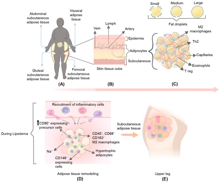
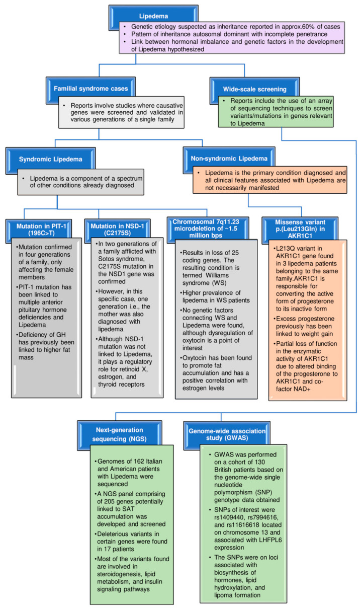
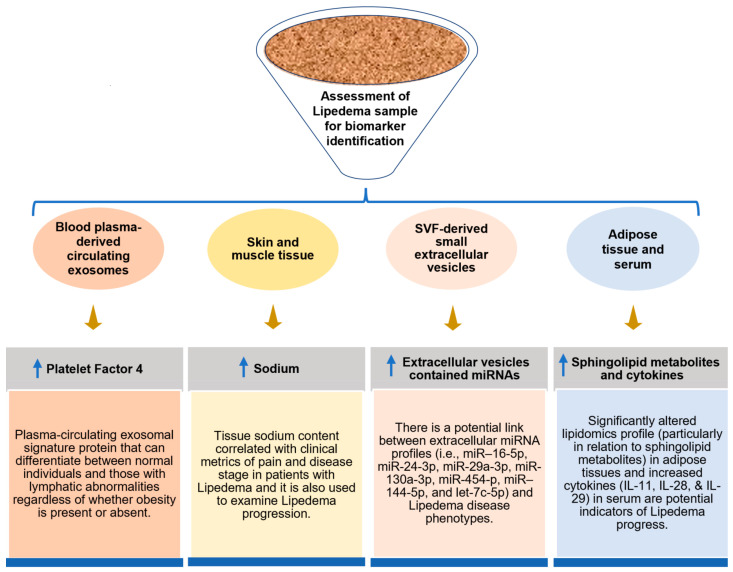
Lipedema is an adipofascial disorder that almost exclusively affects women. Lipedema leads to chronic pain, swelling, and other discomforts due to the bilateral and asymmetrical expansion of subcutaneous adipose tissue. Although various distinctive morphological characteristics, such as the hyperproliferation of fat cells, fibrosis, and inflammation, have been characterized in the progression of lipedema, the mechanisms underlying these changes have not yet been fully investigated. In addition, it is challenging to reduce the excessive fat in lipedema patients using conventional weight-loss techniques, such as lifestyle (diet and exercise) changes, bariatric surgery, and pharmacological interventions. Therefore, lipedema patients also go through additional psychosocial distress in the absence of permanent treatment. Research to understand the pathology of lipedema is still in its infancy, but promising markers derived from exosome, cytokine, lipidomic, and metabolomic profiling studies suggest a condition distinct from obesity and lymphedema. Although genetics seems to be a substantial cause of lipedema, due to the small number of patients involved in such studies, the extrapolation of data at a broader scale is challenging. With the current lack of etiology-guided treatments for lipedema, the discovery of new promising biomarkers could provide potential solutions to combat this complex disease. This review aims to address the morphological phenotype of lipedema fat, as well as its unclear pathophysiology, with a primary emphasis on excessive interstitial fluid, extracellular matrix remodeling, and lymphatic and vasculature dysfunction. The potential mechanisms, genetic implications, and proposed biomarkers for lipedema are further discussed in detail. Finally, we mention the challenges related to lipedema and emphasize the prospects of technological interventions to benefit the lipedema community in the future.

Keywords: adipose tissue; chronic disease; fat disorder; lipedema; lymphedema; obesity.


Conflict of interest statement

The authors declare that the research was conducted without any commercial or financial relationships that could be construed as potential conflicts of interest.

Figures

Figure 1
Figure 1
Representation of subcutaneous adipose tissue (SAT) characteristics pattern in lipedema. (A) The SAT location and deposition in the body; (B) skin tissue cube presenting their residing components; (C) characteristics of lean adipose tissue accompanied by ECM, immune cells, and various forms of typical fat droplets; (D) hypertrophy and hyperplasia in SAT with immune-cell requirements; and (E) inflammation, excessive fluid deposition, and swelling in the upper leg/thigh, which are the common characteristic of individuals with lipedema.
Figure 2
Figure 2
The cycle of events underpinning the lipedema phenotype. (A) Increased expression of genes associated with mitotic clonal expansion (MCE) and cell proliferation leads to hyperplasia. Although increased accumulation of lipids within fat cells leading to hypertrophy has been confirmed in lipedema, the expression of markers associated with such a finding is still under debate. (B) Due to the overgrowth of fat cells, there is a reduced oxygen supply, and extracellular matrix (ECM) remodeling is observed. ECM remodeling includes sodium (Na+) concentration, collagen deposition, and disturbances in the glycocalyx (including proteoglycans and glycosaminoglycan), leading to microangiopathy and fibrosis. (C) Endothelial permeability and paracellular leakage are increased due to the loosening of the tight junctions between endothelial cells (ECs), further deteriorating vascular impairment and causing inflammation. In addition, the ability of the lymphatic capillaries to take up interstitial fluid (IF) is diminished, causing leakage of IF, promoting lymphatic dysfunction and expansion of interstitial space. (D) Pathogenic changes both at the vascular and lymphatic levels contribute to the accumulation of IF, which leads to the deposition of fluid. In addition, the excess IF surrounding the fat cells acts as a source of nutrients, further contributing to the pathological expansion of the fat cells, eventually causing the remodeling of SAT and lipedema phenotype cyclically. Blue: Decreased expression of these genes observed in lipedema; Brown: Increased expression of these genes observed in lipedema; Black: Contradictory findings have been reported for these genes.
Figure 3
Figure 3
The flowchart depicts the holistic view of the current genetic implications for lipedema. Screening and validation of causative genetic factors performed in various generations of a single family are highlighted in familial syndrome cases of patients with lipedema. This is further categorized based on syndromic (described based on the chromosomal structural changes and a mutation in specific genes) and nonsyndromic (development of missense gene variants, i.e., L213Q variant in AKR1C1 gene) conditions. The wide-scale screening included next-generation sequencing (NGS) and genome-wide association studies (GWAS), revealing the variants/mutations in genes found to be linked or relevant to lipedema.
Figure 4
Figure 4
Schematic representation of identified lipedema biomarkers. This image shows the findings of some promising biomarkers from various sources of the lipedema samples and the underlying investigation to find their potential as diagnostic tools for lipedema.

Similar articles

Cited by

References

    1. La Torre Y.S.-D., Wadeea R., Rosas V., Herbst K.L. Lipedema: Friend and Foe. Horm. Mol. Biol. Clin. Investig. 2018;33:1. doi: 10.1515/hmbci-2017-0076. - DOI - PMC - PubMed
    1. Buso G., Depairon M., Tomson D., Raffoul W., Vettor R., Mazzolai L. Lipedema: A Call to Action! Obesity. 2019;27:1567. doi: 10.1002/oby.22597. - DOI - PMC - PubMed
    1. Shavit E., Wollina U., Alavi A. Lipoedema Is Not Lymphoedema: A Review of Current Literature. Int. Wound J. 2018;15:921–928. doi: 10.1111/iwj.12949. - DOI - PMC - PubMed
    1. Herbst K.L. Rare Adipose Disorders (RADs) Masquerading as Obesity. Acta Pharmacol. Sin. 2012;33:155–172. doi: 10.1038/aps.2011.153. - DOI - PMC - PubMed
    1. Buck D.W., Herbst K.L. Lipedema: A Relatively Common Disease with Extremely Common Misconceptions. Plast. Reconstr. Surg. Glob. Open. 2016;4:e1043. doi: 10.1097/GOX.0000000000001043. - DOI - PMC - PubMed

LinkOut - more resources