Monocytic Cell Adhesion to Oxidised Ligands: Relevance to Cardiovascular Disease
- PMID: 36551839
- PMCID: PMC9775297
- DOI: 10.3390/biomedicines10123083
Monocytic Cell Adhesion to Oxidised Ligands: Relevance to Cardiovascular Disease
Abstract
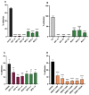
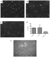
Atherosclerosis, the major cause of vascular disease, is an inflammatory process driven by entry of blood monocytes into the arterial wall. LDL normally enters the wall, and stimulates monocyte adhesion by forming oxidation products such as oxidised phospholipids (oxPLs) and malondialdehyde. Adhesion molecules that bind monocytes to the wall permit traffic of these cells. CD14 is a monocyte surface receptor, a cofactor with TLR4 forming a complex that binds oxidised phospholipids and induces inflammatory changes in the cells, but data have been limited for monocyte adhesion. Here, we show that under static conditions, CD14 and TLR4 are implicated in adhesion of monocytes to solid phase oxidised LDL (oxLDL), and also that oxPL and malondialdehyde (MDA) adducts are involved in adhesion to oxLDL. Similarly, monocytes bound to heat shock protein 60 (HSP60), but this could be through contaminating lipopolysaccharide. Immunohistochemistry on atherosclerotic human arteries demonstrated increased endothelial MDA adducts and HSP60, but endothelial oxPL was not detected. We propose that monocytes could bind to MDA in endothelial cells, inducing atherosclerosis. Monocytes and platelets synergized in binding to oxLDL, forming aggregates; if this occurs at the arterial surface, they could precipitate thrombosis. These interactions could be targeted by cyclodextrins and oxidised phospholipid analogues for therapy.
Keywords: CD14; LDL; TLR4; adhesion; atherosclerosis; monocyte; oxidation; phospholipid; raft; thrombosis.
Conflict of interest statement
The authors declare no conflict of interest.
Figures


Similar articles
-
Attenuation of monocyte adhesion and oxidised LDL uptake in luteolin-treated human endothelial cells exposed to oxidised LDL.Br J Nutr. 2007 Mar;97(3):447-57. doi: 10.1017/S0007114507657894. Br J Nutr. 2007. PMID: 17313705
-
Methyl-β-cyclodextrin suppresses the monocyte-endothelial adhesion triggered by lipopolysaccharide (LPS) or oxidized low-density lipoprotein (oxLDL).Pharm Biol. 2021 Dec;59(1):1036-1044. doi: 10.1080/13880209.2021.1953540. Pharm Biol. 2021. PMID: 34362284 Free PMC article.
-
Co-expression of ICAM-1, VCAM-1, ELAM-1 and Hsp60 in human arterial and venous endothelial cells in response to cytokines and oxidized low-density lipoproteins.Cell Stress Chaperones. 1997 Jun;2(2):94-103. doi: 10.1379/1466-1268(1997)002<0094:ceoive>2.3.co;2. Cell Stress Chaperones. 1997. PMID: 9250400 Free PMC article.
-
Benefits of gliclazide in the atherosclerotic process: decrease in monocyte adhesion to endothelial cells.Metabolism. 2003 Aug;52(8 Suppl 1):13-8. doi: 10.1016/s0026-0495(03)00212-9. Metabolism. 2003. PMID: 12939734 Review.
-
Effects of oxidized low density lipoprotein, lipid mediators and statins on vascular cell interactions.Clin Chem Lab Med. 1999 Mar;37(3):243-51. doi: 10.1515/CCLM.1999.043. Clin Chem Lab Med. 1999. PMID: 10353467 Review.
Cited by
-
Editorial to the Special Issue "Molecular and Cellular Mechanisms of CVD: Focus on Atherosclerosis".Biomedicines. 2024 Sep 23;12(9):2148. doi: 10.3390/biomedicines12092148. Biomedicines. 2024. PMID: 39335661 Free PMC article.
-
Poly-(ADP-ribose) polymerases inhibition by olaparib attenuates activities of the NLRP3 inflammasome and of NF-κB in THP-1 monocytes.PLoS One. 2024 Feb 9;19(2):e0295837. doi: 10.1371/journal.pone.0295837. eCollection 2024. PLoS One. 2024. PMID: 38335214 Free PMC article.
References
-
- Watson A.D., Leitinger N., Navab M., Faull K.F., Hörkkö S., Witztum J.L., Palinski W., Schwenke D., Salomon R.G., Sha W., et al. Structural identification by mass spectrometry of oxidized phospholipids in minimally oxidized low density lipoprotein that induce monocyte/endothelial interactions and evidence for their presence in vivo. J. Biol. Chem. 1997;272:13597–13607. doi: 10.1074/jbc.272.21.13597. - DOI - PubMed
Grants and funding
LinkOut - more resources
Full Text Sources
Research Materials
Miscellaneous

