. 2022 Dec 1;10(12):3091.
doi: 10.3390/biomedicines10123091.

High-Resolution Magic-Angle-Spinning NMR in Revealing Hepatoblastoma Hallmarks

Affiliations

High-Resolution Magic-Angle-Spinning NMR in Revealing Hepatoblastoma Hallmarks

Ljubica Tasic et al. Biomedicines. .

Abstract

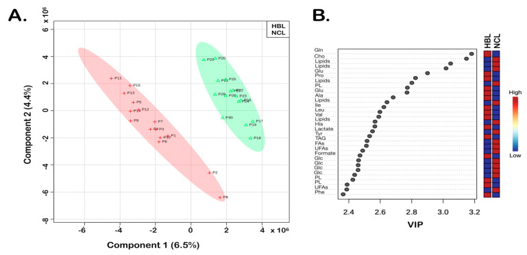
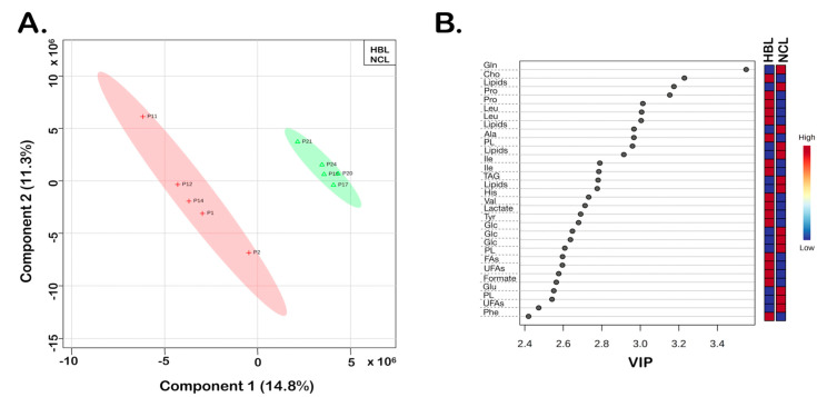
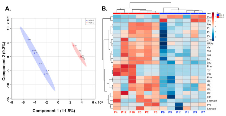
Cancer is one of the leading causes of death in children and adolescents worldwide; among the types of liver cancer, hepatoblastoma (HBL) is the most common in childhood. Although it affects only two to three individuals in a million, it is mostly asymptomatic at diagnosis, so by the time it is detected it has already advanced. There are specific recommendations regarding HBL treatment, and ongoing studies to stratify the risks of HBL, understand the pathology, and predict prognostics and survival rates. Although magnetic resonance imaging spectroscopy is frequently used in diagnostics of HBL, high-resolution magic-angle-spinning (HR-MAS) NMR spectroscopy of HBL tissues is scarce. Using this technique, we studied the alterations among tissue metabolites of ex vivo samples from (a) HBL and non-cancer liver tissues (NCL), (b) HBL and adjacent non-tumor samples, and (c) two regions of the same HBL samples, one more centralized and the other at the edge of the tumor. It was possible to identify metabolites in HBL, then metabolites from the HBL center and the border samples, and link them to altered metabolisms in tumor tissues, highlighting their potential as biochemical markers. Metabolites closely related to liver metabolisms such as some phospholipids, triacylglycerides, fatty acids, glucose, and amino acids showed differences between the tissues.

Keywords: cancer NMR-metabolomics; hepatoblastoma; liver metabolome.


Conflict of interest statement

The authors declare no conflict of interest.

Figures

Figure 1
Figure 1
HR-MAS 1H-NMR spectra of liver tissue samples, from the bottom to the top: non-cancer liver (NCL) sample, hepatoblastoma sample (HBL-C) taken from the center of the tumor, and hepatoblastoma sample taken from the border of the tumor (HBL-B) in 0.0-5.5 ppm. Identification of the tissue metabolites is given in Table 1.
Figure 2
Figure 2
HBL vs. NCL. PLS-DA of the HR-MAS 1H-NMR CPMG data: (A) 2D score plot shows 15 HBL and 15 NCL samples; statistical parameters were as follows: accuracy (0.68), R2 (0.77), and Q2 (0.39). (B) Variable importance in projection (VIP) scores greater than 2.35 were assigned to important metabolites 1–16, as summarized in Table 1, which are discriminatory for HBL vs. NCL in the PLS-DA model. HBL tissue samples are shown with red crosses and the non-cancer liver tissue samples (NCL) are shown in green triangles.
Figure 3
Figure 3
Paired HBL vs. NCL samples, PLS-DA of the HR-MAS 1H-NMR CPMG data: (A) 2D score plot showing five HBL and five NCL samples; statistical parameters were as follows: accuracy (0.48), R2 (0.57), and Q2 (0.03). (B) Variable importance in projection (VIP) scores greater than 2.4 were assigned to important metabolites 1–16 (Table 1). HBL tissue samples are shown in red crosses and the non-cancer liver tissue samples (NCL) are shown with green triangles.
Figure 4
Figure 4
HBL border vs. center, PLS-DA of the HR-MAS 1H-NMR CPMG data in the analysis of the cancer samples from border and center, HBL-B and HBL-C, respectively: (A) 2D score plot; statistical parameters were as follows: accuracy (0.71), R2 (0.87), and Q2 (0.23). (B) Heatmap showing discriminatory metabolites for the cancer samples. HBL-B tissue samples are shown in blue and HBL-C are shown in red.

References

    1. Johnson C., Ivanisevic J., Siuzdak G. Metabolomics: Beyond biomarkers and towards mechanisms. Nat. Rev. Mol. Cell Biol. 2016;17:451–459. doi: 10.1038/nrm.2016.25. - DOI - PMC - PubMed
    1. Wang J., Tian R., Shan Y., Li J., Gao H., Xie C., Ma Y., Wu Y., Ji B., Gu S., et al. Metabolomics study of the metabolic changes in hepatoblastoma cells in response to NTCP/SLC10A1 overexpression. Int. J. Biochem. Cell Biol. 2020;125:105773. doi: 10.1016/j.biocel.2020.105773. - DOI - PubMed
    1. Emwas A.H., Roy R., McKay R.T., Tenori L., Saccenti E., Gowda G.A.N., Raftery D., Alahmari F., Jaremko L., Jaremko M., et al. NMR spectroscopy for metabolomics research. Metabolites. 2019;9:123. doi: 10.3390/metabo9070123. - DOI - PMC - PubMed
    1. Pontes J.G.M., Brasil A.J.M., Cruz G.C.F., de Souza R.N., Tasic L. NMR-based metabolomics strategies: Plants, animals and humans. Anal. Methods. 2017;9:1078. doi: 10.1039/C6AY03102A. - DOI - PubMed
    1. Quintero Escobar M., Costa T.B.B.C., Martins L.G., Costa S.S., VanHelvoort Lengert A., Boldrini E., Morini da Silva S.R., Lopes L.F., Vidal D.O., Krepischi A.C.V., et al. Insights in osteosarcoma by proton nuclear magnetic resonance serum metabonomics. Front. Oncol. 2020;10:506959. doi: 10.3389/fonc.2020.506959. - DOI - PMC - PubMed