

Immunohistochemical Detection of Estrogen Receptor-Beta (ERβ) with PPZ0506 Antibody in Murine Tissue: From Pitfalls to Optimization
- PMID: 36551855
- PMCID: PMC9775465
- DOI: 10.3390/biomedicines10123100
Immunohistochemical Detection of Estrogen Receptor-Beta (ERβ) with PPZ0506 Antibody in Murine Tissue: From Pitfalls to Optimization
Abstract
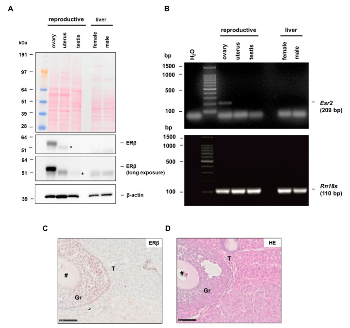
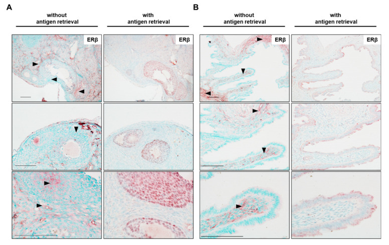
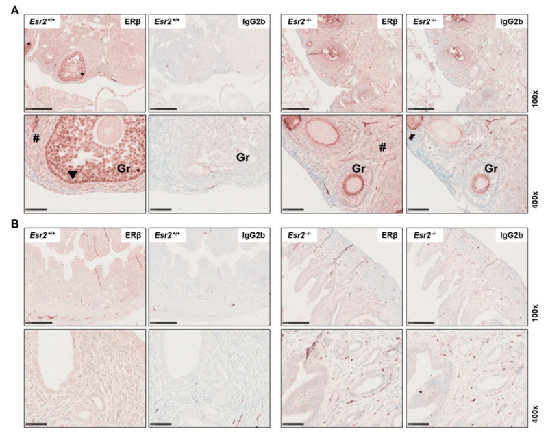
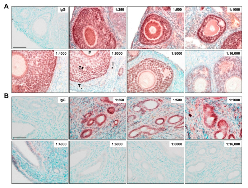
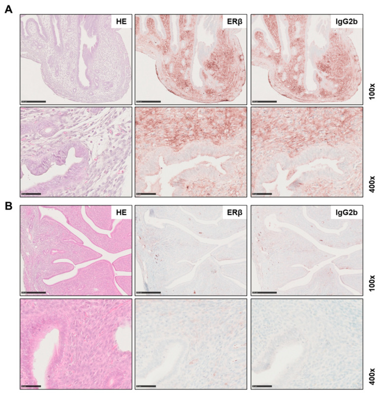
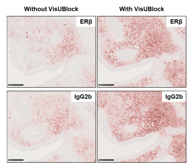
The estrogen receptor beta (ERβ) is physiologically essential for reproductive biology and is implicated in various diseases. However, despite more than 20 years of intensive research on ERβ, there are still uncertainties about its distribution in tissues and cellular expression. Several studies show contrasts between mRNA and protein levels, and the use of knockout strategies revealed that many commercially available antibodies gave false-positive expression results. Recently, a specific monoclonal antibody against human ERβ (PPZ0506) showed cross-reactivity with rodents and was optimized for the detection of rat ERβ. Herein, we established an immunohistochemical detection protocol for ERβ protein in mouse tissue. Staining was optimized on murine ovaries, as granulosa cells are known to strongly express ERβ. The staining results were confirmed by western blot analysis and RT-PCR. To obtain accurate and reliable staining results, different staining conditions were tested in paraffin-embedded tissues. Different pitfalls were encountered in immunohistochemical detection. Strong heat-induced epitope retrieval (HIER) and appropriate antibody dilution were required to visualize specific nuclear expression of ERβ. Finally, the specificity of the antibody was confirmed by using ovaries from Esr2-depleted mice. However, in some animals, strong (non-specific) background staining appeared. These signals could not be significantly alleviated with commercially available additional blocking solutions and are most likely due to estrus-dependent expression of endogenous immunoglobulins. In summary, our study showed that the antibody PPZ0506, originally directed against human ERβ, is also suitable for reliable detection of murine ERβ. An established staining protocol mitigated ambiguities regarding the expression and distribution of ERβ in different tissues and will contribute to an improved understanding of its role and functions in murine tissues in the future.
Keywords: Esr2; PPZ0506; antibody validation; estrogen receptor beta (ERβ); immunohistochemistry; murine tissue; ovary; staining.
Conflict of interest statement
The authors declare no conflict of interest. The funders had no role in the design of the study; in the collection, analyses, or interpretation of data; in the writing of the manuscript, or in the decision to publish the results.
Figures
Similar articles
-
Optimized Mouse-on-mouse Immunohistochemical Detection of Mouse ESR2 Proteins with PPZ0506 Monoclonal Antibody.Acta Histochem Cytochem. 2022 Oct 28;55(5):159-168. doi: 10.1267/ahc.22-00043. Epub 2022 Oct 25. Acta Histochem Cytochem. 2022. PMID: 36405553 Free PMC article.
-
Optimization of immunohistochemical detection of rat ESR2 proteins with well-validated monoclonal antibody PPZ0506.Mol Cell Endocrinol. 2021 Mar 1;523:111145. doi: 10.1016/j.mce.2020.111145. Epub 2021 Jan 2. Mol Cell Endocrinol. 2021. PMID: 33400952
-
Applicability of Anti-Human Estrogen Receptor β Antibody PPZ0506 for the Immunodetection of Rodent Estrogen Receptor β Proteins.Int J Mol Sci. 2019 Dec 13;20(24):6312. doi: 10.3390/ijms20246312. Int J Mol Sci. 2019. PMID: 31847265 Free PMC article.
-
The importance of ERbeta signalling in the ovary.J Endocrinol. 2010 Apr;205(1):15-23. doi: 10.1677/JOE-09-0379. Epub 2009 Dec 17. J Endocrinol. 2010. PMID: 20019181 Review.
-
The estrogen receptor beta subtype: a novel mediator of estrogen action in neuroendocrine systems.Front Neuroendocrinol. 1998 Oct;19(4):253-86. doi: 10.1006/frne.1998.0170. Front Neuroendocrinol. 1998. PMID: 9799586 Review.
Cited by
-
Expression Analysis of Lipocalin 2 (LCN2) in Reproductive and Non-Reproductive Tissues of Esr1-Deficient Mice.Int J Mol Sci. 2023 May 25;24(11):9280. doi: 10.3390/ijms24119280. Int J Mol Sci. 2023. PMID: 37298232 Free PMC article.
-
Divergent features of ERβ isoforms in triple negative breast cancer: progress and implications for further research.Front Cell Dev Biol. 2023 Oct 23;11:1240386. doi: 10.3389/fcell.2023.1240386. eCollection 2023. Front Cell Dev Biol. 2023. PMID: 37936981 Free PMC article. Review.
-
Ovarian ERβ cistrome and transcriptome reveal chromatin interaction with LRH-1.BMC Biol. 2023 Nov 29;21(1):277. doi: 10.1186/s12915-023-01773-1. BMC Biol. 2023. PMID: 38031019 Free PMC article.
-
Ovaries of estrogen receptor 1-deficient mice show iron overload and signs of aging.Front Endocrinol (Lausanne). 2024 Feb 23;15:1325386. doi: 10.3389/fendo.2024.1325386. eCollection 2024. Front Endocrinol (Lausanne). 2024. PMID: 38464972 Free PMC article.
-
Isolation of Bovine and Human Milk Extracellular Vesicles.Biomedicines. 2023 Oct 6;11(10):2715. doi: 10.3390/biomedicines11102715. Biomedicines. 2023. PMID: 37893089 Free PMC article.
References
Grants and funding
LinkOut - more resources
Full Text Sources
