Review
. 2022 Dec 8;10(12):3186.
doi: 10.3390/biomedicines10123186.

Axonal Regeneration: Underlying Molecular Mechanisms and Potential Therapeutic Targets

Affiliations
Review

Axonal Regeneration: Underlying Molecular Mechanisms and Potential Therapeutic Targets

Rabia Akram et al. Biomedicines. .

Abstract

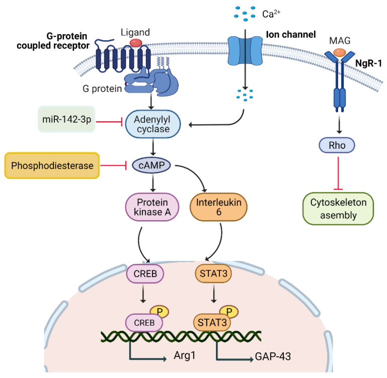
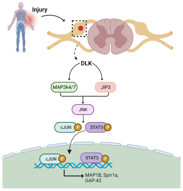
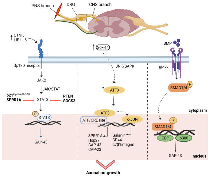
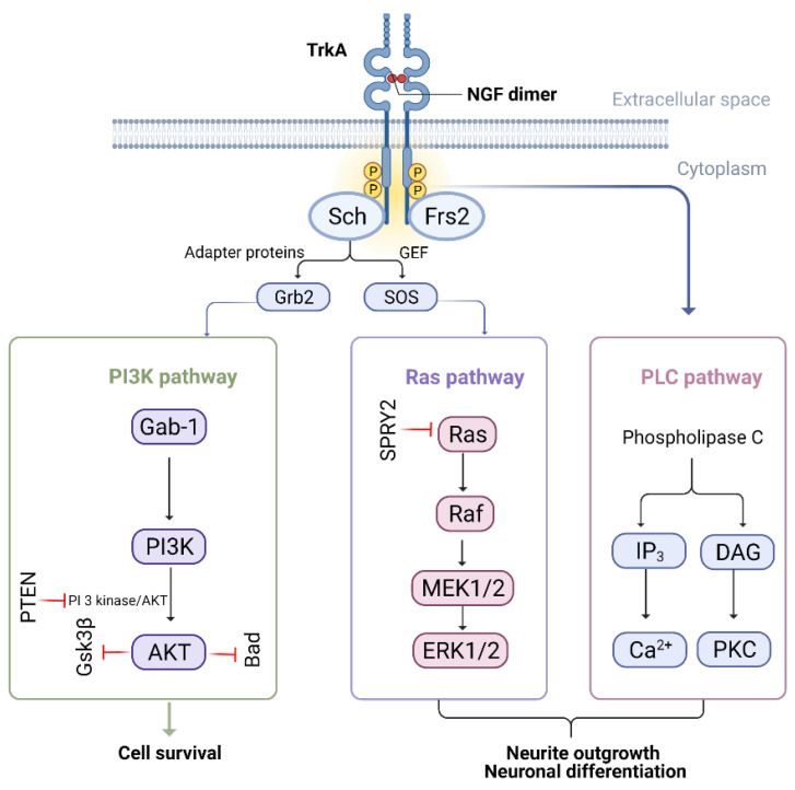
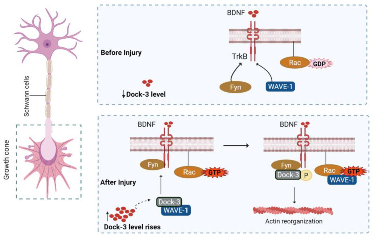
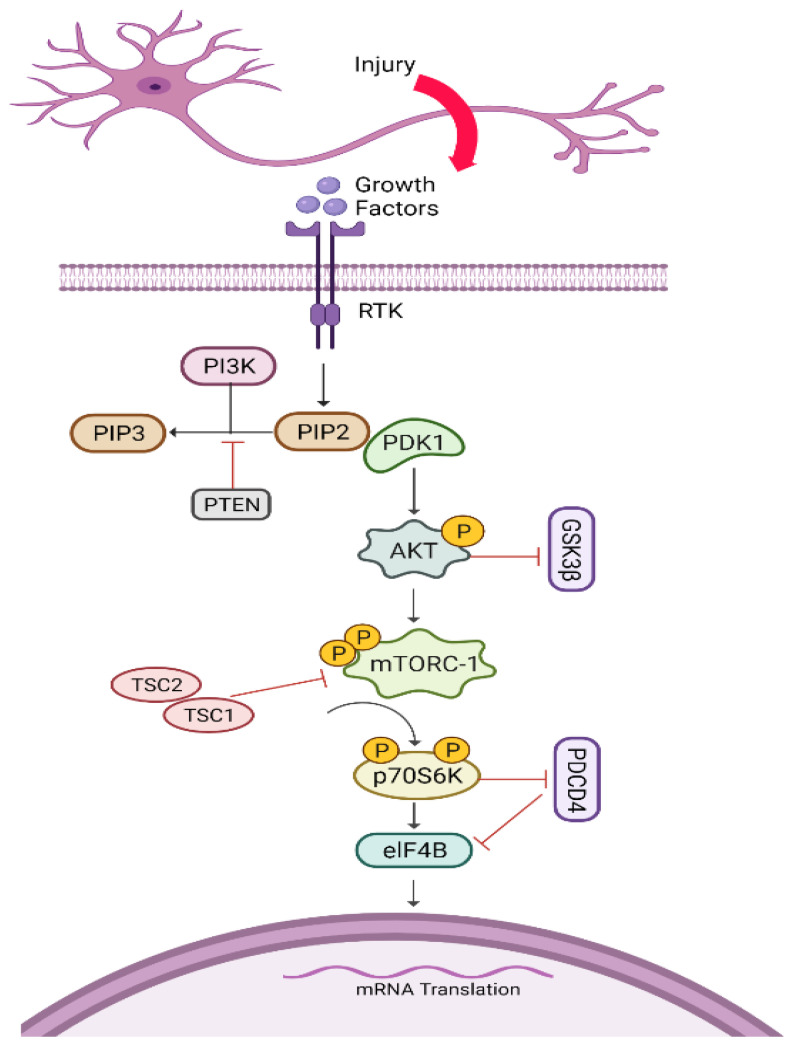
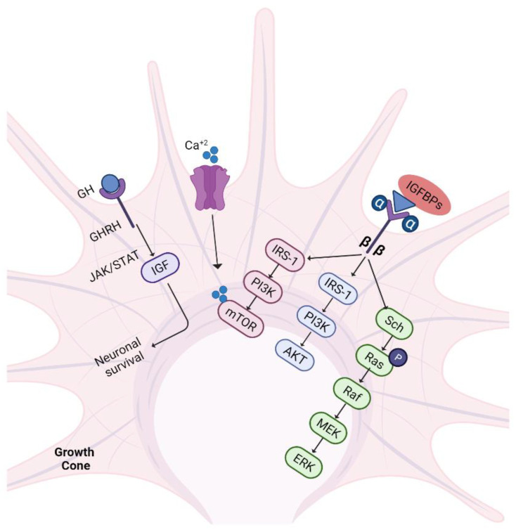
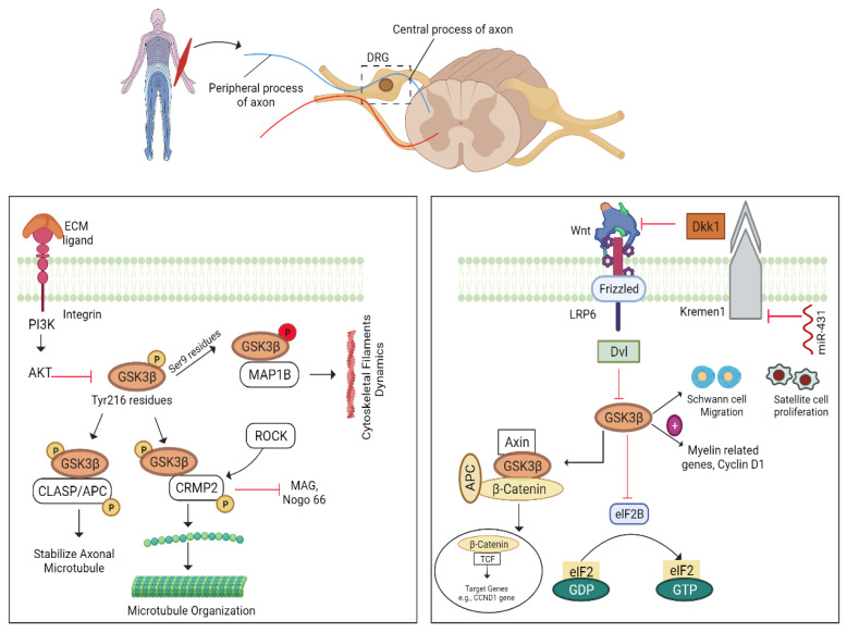
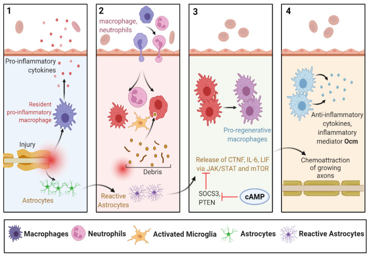
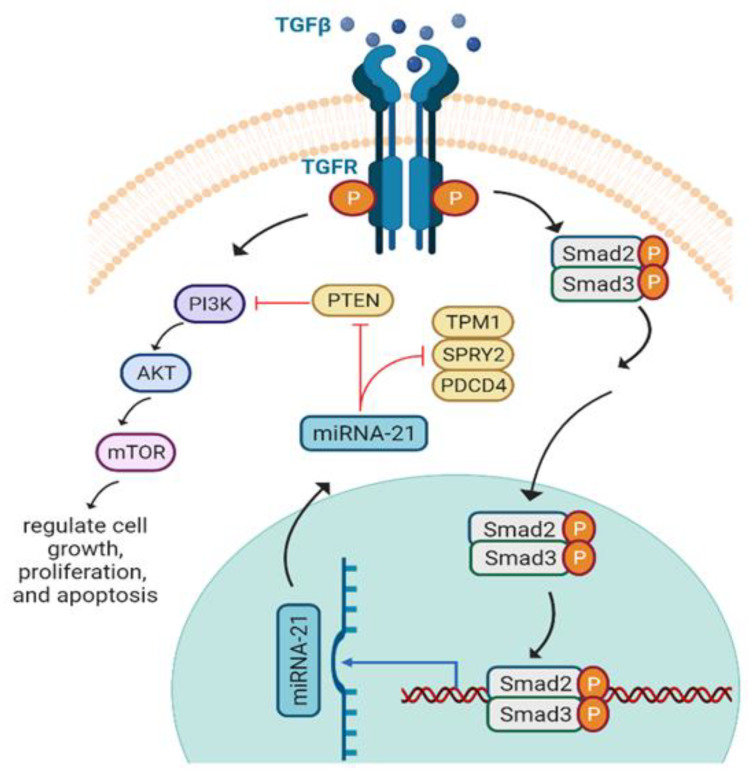
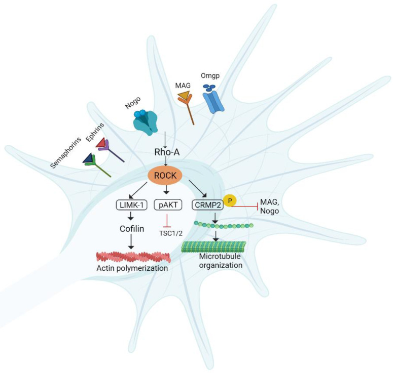
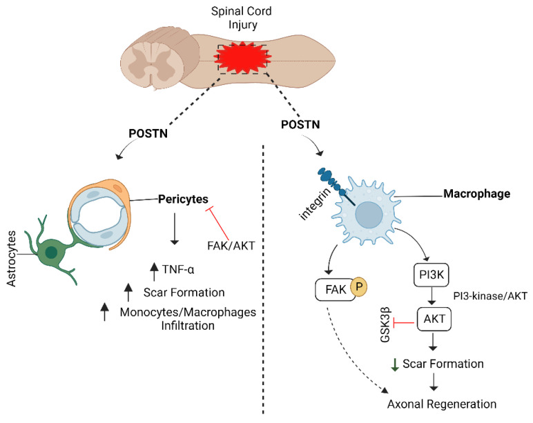
Axons in the peripheral nervous system have the ability to repair themselves after damage, whereas axons in the central nervous system are unable to do so. A common and important characteristic of damage to the spinal cord, brain, and peripheral nerves is the disruption of axonal regrowth. Interestingly, intrinsic growth factors play a significant role in the axonal regeneration of injured nerves. Various factors such as proteomic profile, microtubule stability, ribosomal location, and signalling pathways mark a line between the central and peripheral axons' capacity for self-renewal. Unfortunately, glial scar development, myelin-associated inhibitor molecules, lack of neurotrophic factors, and inflammatory reactions are among the factors that restrict axonal regeneration. Molecular pathways such as cAMP, MAPK, JAK/STAT, ATF3/CREB, BMP/SMAD, AKT/mTORC1/p70S6K, PI3K/AKT, GSK-3β/CLASP, BDNF/Trk, Ras/ERK, integrin/FAK, RhoA/ROCK/LIMK, and POSTN/integrin are activated after nerve injury and are considered significant players in axonal regeneration. In addition to the aforementioned pathways, growth factors, microRNAs, and astrocytes are also commendable participants in regeneration. In this review, we discuss the detailed mechanism of each pathway along with key players that can be potentially valuable targets to help achieve quick axonal healing. We also identify the prospective targets that could help close knowledge gaps in the molecular pathways underlying regeneration and shed light on the creation of more powerful strategies to encourage axonal regeneration after nervous system injury.

Keywords: axonal regeneration; cyclic adenosine monophosphate; microRNAs; nerve injury; neurotrophic factors; regeneration-associated genes.


Conflict of interest statement

The authors declare no conflict of interest.

Figures

Figure 1
Figure 1
cAMP pathway. Peripheral nerve injury triggers calcium release that upregulates cAMP level via enhancing AC9 expression and regulates three pathways in DRG neurons to promote neurite outgrowth. miR-142-3p, PKA, Rho and phosphodiesterase are the target sites of AC9, Rho, cytoskeletal assembly, and cAMP, respectively, and can be used as therapeutic targets to enhance neurite outgrowth. cAMP: cycline adenosine monophosphate; AC9: adenylyl cyclase 9; DRG: dorsal root ganglion; PKA: protein kinase A; MAG: myelin-associated glycoproteins; NgR1: NOGO receptor; CREB: cAMP response element-binding protein; STAT3: signal transducer and activator of transcription 3; Gap-43: growth-associated proteins; Arg-1: arginase-1.
Figure 2
Figure 2
DLK/JNK/MAPK pathway. DLK acts on JIP3 and MAP2k4/7 to activate JNK-mediated STAT3 and cJUN to enhance axon regeneration. DLK: dual leucine zipper kinase; JIP3: JNK-interacting protein 3; MAP2k4/7: mitogen-activated protein kinase; JNK: c-Jun N terminal kinase; STAT3: signal transducer and activator of transcription 3; Map1b: microtubule-associated proteins; Sprr1a: small proline-rich protein 1A; Gap-43: growth-associated proteins.
Figure 3
Figure 3
Transcriptional factors’ mediated pathways. Following injury, different intrinsic mechanisms are stimulated and mediated by regeneration associated transcriptional factors. First, injury upregulates the expression of IL-6, LIF, and CTNF in DRG neurons that interfere with Gp130 receptors to activate the JAK/STAT pathway to promote axon growth. Similarly, ATF3 increases upon injury and undergoes JNK signaling to activate AP-1 and c-JUN-mediated RAGs to increase axon outgrowth, respectively. Moreover, phosphorylation of SMAD 1 and 4 causes p300 and CBP recruitment and enhances Gap-43 expression to promote axon regeneration. IL-6: interleukin 6; LIF: leukemia inhibitory factor; CTNF: ciliary neurotrophic factor; DRG: dorsal root ganglion; ATF3: activating transcription factor 3; JNK; AP-1: activator protein-1; Gap-43: growth-associated protein; SMAD: protein; p300/CBP: co-activating proteins; BMP: bone morphogenic proteins; BMPR: bone morphogenic proteins receptors; Sprr1a/Hsp27/Gap-43/Cap-23/Galanin/α7βintegrin: regeneration-associated genes.
Figure 4
Figure 4
NGF-mediated pathways. Following NGF binding to tropomyosin, receptor kinase activates Ras/ERK and PI3K/AKT, which subsequently leads to increase neuronal survival and, ultimately, axonal growth. TrkA: tyrosine kinase receptor; NGF: nerve growth factor; GEF: guanine nucleotide exchange factor; SOS/Grb2: adaptor proteins; Frs2: fibroblast growth factor receptor substrate 2; ERK: extracellular signal-regulated kinase; PI3K: phosphatidylinositol-3-kinase; AKT: protein kinase B; DAG: diacyl glycerol; PKC: protein kinase C; IP3: inositol triphosphate; Ras/Raf/MEK: protein kinases; Gab-1: GRB2-associated-binding protein 1.
Figure 5
Figure 5
Dock-mediated BDNF signaling. Upon injury, Dock-3 expression increases at the injury site that recruits WAVE-1 to form a complex with Fyn. This complex interacts with Trk-B receptors to modulate Rac-GDP to Rac-GTP and mediates actin organization for axonal growth. BDNF: brain-derived neurotrophic factor; TrkB: tyrosine kinase receptor; GDP: guanosine diphosphate; GTP: guanosine triphosphate; Dock3/Rac/Fyn/WAVE-1: protein.
Figure 6
Figure 6
AKT/mTORC1/p70S6K pathway. After nerve injury, several growth factors participate to initiate a cascade of reactions for axonal growth. They bind to receptor tyrosine kinase and activate PI3K/AKT-mediated mTORC1, which further phosphorylates S6. pS6 stimulates elG4B to prevent muscle atrophy and thus increases axon regeneration. RTK: receptor tyrosine kinase; PI3K: phosphatidylinositol-3-kinase; PIP3: phosphatidylinositol (3,4,5)-trisphosphate; PIP2: phosphatidylinositol 4,5-bisphosphate; PDK1: pyruvate dehydrogenase kinase 1; PTEN: phosphatase and tensin homolog; AKT: protein kinase B; Gsk3β: glycogen synthase kinase; mTORC-1: mammalian target of rapamycin; TSC1: hamartin; TSC2: tuberin; Pdcd4: programmed cell death 4 gene.
Figure 7
Figure 7
IGF-1/GH pathway. Nerve trauma triggers the release of IGF-1 at the injury site. IGF-1 binds to IGF-1R with the help of binding proteins and increases axonal outgrowth by acting in 3 different ways. Concomitantly, GH also activates IGF by JAK/STAT signalling and promotes neuronal survival. GH: growth hormone; GHRH: growth hormone receptor hormones; JAK/STAT: Janus kinase/signal transducer and activator of transcription; IGF: insulin-like growth factors; IGFBPs: IGF binding proteins; IRS-1: insulin receptor substrate-1; PI3K: phosphatidylinositol-3-kinase; AKT: protein kinase B; mTOR: mammalian target of rapamycin.
Figure 8
Figure 8
GSK3β mediated pathways. GSK3β mediates CLASP/APC signalling and Wnt signalling to stabilize growth cone and axon regeneration. ECM: extracellular matrix proteins; PI3K: phosphatidylinositol-3-kinase; AKT: protein kinase B; GSK3β: glycogen synthase kinase; CLASP/APC: clip-associated proteins/tumor suppressor protein; CRMP2: collapsin response mediator protein 2; Map1b: microtubule-associated proteins; ROCK: Rho-associated kinases; MAG/Nogo; myelin inhibitors; Wnt: protein; Dkk1: Dickkopf-1; Dvl: dishevelled proteins; GDP: guanosine diphosphate; GTP: guanosine triphosphate.
Figure 9
Figure 9
Regeneration by astrocytes/inflammatory mediators. Injury and inflammatory stimuli upon CNS contusion/trauma trigger different pathways and cytokines release. Migration of macrophages and neutrophils toward the injury site, as well as morphological changes of astrocytes promote axonal regeneration. IL-6: interleukin 6; LIF: leukemia inhibitory factor; CTNF: ciliary neurotrophic factor; cAMP: cyclic adenosine monophosphate: PTEN: phosphatase and tensin homolog; SOCS3: suppressor of cytokine signalling 3; Ocm: oncomodulin.
Figure 10
Figure 10
mi-RNA-21 signaling and axonal growth. Following trauma, TGF-β1 binds to particular TGF receptors and stimulates PI3K/AKT/mTOR signalling as well as phosphorylating and translocating SMAD2 and SMAD3 into the nucleus where it modulates miR-21 expression and allows it to move to the cytoplasm and inhibits growth suppressors such as PTEN, Pdcd4, Spry2, and Tpm1 to regulate activation of astrocytes and axonal growth. TGFβ: transforming growth factor; TGFR: transforming growth factor receptors; Smad; PI3K: phosphatidylinositol-3-kinase; PTEN: phosphatase and tensin homolog; AKT: protein kinase B; mTOR: mammalian target of rapamycin; Pdcd4: programmed cell death 4 gene; Tpm1: tropomyosin 1; Spry2: sprouty RTK signalling antagonist 2.
Figure 11
Figure 11
Integrin/FAK pathway. After nerve damage, integrin binds to different extracellular matrix ligands to activate FAK and PI3K/AKT, and Src. At the same time, Nogo and MAG not only bind to their respective receptors but also interfere with integrin to prevent activation of FAK. FAK: focal adhesion kinase; PI3K: phosphatidylinositol-3-kinase; PTEN: phosphatase and tensin homolog; AKT: protein kinase B; Gsk3β: glycogen synthase kinase; MAG: myelin-associated glycoproteins; NgR: Nogo receptor; Rho-A/Src: protein.
Figure 12
Figure 12
RhoA/ROCK/LIMK pathway. Different myelin inhibitors activate RhoA and Rho-associated kinases (ROCK), which in turn activate LIMK-1, cofilin, CRMP2, and AKT to control axonal development. RhoA and ROCK inactivation enhances cytoskeletal actin filament formation and prevents axonal degeneration to boost growth cone stability. TSC1/2 (hamartin–tuberin); AKT: protein kinase B; MAG: myelin-associated glycoproteins; Omgp: oligodendrocyte myelin glycoprotein; CRMP2: collapsin response mediator protein 2; LIMK: LIM domain kinase.
Figure 13
Figure 13
POSTN/integrin pathway. POSTN binds to pericytes and increases scar formation that hinders CNS neurons’ regeneration capacity. On the other side, POSTN binds to integrin receptors and activates PI3K/AKT and FAK signalling. AKT increases monocyte/macrophage migration at the injury site and prevents the formation of scars to increase axon regeneration in CNS neurons. POSTN: periostin; PI3K: phosphatidylinositol-3-kinase; AKT: protein kinase B; FAK: focal adhesion kinase; CNS: central nervous system.

References

    1. Lingor P., Koch J.C., Tönges L., Bähr M. Axonal Degeneration as a Therapeutic Target in the CNS. Cell Tissue Res. 2012;349:289–311. doi: 10.1007/s00441-012-1362-3. - DOI - PMC - PubMed
    1. Sutherland T.C., Geoffroy C.G. The Influence of Neuron-Extrinsic Factors and Aging on Injury Progression and Axonal Repair in the Central Nervous System. Front. Cell Dev. Biol. 2020;8:190. doi: 10.3389/fcell.2020.00190. - DOI - PMC - PubMed
    1. Farah M.H., Pan B.H., Hoffman P.N., Ferraris D., Tsukamoto T., Nguyen T., Wong P.C., Price D.L., Slusher B.S., Griffin J.W. Reduced BACE1 Activity Enhances Clearance of Myelin Debris and Regeneration of Axons in the Injured Peripheral Nervous System. J. Neurosci. 2011;31:5744–5754. doi: 10.1523/JNEUROSCI.6810-10.2011. - DOI - PMC - PubMed
    1. Mahar M., Cavalli V. Intrinsic Mechanisms of Neuronal Axon Regeneration. Nat. Rev. Neurosci. 2018;19:323–337. doi: 10.1038/s41583-018-0001-8. - DOI - PMC - PubMed
    1. Hussain G., Wang J., Rasul A., Anwar H., Qasim M., Zafar S., Aziz N., Razzaq A., Hussain R., de Aguilar J.L.G., et al. Current Status of Therapeutic Approaches against Peripheral Nerve Injuries: A Detailed Story from Injury to Recovery. Int. J. Biol. Sci. 2020;16:116–134. doi: 10.7150/ijbs.35653. - DOI - PMC - PubMed

LinkOut - more resources