

Antimicrobial and Immunomodulatory Potential of Cow Colostrum Extracellular Vesicles (ColosEVs) in an Intestinal In Vitro Model
- PMID: 36552020
- PMCID: PMC9775086
- DOI: 10.3390/biomedicines10123264
Antimicrobial and Immunomodulatory Potential of Cow Colostrum Extracellular Vesicles (ColosEVs) in an Intestinal In Vitro Model
Abstract
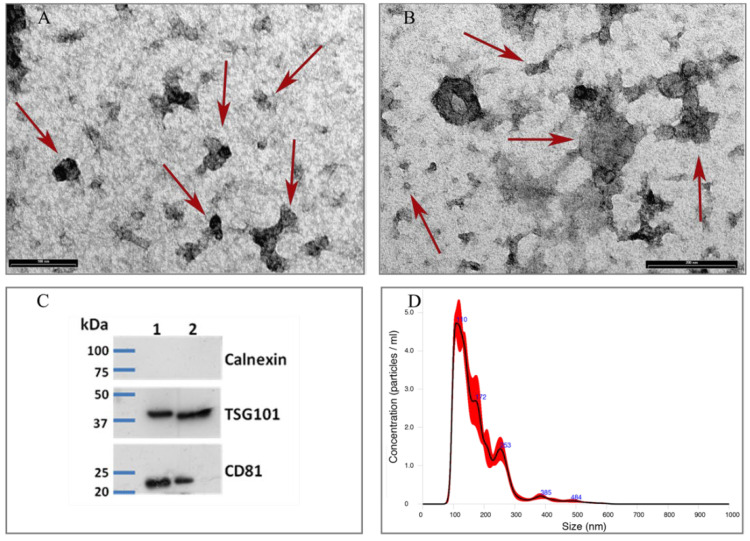
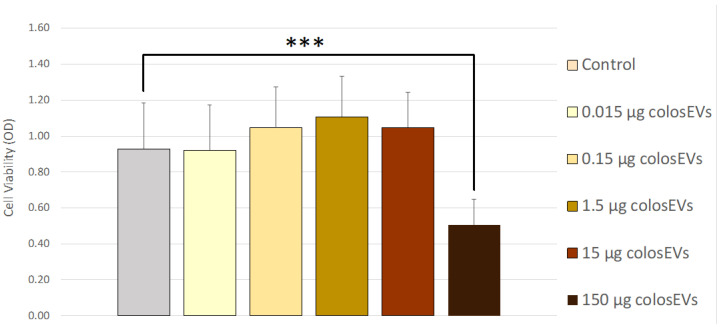
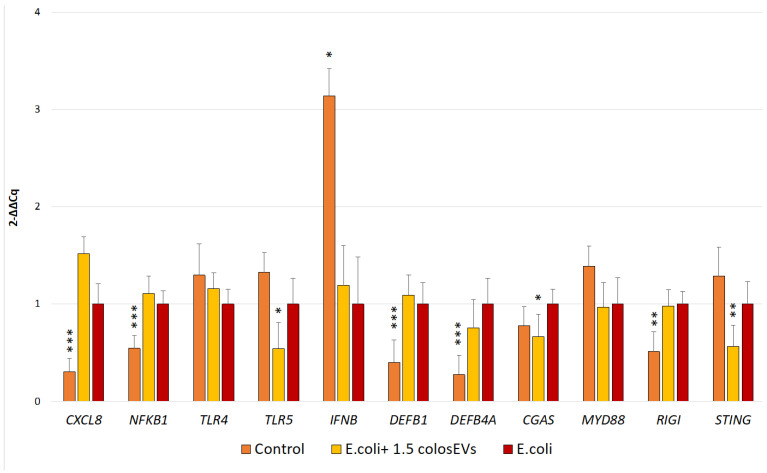
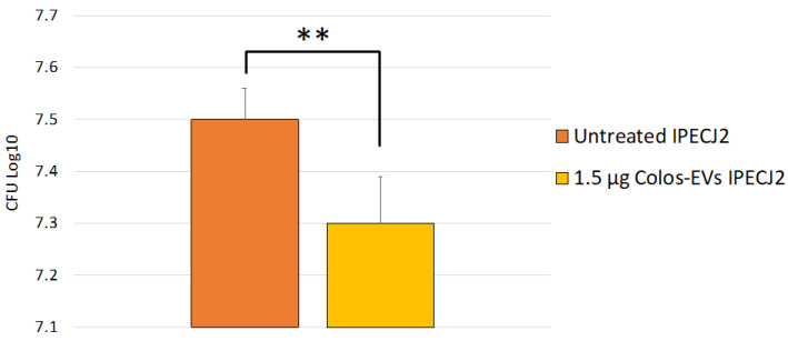
Extracellular Vesicles (EVs) are nano-sized double-lipid-membrane-bound structures, acting mainly as signalling mediators between distant cells and, in particular, modulating the immune response and inflammation of targeted cells. Milk and colostrum contain high amounts of EVs that could be exploited as alternative natural systems in antimicrobial fighting. The aim of this study is to evaluate cow colostrum-derived EVs (colosEVs) for their antimicrobial, anti-inflammatory and immunomodulating effects in vitro to assess their suitability as natural antimicrobial agents as a strategy to cope with the drug resistance problem. ColosEVs were evaluated on a model of neonatal calf diarrhoea caused by Escherichia coli infection, a livestock disease where antibiotic therapy often has poor results. Colostrum from Piedmontese cows was collected within 24 h of calving and colosEVs were immediately isolated. IPEC-J2 cell line was pre-treated with colosEVs for 48 h and then infected with EPEC/NTEC field strains for 2 h. Bacterial adherence and IPEC-J2 gene expression analysis (RT-qPCR) of CXCL8, DEFB1, DEFB4A, TLR4, TLR5, NFKB1, MYD88, CGAS, RIGI and STING were evaluated. The colosEVs pre-treatment significantly reduced the ability of EPEC/NTEC strains to adhere to cell surfaces (p = 0.006), suggesting a role of ColosEVs in modulating host−pathogen interactions. Moreover, our results showed a significant decrease in TLR5 (p < 0.05), CGAS (p < 0.05) and STING (p < 0.01) gene expression in cells that were pre-treated with ColosEVs and then infected, thus highlighting a potential antimicrobial activity of ColosEVs. This is the first preliminarily study investigating ColosEV immunomodulatory and anti-inflammatory effects on an in vitro model of neonatal calf diarrhoea, showing its potential as a therapeutic and prophylactic tool.
Keywords: Extracellular Vesicles; antimicrobial; colibacillosis; coloEVs; colostrum.
Conflict of interest statement
The authors declare no conflict of interest.
Figures
Similar articles
-
Comparison of colostrum and milk extracellular vesicles small RNA cargo in water buffalo.Sci Rep. 2024 Aug 3;14(1):17991. doi: 10.1038/s41598-024-67249-6. Sci Rep. 2024. PMID: 39097641 Free PMC article.
-
Toll-like Receptors and Cytokine Modulation by Goat Milk Extracellular Vesicles in a Model of Intestinal Inflammation.Int J Mol Sci. 2023 Jul 4;24(13):11096. doi: 10.3390/ijms241311096. Int J Mol Sci. 2023. PMID: 37446274 Free PMC article.
-
In vitro evaluation of immunomodulatory activities of goat milk Extracellular Vesicles (mEVs) in a model of gut inflammation.Res Vet Sci. 2022 Dec 20;152:546-556. doi: 10.1016/j.rvsc.2022.09.021. Epub 2022 Sep 24. Res Vet Sci. 2022. PMID: 36179548
-
Flood Control: How Milk-Derived Extracellular Vesicles Can Help to Improve the Intestinal Barrier Function and Break the Gut-Joint Axis in Rheumatoid Arthritis.Front Immunol. 2021 Jul 28;12:703277. doi: 10.3389/fimmu.2021.703277. eCollection 2021. Front Immunol. 2021. PMID: 34394100 Free PMC article. Review.
-
Invited review: MicroRNAs in bovine colostrum-Focus on their origin and potential health benefits for the calf.J Dairy Sci. 2020 Jan;103(1):1-15. doi: 10.3168/jds.2019-16959. Epub 2019 Oct 31. J Dairy Sci. 2020. PMID: 31677833 Review.
Cited by
-
Therapeutic Potential of Bovine Milk-Derived Extracellular Vesicles.Int J Mol Sci. 2024 May 19;25(10):5543. doi: 10.3390/ijms25105543. Int J Mol Sci. 2024. PMID: 38791583 Free PMC article. Review.
-
Feeding a Saccharomyces cerevisiae Fermentation Product to Mares in Late Gestation Alters the Biological Activity of Colostrum.Animals (Basel). 2024 Aug 24;14(17):2459. doi: 10.3390/ani14172459. Animals (Basel). 2024. PMID: 39272244 Free PMC article.
-
Olive Mill Waste-Water Extract Enriched in Hydroxytyrosol and Tyrosol Modulates Host-Pathogen Interaction in IPEC-J2 Cells.Animals (Basel). 2024 Feb 7;14(4):564. doi: 10.3390/ani14040564. Animals (Basel). 2024. PMID: 38396532 Free PMC article.
-
Single cell transcriptomic analysis reveals tumor immune infiltration by macrophage cells gene signature in lung adenocarcinoma.Discov Oncol. 2025 Mar 3;16(1):261. doi: 10.1007/s12672-025-01834-7. Discov Oncol. 2025. PMID: 40029500 Free PMC article.
-
Extracellular vesicles in dairy cattle: research progress and prospects for practical applications.J Anim Sci Biotechnol. 2025 Aug 4;16(1):110. doi: 10.1186/s40104-025-01242-5. J Anim Sci Biotechnol. 2025. PMID: 40754626 Free PMC article. Review.
References
-
- McGrath B.A., Fox P.F., McSweeney P.L.H., Kelly A.L. Composition and Properties of Bovine Colostrum: A Review. Dairy Sci. Technol. 2016;96:133–158. doi: 10.1007/s13594-015-0258-x. - DOI
-
- Ma T., Li W., Chen Y., Cobo E.R., Windeyer C., Gamsjäger L., Diao Q., Tu Y., Guan L.L. Assessment of MicroRNA Profiles in Small Extracellular Vesicles Isolated from Bovine Colostrum with Different Immunoglobulin G Concentrations. JDS Commun. 2022;3:328–333. doi: 10.3168/jdsc.2022-0225. - DOI - PMC - PubMed
Grants and funding
LinkOut - more resources
Full Text Sources
Research Materials
Miscellaneous
