

Heterogeneity of White Matter Hyperintensity and Cognitive Impairment in Patients with Acute Lacunar Stroke
- PMID: 36552134
- PMCID: PMC9776102
- DOI: 10.3390/brainsci12121674
Heterogeneity of White Matter Hyperintensity and Cognitive Impairment in Patients with Acute Lacunar Stroke
Abstract
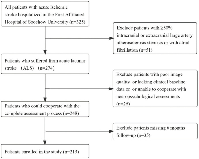
Background: The severity of white matter hyperintensity (WMH) in patients with acute lacunar stroke (ALS) may be not completely parallel to cognitive impairment. Controversies persist about the effects of WMH on cognitive dysfunction. It is vital to explore whether the association may be affected by certain factors and whether a subsequent subgroup analysis is necessary. The aim of this study was to evaluate the relationship between WMH and cognitive impairment in acute lacunar stroke patients and the possible causal factors. Methods: We continuously enrolled patients with ALS who were hospitalized at the First Affiliated Hospital of Soochow University between October 2017 and June 2022. The cognitive function of all patients was assessed by using the Montreal Cognitive Assessment (MoCA) scale 14 ± 2 days after the onset of AIS, and the results were adjusted to the education level. The MoCA scale was reevaluated at the 6-month (day 182 ± 7) follow-up by outpatient visit or video. Demographic and clinical data were collected. The manifestations of chronic cerebral small-vessel disease (CSVD), including the total Fazekas score and total CSVD burden score, were assessed with an MRI scan. A mismatch refers to an inconsistency between the severity of WMH and cognitive dysfunction. A Type 1 mismatch refers to cognitive impairment with mild WMH (total Fazekas score = 0−1), and a Type 2 mismatch refers to severe WMH (total Fazekas score = 5−6) in patients with normal cognitive function. Results: Among 213 enrolled ALS patients, 66 patients (31.0%) had cognitive dysfunction, and 40 patients (18.8%) had mismatches. Twenty-seven cases (12.7%) were Type 1 mismatched, and seventeen cases (8.0%) were Type 2 mismatched. Age, gender, fibrinogen and cerebral infarction history were independent risk factors for cognitive impairment in ALS patients. Imaging features, including moderate to severe WMH, deep WMH and the total CSVD burden score, were also independently associated with cognitive impairment. The patients in the mismatched group were older, had more severe deep WMH and had a higher occurrence of depression (p < 0.05). The NIHSS score, depression and microbleeds were significantly different between the Type 1 mismatched group and the matched group (p = 0.018, p = 0.012 and p = 0.047). Patients in the Type 2 mismatched group were male (p = 0.04), had a lower level of fibrinogen (p = 0.005), a lower incidence of CMBs (p = 0.003), a lower total CSVD burden score (p = 0.017), more severe paraventricular WMH (p = 0.035) and milder deep WMH (p = 0.026). Conclusions: Our study examined a homogeneous study cohort of recruited patients with symptomatic ALS. We found heterogeneity between WMH and cognitive function in ALS patients. Despite a similar WMH severity, some baseline clinical features and other conventional CSVD imaging characteristics may account for this heterogeneity phenomenon. Our findings provide data for the early diagnosis and prevention of cognitive impairment in ALS patients and suggest that the severity of WMH is not completely parallel to cognitive impairment. The white matter microstructural injury and remote WMH effects may account for the mismatch phenomenon. More attention should be paid to understanding the underlying mechanisms and finding new imaging markers.
Keywords: acute lacunar stroke; cerebral small-vessel disease; cognitive impairment; heterogeneity; white matter hyperintensity.
Conflict of interest statement
The authors declare no conflict of interest.
Figures
Similar articles
-
Heterogeneity of White Matter Hyperintensities in Cognitively Impaired Patients With Cerebral Small Vessel Disease.Front Immunol. 2021 Dec 9;12:803504. doi: 10.3389/fimmu.2021.803504. eCollection 2021. Front Immunol. 2021. PMID: 34956241 Free PMC article.
-
Regional white matter hyperintensity volume predicts persistent cognitive impairment in acute lacunar infarct patients.Front Neurol. 2023 Oct 10;14:1265743. doi: 10.3389/fneur.2023.1265743. eCollection 2023. Front Neurol. 2023. PMID: 37881309 Free PMC article.
-
Arteriolosclerosis CSVD: a common cause of dementia and stroke and its association with cognitive function and total MRI burden.Front Aging Neurosci. 2023 Jul 14;15:1163349. doi: 10.3389/fnagi.2023.1163349. eCollection 2023. Front Aging Neurosci. 2023. PMID: 37520130 Free PMC article.
-
Effects of white matter hyperintensity on cognitive function in PD patients: a meta-analysis.Front Neurol. 2023 Aug 9;14:1203311. doi: 10.3389/fneur.2023.1203311. eCollection 2023. Front Neurol. 2023. PMID: 37621858 Free PMC article.
-
Impact of obstructive sleep apnea on silent cerebral small vessel disease: a systematic review and meta-analysis.Sleep Med. 2020 Apr;68:80-88. doi: 10.1016/j.sleep.2019.11.1262. Epub 2019 Dec 16. Sleep Med. 2020. PMID: 32028230
Cited by
-
Dynamic functional network connectivity in patients with a mismatch between white matter hyperintensity and cognitive function.Front Aging Neurosci. 2024 Jul 17;16:1418173. doi: 10.3389/fnagi.2024.1418173. eCollection 2024. Front Aging Neurosci. 2024. PMID: 39086757 Free PMC article.
-
Evidence-based evaluation of adjuvant therapy with Chinese medicine for cerebral small vessel disease: A systematic review and meta-analysis.Medicine (Baltimore). 2023 Dec 29;102(52):e36221. doi: 10.1097/MD.0000000000036221. Medicine (Baltimore). 2023. PMID: 38206688 Free PMC article.
References
-
- Wardlaw J.M., Smith E.E., Biessels G.J., Cordonnier C., Fazekas F., Frayne R., Lindley R.I., O’Brien J.T., Barkhof F., Benavente O.R., et al. Neuroimaging standards for research into small vessel disease and its contribution to ageing and neurodegeneration. Lancet Neurol. 2013;12:822–838. doi: 10.1016/S1474-4422(13)70124-8. - DOI - PMC - PubMed
Grants and funding
LinkOut - more resources
Full Text Sources
Miscellaneous
