. 2022 Dec 17;12(12):1732.
doi: 10.3390/brainsci12121732.

Electroacupuncture Alleviates Neuroinflammation by Inhibiting the HMGB1 Signaling Pathway in Rats with Sepsis-Associated Encephalopathy

Affiliations

Electroacupuncture Alleviates Neuroinflammation by Inhibiting the HMGB1 Signaling Pathway in Rats with Sepsis-Associated Encephalopathy

Yueyang Xin et al. Brain Sci. .

Abstract

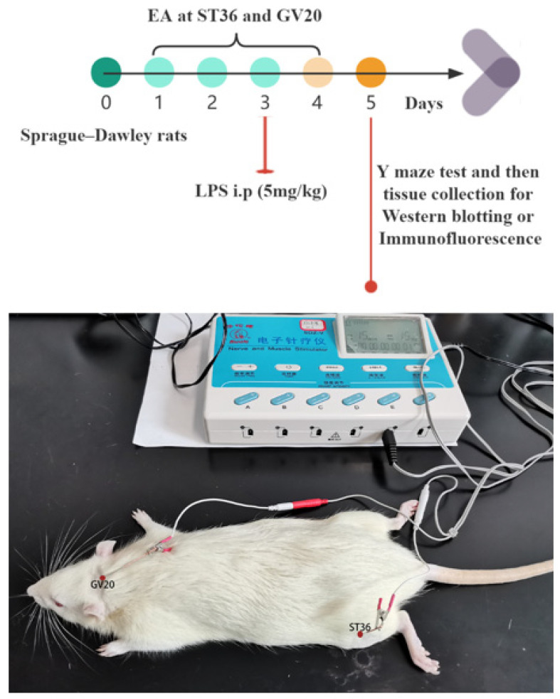
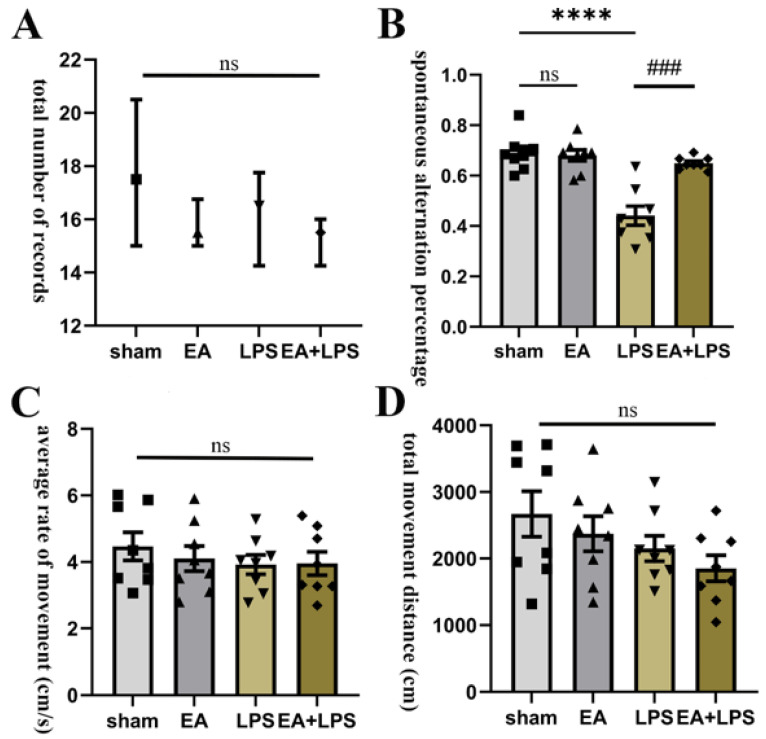
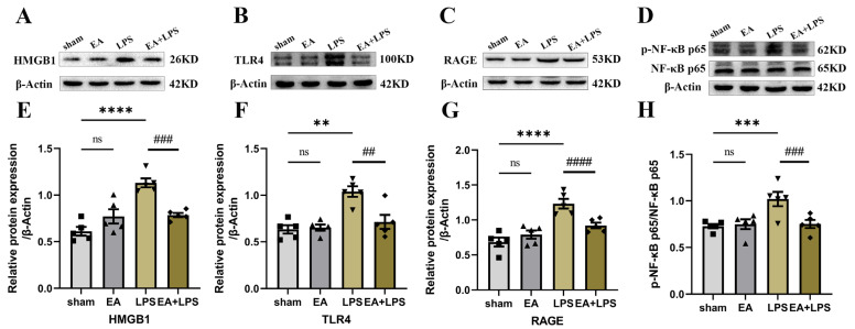
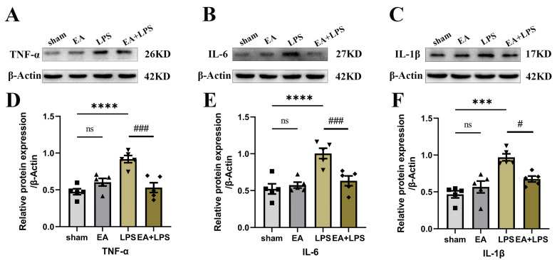
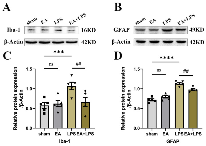
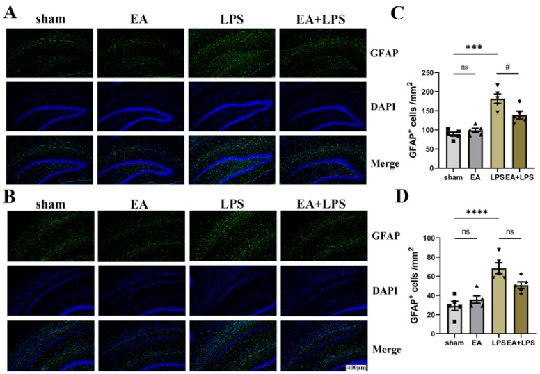
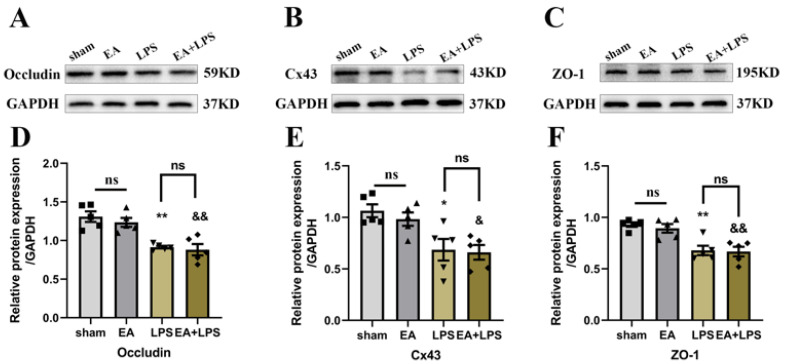
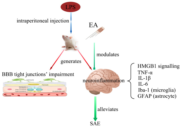
Sepsis-Associated Encephalopathy (SAE) is common in sepsis patients, with high mortality rates. It is believed that neuroinflammation is an important mechanism involved in SAE. High mobility group box 1 protein (HMGB1), as a late pro-inflammatory factor, is significantly increased during sepsis in different brain regions, including the hippocampus. HMGB1 causes neuroinflammation and cognitive impairment through direct binding to advanced glycation end products (RAGE) and Toll-like receptor 4 (TLR4). Electroacupuncture (EA) at Baihui (GV20) and Zusanli (ST36) is beneficial for neurological diseases and experimental sepsis. Our study used EA to treat SAE induced by lipopolysaccharide (LPS) in male Sprague-Dawley rats. The Y maze test was performed to assess working memory. Immunofluorescence (IF) and Western blotting (WB) were used to determine neuroinflammation and the HMGB1 signaling pathway. Results showed that EA could improve working memory impairment in rats with SAE. EA alleviated neuroinflammation by downregulating the hippocampus's HMGB1/TLR4 and HMGB1/RAGE signaling, reducing the levels of pro-inflammatory factors, and relieving microglial and astrocyte activation. However, EA did not affect the tight junctions' expression of the blood-brain barrier (BBB) in the hippocampus.

Keywords: HMGB1; electroacupuncture; neuroinflammation; sepsis-associated encephalopathy.


Conflict of interest statement

The authors declare no conflict of interest.

Figures

Figure 1
Figure 1
Experimental design and EA diagram. EA treatment was given at 15 Hz and an intensity of 1 mA for 20 min once a day from day 1 to day 4. Abbreviations: EA, electroacupuncture; LPS, lipopolysaccharide; GV20, Baihui; ST36, Zusanli; i.p., intraperitoneal injection.
Figure 2
Figure 2
EA improved working memory tested by Y maze: n = 8 per group. (A) The total number of records in each group. (B) The spontaneous alternation percentage in each group. (C) The average rate of movement in each group. (D) The total movement distance in each group. Data are presented as median and interquartile range and means ± SEM. Compared with the sham group, **** p < 0.0001; compared with the LPS group, ### p < 0.001. ■, ▲, ▼, ◆: Represents the individual value of the rats in each group. Abbreviations: EA, electroacupuncture; LPS, lipopolysaccharide; ns, no significant difference.
Figure 3
Figure 3
EA modulated HMGB1 signaling in the hippocampus: n = 5 per group. (A) Representative blots of HMGB1. (B) Representative blots of TLR4. (C) Representative blots of RAGE. (D) Representative blots of p-NF-κB p65 and NF-κB p65. (E) Quantification of the expression level of HMGB1. (F) Quantification of the expression level of TLR4. (G) Quantification of the expression level of RAGE. (H) Quantification of the ratio of p-NF-κB p65 and NF-κB p65. Data are presented as means ± SEM. Compared with the sham group, ** p < 0.01, *** p < 0.001, and **** p < 0.0001; compared with the LPS group, ## p < 0.01, ### p < 0.001, and #### p < 0.0001. ■, ▲, ▼, ◆: Represents the individual value of the rats in each group. Abbreviations: HMGB1, high mobility group box 1 protein; NF-κB, nuclear factor-κB; p- NF-κB, phosphorylated-NF-κB; RAGE, the receptor for advanced glycation end products; TLR4, Toll-like receptor 4; EA, electroacupuncture; LPS: lipopolysaccharide; ns: no significant difference.
Figure 4
Figure 4
EA decreased pro-inflammatory cytokines in the hippocampus: n = 5 per group. (A) Representative blots of TNF-α. (B) Representative blots of IL-6. (C) Representative blots of IL-1β. (D) Quantification of the expression level of TNF-α. (E) Quantification of the expression level of IL-6. (F) Quantification of the expression level of IL-1β. Data are presented as means ± SEM. Compared with the sham group, *** p < 0.001, **** p < 0.0001; compared with the LPS group, # p < 0.05, ### p < 0.001. ■, ▲, ▼, ◆: Represents the individual value of the rats in each group. Abbreviations: IL-1β, interleukin-1β; IL-6, interleukin-6; TNF-α, tumor necrosis factor α; EA: electroacupuncture; LPS, lipopolysaccharide; ns, no significant difference.
Figure 5
Figure 5
EA suppressed the expression level of Iba-1 and GFAP in the hippocampus: n = 5 per group. (A) Representative blots of Iba-1. (B) Representative blots of GFAP. (C) Quantification of the expression level of Iba-1. (D) Quantification of the expression level of GFAP. Data are presented as means ± SEM. Compared with the sham group, *** p < 0.001, **** p < 0.0001; compared with the LPS group, ## p < 0.01. ■, ▲, ▼, ◆: Represents the individual value of the rats in each group. Abbreviations: GFAP, glial fibrillary acidic protein; Iba-1, ionized calcium-binding adapter molecule 1; EA, electroacupuncture; LPS, lipopolysaccharide; ns, no significant difference.
Figure 6
Figure 6
EA alleviated the activation of microglia in the DG and CA1 regions, n = 5 per group. (A) Representative IF images of activated microglia in the DG region. (B) Representative IF images of activated microglia in the CA1 region. (C) The statistical results of Iba-1+ cell count in the DG region. (D) The statistical results of Iba-1+ cell count in the CA1 region. Magnification: 20×, scale bar = 400 µm. Data are presented as means ± SEM. Compared with the control group, ** p < 0.01, *** p < 0.001; compared with the LPS group, ## p < 0.01, # p < 0.05. ■, ▲, ▼, ◆: Represents the individual value of the rats in each group. Abbreviations: DAPI, 4′,6-diamidino-2-phenylindole; DG, dentate gyrus; Iba-1, ionized calcium-binding adapter molecule 1; EA, electroacupuncture; LPS, lipopolysaccharide; ns, no significant difference.
Figure 7
Figure 7
EA affected the activation of astrocytes in the DG and CA1 region: n = 5 per group. (A) Representative IF images of activated astrocytes in the DG region. (B) Representative IF images of activated astrocytes in the CA1 region. (C) The statistical results of GFAP+ cell count in the DG region. (D) The statistical results of GFAP+ cell count in the CA1 region. Magnification: 20×, scale bar = 400 µm. Data are presented as means ± SEM. Compared with the control group, *** p < 0.001, **** p < 0.0001; compared with the LPS group, # p < 0.05. ■, ▲, ▼, ◆: Represents the individual value of the rats in each group. Abbreviations: DAPI, 4′,6-diamidino-2-phenylindole; DG, dentate gyrus; GFAP, glial fibrillary acidic protein; EA, electroacupuncture; LPS, lipopolysaccharide; ns, no significant difference.
Figure 8
Figure 8
EA was ineffective in the junction proteins of the BBB: n = 5 per group. (A) Representative blots of Occludin. (B) Representative blots of Cx43. (C) Representative blots of ZO-1. (D) Quantification of the expression level of Occludin. (E) Quantification of the expression level of Cx43. (F) Quantification of the expression level of ZO-1. Data are presented as means ± SEM. Compared with the sham group, ** p < 0.01, * p < 0.05, && p < 0.01, & p < 0.05. ■, ▲, ▼, ◆: Represents the individual value of the rats in each group. Abbreviations: Cx43, connexin 43; ZO-1, zonula occludens-1; GAPDH, glyceraldehyde 3-phosphate dehydrogenase; EA, electroacupuncture; LPS, lipopolysaccharide; ns, no significant difference.
Figure 9
Figure 9
Working model: intraperitoneal injection of LPS to induce SAE impairs BBB’s tight junctions and evokes neuroinflammation in the hippocampus. The expression levels of HMGB1 signaling are enhanced, microglia and astrocytes are activated, and the content of pro-inflammatory factors is elevated. EA can modulate the HMGB1 signaling and relieve the intensity of neuroinflammation in the hippocampus. However, EA does not affect the tight junctions’ impairment of BBB. Abbreviations: BBB, blood–brain barrier; EA, electroacupuncture; GFAP, glial fibrillary acidic protein; HMGB1, high mobility group box 1 protein; Iba-1, ionized calcium-binding adapter molecule 1; IL-1β, interleukin-1β; IL-6, interleukin-6; LPS, lipopolysaccharide; TNF-α, tumor necrosis factor α; SAE: sepsis-associated encephalopathy.

Similar articles

Cited by

References

    1. Singer M., Deutschman C.S., Seymour C.W., Shankar-Hari M., Annane D., Bauer M., Bellomo R., Bernard G.R., Chiche J.D., Coopersmith C.M., et al. The Third International Consensus Definitions for Sepsis and Septic Shock (Sepsis-3) JAMA. 2016;315:801–810. doi: 10.1001/jama.2016.0287. - DOI - PMC - PubMed
    1. Zhao L., An R., Yang Y., Yang X., Liu H., Yue L., Li X., Lin Y., Reiter R.J., Qu Y. Melatonin alleviates brain injury in mice subjected to cecal ligation and puncture via attenuating inflammation, apoptosis, and oxidative stress: The role of SIRT1 signaling. J. Pineal Res. 2015;59:230–239. doi: 10.1111/jpi.12254. - DOI - PubMed
    1. Tauber S.C., Djukic M., Gossner J., Eiffert H., Bruck W., Nau R. Sepsis-associated encephalopathy and septic encephalitis: An update. Expert Rev. Anti-Infect Ther. 2021;19:215–231. doi: 10.1080/14787210.2020.1812384. - DOI - PubMed
    1. Catarina A.V., Branchini G., Bettoni L., De Oliveira J.R., Nunes F.B. Sepsis-Associated Encephalopathy: From Pathophysiology to Progress in Experimental Studies. Mol. Neurobiol. 2021;58:2770–2779. doi: 10.1007/s12035-021-02303-2. - DOI - PubMed
    1. Li Y., Yin L., Fan Z., Su B., Chen Y., Ma Y., Zhong Y., Hou W., Fang Z., Zhang X. Microglia: A Potential Therapeutic Target for Sepsis-Associated Encephalopathy and Sepsis-Associated Chronic Pain. Front. Pharmacol. 2020;11:600421. doi: 10.3389/fphar.2020.600421. - DOI - PMC - PubMed

LinkOut - more resources