. 2022 Nov 29;11(12):1739.
doi: 10.3390/biology11121739.

Alleviation of Cognitive and Physical Fatigue with Enzymatic Porcine Placenta Hydrolysate Intake through Reducing Oxidative Stress and Inflammation in Intensely Exercised Rats

Affiliations

Alleviation of Cognitive and Physical Fatigue with Enzymatic Porcine Placenta Hydrolysate Intake through Reducing Oxidative Stress and Inflammation in Intensely Exercised Rats

Min Ju Kim et al. Biology (Basel). .

Abstract

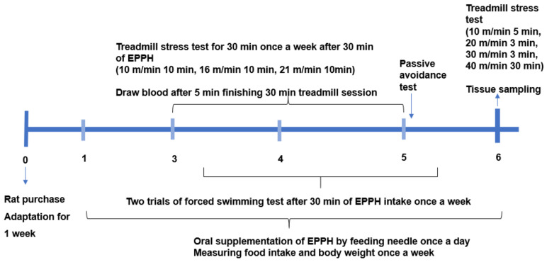
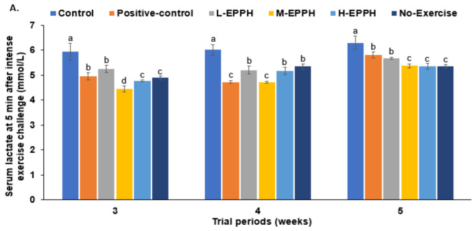
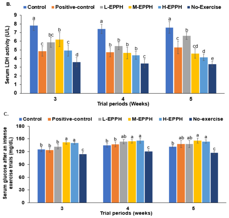
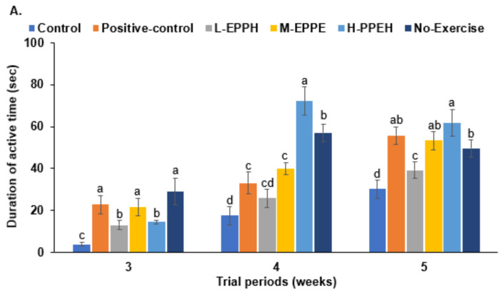
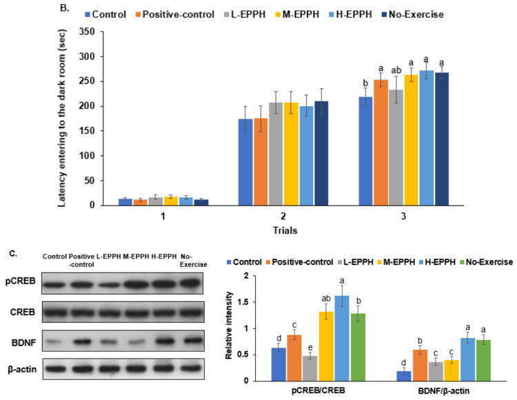
Intense exercise is reported to induce physical and cognitive fatigue, but few studies have focused on treatments to alleviate fatigue. We hypothesized that the oral supplementation of enzymatic porcine placenta hydrolysate (EPPH) prepared using protease enzymes could alleviate exercise-induced fatigue in an animal model. The objectives of the study were to examine the hypothesis and the action mechanism of EPPH in relieving physical and cognitive fatigue. Fifty male Sprague−Dawley rats aged 8 weeks (body weight: 201 g) were classified into five groups, and rats in each group were given oral distilled water, EPPH (5 mg nitrogen/mL) at doses of 0.08, 0.16, or 0.31 mL/kg body weight (BW)/day, or glutathione (100 mg/kg BW/day) by a feeding needle for 5 weeks, which were named as the control, L-EPPH, M-EPPH, H-EPPH, or positive-control groups, respectively. Ten additional rats had no intense exercise with water administration and were designated as the no-exercise group. After 2 weeks, the rats were subjected to intense exercise and forced swimming trial for 30 min once per week for an additional 4 weeks. At 5 min after the intense exercise, lactate concentrations and lactate dehydrogenase (LDH) activity in the serum and the gastrocnemius muscle were higher in the control group, whereas M-EPPH and H-EPPH treatments suppressed the increase better than in the positive-control (p < 0.05). Intense exercise decreased glycogen content in the liver and gastrocnemius muscle, and M-EPPH and H-EPPH inhibited the decrement (p < 0.05). Moreover, lipid peroxide contents in the gastrocnemius muscle and liver were higher in the control group than in the M-EPPH, H-EPPH, positive-control, and no-exercise groups (p < 0.05). However, antioxidant enzyme activities such as superoxide dismutase (SOD) and glutathione peroxidase (GSH-Px) were opposite to the lipid peroxide contents. Hypothalamic corticosterone and hippocampal mRNA expressions of tumor necrosis factor (TNF)-α and IL-1β were higher. However, hippocampal brain-derived neurotrophic factor (BDNF) mRNA expression and protein contents were lower in the control group than in the positive-control group. M-EPPH, H-EPPH, and positive-control suppressed the changes via activating hippocampal cAMP response element-binding protein phosphorylation, and H-EPPH showed better activity than in the positive-control (p < 0.05). In conclusion, EPPH (0.16−0.31 mL/kg BW) intake reduced exercise-induced physical and cognitive fatigue in rats and could potentially be developed as a therapeutic agent for relieving fatigue in humans.

Keywords: cognitive fatigue; exercise-induced fatigue; hypothalamus–pituitary–adrenaline axis; lactate; porcine placenta enzyme hydrolysates.


Conflict of interest statement

The authors declare no conflict of interest.

Figures

Figure 1
Figure 1
Experimental design. Rats had oral supplementation of EPPH (0.08, 0.16, and 0.31 mL/kg body weight) by a feeding needle once per day for two weeks, and from the third week, rats were given intense exercise using a treadmill protocol for 30 min after 30 min of being given EPPH. However, rats in the no-exercise group did not have intense exercise. The treadmill exercise was conducted for two weeks according to the protocol 10 m/min for 10 min, 16 m/min for 10 min, and 21 m/min for 10 min. Two days after the treadmill exercise, they were given a forced swimming test 30 min after EPPH supplementation. In the fourth week after starting the treadmill, the final treadmill test was performed according to the protocol of 10 m/min for 5 min, 20 m/min for 3 min, 30 m/min for 3 min, 40 m/min for 30 min, and after 5 min, rats were sacrificed for collecting their tissues.
Figure 2
Figure 2
Fasting serum glucose concentrations before exercise-induced fatigue during the experimental periods. Dots and error bars in each period represent means ± standard deviation (SD; n = 10). The rats with exercise-induced fatigue had oral administration of distilled water for the control group, porcine placenta enzyme hydrolysates (EPPH) dissolved in distilled water for experimental groups, and glutathione (100 mg/kg body weight) for the positive-control group. EPPH (0.08, 0.16, and 0.32 mL/kg body weight) was orally provided with a feeding needle for the L-EPPH, M-EPPH, and H-EPPH groups, respectively, for 6 weeks. The rats were designated as the no-exercise group without exercise-induced fatigue and oral administration of distilled water. * Significantly different among groups in one-way ANOVA at p < 0.05.
Figure 3
Figure 3
Metabolites related to exercise-induced fatigue at 5 min after intense exercise; (A) serum lactate concentrations; (B) LDH activity in the serum; (C) serum glucose concentrations. Bars and error bars represent means ± standard deviation (SD; n = 10). The rats with exercise-induced fatigue had oral administration of distilled water for the control group, porcine placenta enzyme hydrolysates (EPPH) dissolved in distilled water for experimental groups, and glutathione (100 mg/kg body weight) for the positive-control group. EPPH (0.08, 0.16, and 0.32 mL/kg body weight) was orally provided with a feeding needle for the L-EPPH, M-EPPH, and H-EPPH groups, respectively, for 6 weeks. The rats that had no exercise-induced fatigue and oral administration of distilled water were designated the no-exercise group. a,b,c,d Different letters on the bars indicate significant differences among the groups by Tukey test at p < 0.05.
Figure 3
Figure 3
Metabolites related to exercise-induced fatigue at 5 min after intense exercise; (A) serum lactate concentrations; (B) LDH activity in the serum; (C) serum glucose concentrations. Bars and error bars represent means ± standard deviation (SD; n = 10). The rats with exercise-induced fatigue had oral administration of distilled water for the control group, porcine placenta enzyme hydrolysates (EPPH) dissolved in distilled water for experimental groups, and glutathione (100 mg/kg body weight) for the positive-control group. EPPH (0.08, 0.16, and 0.32 mL/kg body weight) was orally provided with a feeding needle for the L-EPPH, M-EPPH, and H-EPPH groups, respectively, for 6 weeks. The rats that had no exercise-induced fatigue and oral administration of distilled water were designated the no-exercise group. a,b,c,d Different letters on the bars indicate significant differences among the groups by Tukey test at p < 0.05.
Figure 4
Figure 4
Brain fatigue; (A) the active time during forced swimming; (B) latency entering the dark room; (C) hippocampal Western blots. Bars and error bars represent means ± standard deviation (SD; n = 10). The rats with exercise-induced fatigue had oral administration of distilled water for the control group, porcine placenta enzyme hydrolysates (EPPH) dissolved in distilled water for experimental groups, and glutathione (100 mg/kg body weight) for the positive-control group. EPPH (0.08, 0.16, and 0.32 mL/kg body weight) was orally provided with a feeding needle for the L-EPPH, M-EPPH, and H-EPPH groups, respectively, for 6 weeks. The rats that had no exercise-induced fatigue and oral administration of distilled water were designated the no-exercise group. a,b,c,d Different letters on the bars indicate significant differences among the groups by Tukey test at p < 0.05.
Figure 4
Figure 4
Brain fatigue; (A) the active time during forced swimming; (B) latency entering the dark room; (C) hippocampal Western blots. Bars and error bars represent means ± standard deviation (SD; n = 10). The rats with exercise-induced fatigue had oral administration of distilled water for the control group, porcine placenta enzyme hydrolysates (EPPH) dissolved in distilled water for experimental groups, and glutathione (100 mg/kg body weight) for the positive-control group. EPPH (0.08, 0.16, and 0.32 mL/kg body weight) was orally provided with a feeding needle for the L-EPPH, M-EPPH, and H-EPPH groups, respectively, for 6 weeks. The rats that had no exercise-induced fatigue and oral administration of distilled water were designated the no-exercise group. a,b,c,d Different letters on the bars indicate significant differences among the groups by Tukey test at p < 0.05.

Similar articles

Cited by

References

    1. Cordeiro L.M.S., Rabelo P.C.R., Moraes M.M., Teixeira-Coelho F., Coimbra C.C., Wanner S.P., Soares D.D. Physical exercise-induced fatigue: The role of serotonergic and dopaminergic systems. Braz. J. Med. Biol. Res. 2017;50:e6432. doi: 10.1590/1414-431x20176432. - DOI - PMC - PubMed
    1. Dotan R., Woods S., Contessa P. On the reliability and validity of central fatigue determination. Eur. J. Appl. Physiol. 2021;121:2393–2411. doi: 10.1007/s00421-021-04700-w. - DOI - PubMed
    1. Gentile D., Beeler D., Wang X.S., Ben-Ayre E., Zick S.M., Bao T., Carlson L.E., Ghelman R., Master V., Tripathy D., et al. Cancer-Related Fatigue Outcome Measures in Integrative Oncology: Evidence for Practice and Research Recommendations. Oncology. 2022;36:276–287. - PubMed
    1. Sapra A., Bhandari P. StatPearls. StatPearls Publishing; Treasure Island, FL, USA: 2022. Chronic Fatigue Syndrome. - PubMed
    1. Flockhart M., Nilsson L.C., Tais S., Ekblom B., Apró W., Larsen F.J. Excessive exercise training causes mitochondrial functional impairment and decreases glucose tolerance in healthy volunteers. Cell Metab. 2021;33:957–970.e956. doi: 10.1016/j.cmet.2021.02.017. - DOI - PubMed

LinkOut - more resources