Review
. 2022 Dec 14;11(12):1822.
doi: 10.3390/biology11121822.

The Contribution of the Locus Coeruleus-Noradrenaline System Degeneration during the Progression of Alzheimer's Disease

Affiliations
Review

The Contribution of the Locus Coeruleus-Noradrenaline System Degeneration during the Progression of Alzheimer's Disease

Dilek Mercan et al. Biology (Basel). .

Abstract

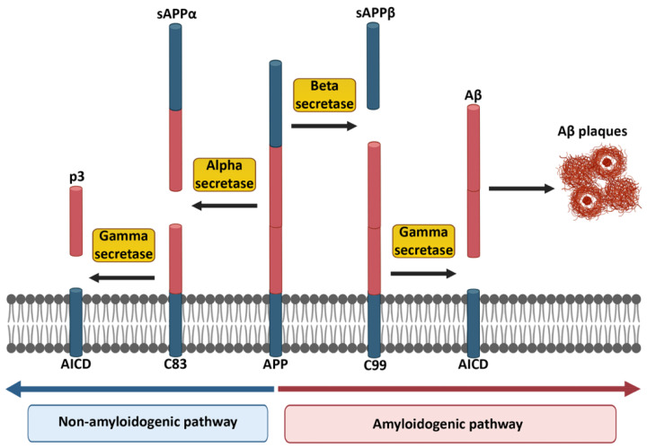
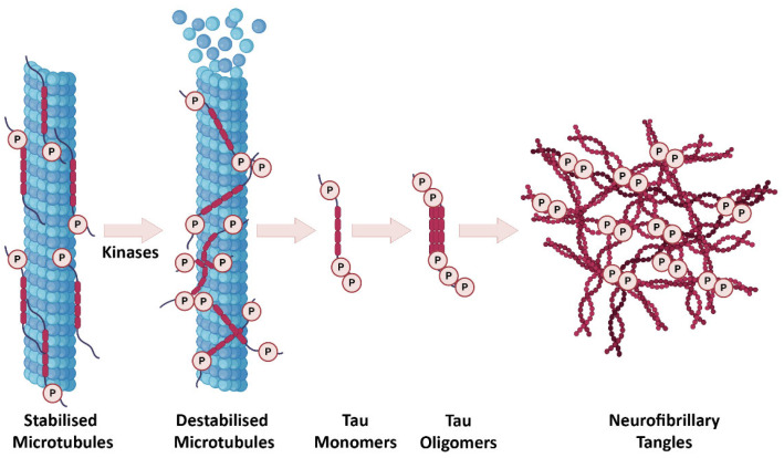
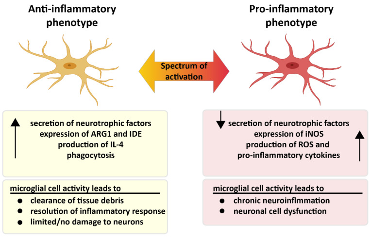
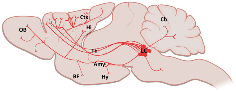
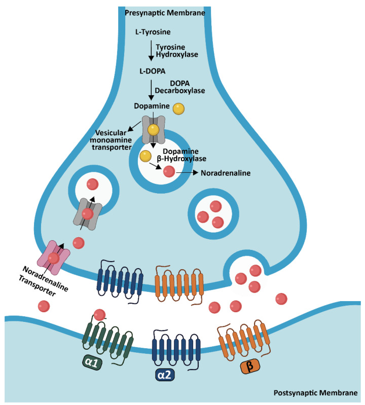
Alzheimer's disease (AD), which is characterized by extracellular accumulation of amyloid-beta peptide and intracellular aggregation of hyperphosphorylated tau, is the most common form of dementia. Memory loss, cognitive decline and disorientation are the ultimate consequences of neuronal death, synapse loss and neuroinflammation in AD. In general, there are many brain regions affected but neuronal loss in the locus coeruleus (LC) is one of the earliest indicators of neurodegeneration in AD. Since the LC is the main source of noradrenaline (NA) in the brain, degeneration of the LC in AD leads to decreased NA levels, causing increased neuroinflammation, enhanced amyloid and tau burden, decreased phagocytosis and impairment in cognition and long-term synaptic plasticity. In this review, we summarized current findings on the locus coeruleus-noradrenaline system and consequences of its dysfunction which is now recognized as an important contributor to AD progression.

Keywords: Alzheimer’s disease; cognition; locus coeruleus; neurodegeneration; neuroinflammation; noradrenaline; norepinephrine.


Conflict of interest statement

The authors declare no conflict of interest.

Figures

Figure 1
Figure 1
Non-amyloidogenic and amyloidogenic pathways in the cleavage of APP. The red part of APP indicates the potentially harmful Aβ portion, which appears at the end of the amyloidogenic pathway. Created partially with BioRender.com (accessed on 28 June 2021).
Figure 2
Figure 2
The progression of tau pathology in AD. In a healthy brain, tau is involved in regulation of microtubule stabilization. After tau hyperphosphorylation, microtubules are destabilized and start to disassemble. Tau monomers and oligomers subsequently occur intraneuronally and eventually formation of NFTs takes place. Created partially with BioRender.com (accessed on 28 June 2021).
Figure 3
Figure 3
Two different faces of microglia. Pro- and anti-inflammatory microglia display different characteristics in the presence of microglial activation. While microglia are neuroprotective in the anti-inflammatory state, microglia in the pro-inflammatory condition trigger chronic inflammation, which leads to neuronal dysfunction. Created partially with BioRender.com (accessed on 28 June 2021).
Figure 4
Figure 4
A sagittal schematic illustration showing the LC projections in the brain (Amy: Amygdala; BF: Basal Forebrain; Cb: Cerebellum; Ctx: Cortex; Hi: Hippocampus; Hy: Hypothalamus; LC: Locus Coeruleus; Th: Thalamus, OB: Olfactory Bulb). Created partially with BioRender.com (accessed on 28 June 2021).
Figure 5
Figure 5
NA synthesis in the brain. NA synthesis is performed from dopamine through DBH in the LC and transported into synaptic vesicles by a vesicular monoamine transporter. After NA is released into the synaptic cleft, it binds to specific adrenergic receptors in order to activate the signaling for the specific task. Created partially with BioRender.com (accessed on 28 June 2021).

References

    1. World Alzheimer Reports. [(accessed on 20 December 2019)]. Available online: https://www.alz.co.uk/research/world-report.
    1. Kawas C., Gray S., Brookmeyer R., Fozard J., Zonderman A. Age-specific incidence rates of Alzheimer’s disease. Neurology. 2000;54:2072–2077. doi: 10.1212/WNL.54.11.2072. - DOI - PubMed
    1. Huang H.-C., Jiang Z.-F. Accumulated Amyloid-β Peptide and Hyperphosphorylated Tau Protein: Relationship and Links in Alzheimer’s Disease. J. Alzheimer’s Dis. 2009;16:15–27. doi: 10.3233/JAD-2009-0960. - DOI - PubMed
    1. Yankner B.A., Mesulam M.-M. β-Amyloid and the Pathogenesis of Alzheimer’s Disease. N. Engl. J. Med. 1991;325:1849–1857. - PubMed
    1. Roher A.E., Esh C.L., Kokjohn T.A., Castaño E.M., Van Vickle G.D., Kalback W.M., Patton R.L., Luehrs D.C., Daugs I.D., Kuo Y., et al. Amyloid beta peptides in human plasma and tissues and their significance for Alzheimer’s disease. Alzheimer’s Dement. 2009;5:18–29. doi: 10.1016/j.jalz.2008.10.004. - DOI - PMC - PubMed

LinkOut - more resources