Management of Vaginal Hyperplasia in Bitches by Bühner Suture
- PMID: 36552425
- PMCID: PMC9774832
- DOI: 10.3390/ani12243505
Management of Vaginal Hyperplasia in Bitches by Bühner Suture
Abstract
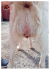
Vaginal hyperplasia in bitch is an exaggerated response of the vaginal mucosa to estrogens during the proestral-estral phase of the cycle that can protrude through the vulvar lips. The present study refers to the management of vaginal hyperplasia in bitches by Bühner vulvar suture. Fourteen private-owners animals were refereed for spontaneous vaginal hyperplasia and complete protrusion of the mucosa, without ischemic or necrotic areas, which occurred during the proestral-estral phase. Under general anesthesia, prolapsed mass was cleaned with 50% glucose solution to reduce oedema and gently repositioned; the Bühner suture was applied using a Gerlach needle and a sterile vaginal suture tape, maintaining a minimal opening to allow urination in order to avoid possible recurrence in the same estrus. None of the bitches showed recurrence during the current cycle, proving the effectiveness of the Bühner suture. To prevent the possible recurrence of vaginal hyperplasia at the subsequent estrus, all the bitches underwent an ovariectomy 2 months later, when the Bühner suture was removed. In conclusion, the Bühner suture proved to be useful for the conservative treatment of vaginal hyperplasia in medium- or large-sized bitches. However, this approach should be considered only for cases in which the prolapsed mass does not show trauma, ulceration, ischemic or necrotic areas.
Keywords: canine reproduction; estrogens; proestral–estral disease; vulvar suture.
Conflict of interest statement
The authors declare no conflict of interest.
Figures


Similar articles
-
Oestrogen and progestagen treated ovariectomized bitches: a model for the study of uterine function.J Reprod Fertil Suppl. 2001;57:45-54. J Reprod Fertil Suppl. 2001. PMID: 11787189
-
Matrix metalloproteinases (MMPs) in the endometrium of bitches.Reproduction. 2002 Mar;123(3):467-77. Reproduction. 2002. PMID: 11882024
-
A model for the study of cystic endometrial hyperplasia in bitches.J Reprod Fertil Suppl. 2001;57:407-14. J Reprod Fertil Suppl. 2001. PMID: 11787183
-
Clinical approach to vaginal/vestibular masses in the bitch.Vet Clin North Am Small Anim Pract. 1991 May;21(3):509-21. doi: 10.1016/s0195-5616(91)50057-7. Vet Clin North Am Small Anim Pract. 1991. PMID: 1858246 Review.
-
Endometrial safety of low-dose vaginal estrogens in menopausal women: a systematic evidence review.Menopause. 2019 Jul;26(7):800-807. doi: 10.1097/GME.0000000000001315. Menopause. 2019. PMID: 30889085 Free PMC article.
References
-
- Kumar S., Pandey A.K., Singh G., Arora N., Potliya S., Kumar K., Ahmad S.Q., Kumar S. Management of Vaginal Hyperplasia in a Bitch. Haryana Vet. 2014;53:76–77.
LinkOut - more resources
Full Text Sources

