. 2022 Dec 13;12(24):3523.
doi: 10.3390/ani12243523.

Pathological and Molecular Characterization of a Duck Plague Outbreak in Southern China in 2021

Affiliations

Pathological and Molecular Characterization of a Duck Plague Outbreak in Southern China in 2021

Zhipeng Liang et al. Animals (Basel). .

Abstract

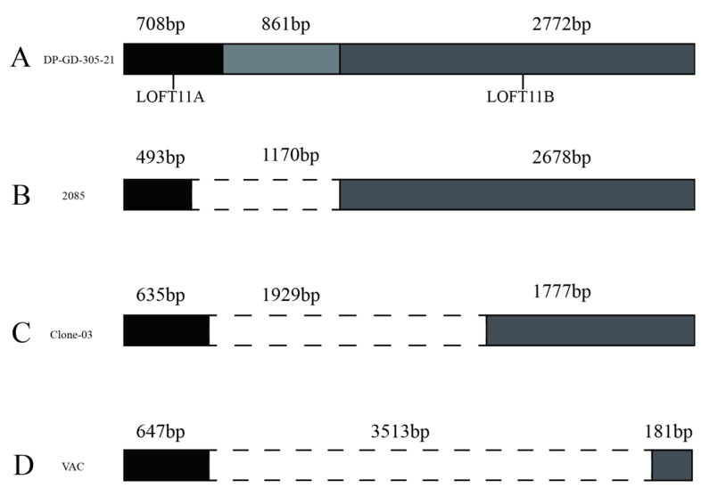
Duck plague (DP) is a highly contagious viral disease in ducks caused by the duck plague virus (DPV). The DPV, a member of Herpesviridae, poses a severe threat to the waterfowl farming industry worldwide. In this study, we reported a recent outbreak of DPV in domestic laying ducks at 310 days of age from southern China in December 2021. The gross lesion, histopathologic examination, molecular detection, and genetic characterization studies of DPV are described here. As a result, gross lesions such as an enlarged congestive spleen and liver were observed. Liver with vacuolar degeneration and small vacuoles and spleen with hemosiderosis were remarkable microscopic findings. Our results suggested that the liver had the highest viral load, followed by the trachea, pancreas, kidney, brain, spleen, and heart. In addition, DPV was successfully isolated in chicken embryo fibroblast cell culture and designated as DP-GD-305-21. The UL2, UL12, UL41, UL47, and LORF11 genes of DP-GD-305-21 shared a high nucleotide homology with the Chinese virulent (CHv) strain and the Chinese variant (CV) strain. In conclusion, this study reports the isolation and molecular characterization of DPV from a recent outbreak in southern China. Our results contributed to the understanding of the pathological and molecular characterization of currently circulating DPV in China.

Keywords: Herpesviridae; LORF11; UL2; duck plague; duck plague virus.


Conflict of interest statement

The authors declare no conflict of interest.

Figures

Figure 1
Figure 1
Gross lesions of ducks infected with DPV at autopsy. (A): Presence of bold clots related to the pericardial sac and hepatic capsule; (B): Enlarged and congested liver; (C): Spotted hemorrhage in the heart; (D): Enlarged and congested brain; (E): Spotted hyperemia of the trachea; (F): Enlarged and severely congested spleen.
Figure 2
Figure 2
Histopathological images of duck tissue sections stained with hematoxylin and eosin (H&E) (magnification, ×200). The scale bar included in each image presents a length of 100 μm. (A): Extensive vacuolar degeneration and small vacuoles were detected in hepatocytes; (B): Lymphoid depletion, focal necrosis, and multifocal fibrinoid necrotic foci were observed in spleen; (C): Detached epithelial cells and damaged bronchial wall were observed in the tracheal mucosa; (D): Generalized tubular atrophy and inflammatory cell infiltration in the interstitium were observed in kidney; (E): Cardiomyocytes were tightly packed and surrounded by inflammatory cell infiltration in heart; (F): Necrosis of acinar and islet structures were detected in pancreatic tissue.
Figure 3
Figure 3
Cytopathic effects on chicken embryo fibroblasts from homogenate duck-infected DPV tissue samples. (A): Negative control (100×); (B): Infected CEF cells showing vacuolation and syncytia (400×).
Figure 4
Figure 4
Levels of DPV viral load at the time of initial sampling. The collected tissues are shown at the bottom. Viral loads were determined by quantitative Real-time PCR. Viral loads are expressed as mean ± SD. Differences between groups were determined using two-way ANOVA. ****, p < 0.0001; ns, p > 0.05.
Figure 5
Figure 5
Gene structure types of UL2. (A): Strains with a 1002 bp coding region, such as the DP-GD-305-21 and CHv strain; (B): Attenuated strain with 528 bp deletion 2; (C): Strains with a 528 bp deletion and three 1 bp insertions, including VAC and IVRI-2016.
Figure 6
Figure 6
Gene structure types of LOFT11. (A): A coding region of 4341 bp for the LOFT11 gene was observed in strains such as DP-GD-305-21; (B): Strains such as the 21 have a 1170 bp deletion in the coding region of LORF11; (C): A 1929 bp deletion was found in the LOFT11 gene of strains such as the ‘Clone-03’; (D): A 3513 bp deletion was found in the LOFT11 gene of strains such as the VAC strain.
Figure 7
Figure 7
The secondary structure, hydrophilicity, flexible regions, antigenic index, and surface probability of the DP-GD-305-21 proteins. (A): UL2 protein; (B): UL12 protein; (C): UL47 protein; (D): UL41 protein; (E): LORF11 protein.

Similar articles

Cited by

References

    1. Leibovitz L., Hwang J. Duck plague on the American continent. Avian Dis. 1968;12:361–378. doi: 10.2307/1588237. - DOI - PubMed
    1. Salguero F.J., Sanchez-Cordon P.J., Nunez A., Gomez-Villamandos J.C. Histopathological and ultrastructural changes associated with herpesvirus infection in waterfowl. Avian Pathol. 2002;31:133–140. doi: 10.1080/03079450120118612. - DOI - PubMed
    1. Sarmah H., Shah M., Pathak M., Barman N.N., Koul M., Gupta A., Sahariah P.J., Neher S., Das S.K., Gogoi S.M., et al. Pathodynamics of Circulating Strains of Duck Enteritis Virus: A Step Forward to Understand Its Pathogenesis. Avian Dis. 2020;64:166–173. doi: 10.1637/0005-2086-64.2.166. - DOI - PubMed
    1. Pazhanivel N., Rajeswar J., Ramprabhu R., Manoharan S., Bala M.A., Balachandran C., Kumanan K., Prathaban S., Saahithya R. Duck plague outbreak in a Chara-Chemballi duck farm. Iran. J. Vet. Res. 2019;20:308–312. - PMC - PubMed
    1. Li N., Hong T., Li R., Guo M., Wang Y., Zhang J., Liu J., Cai Y., Liu S., Chai T., et al. Pathogenicity of duck plague and innate immune responses of the Cherry Valley ducks to duck plague virus. Sci. Rep. 2016;6:32183. doi: 10.1038/srep32183. - DOI - PMC - PubMed