

Co-Expression of T- and B-Cell Markers in a Canine Intestinal Lymphoma: A Case Report
- PMID: 36552451
- PMCID: PMC9774803
- DOI: 10.3390/ani12243531
Co-Expression of T- and B-Cell Markers in a Canine Intestinal Lymphoma: A Case Report
Abstract
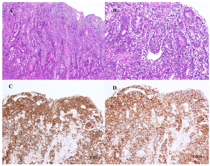
An 8-year-old female neutered Labrador retriever was presented for a second opinion consultation due to vomiting and lethargy, having failed to respond to symptomatic therapy. Blood analysis revealed hyperbilirubinemia and hypoalbuminemia, associated with hypocobalaminemia. An abdominal ultrasound identified diffused bowel thickening and hypoechoic hepatomegaly. An ultrasound-guided liver fine-needle aspiration was performed for cytology and also for cell block immunocytochemistry. Gastric and duodenal biopsies were collected by gastroduodenoscopy. Liver cytology showed numerous lymphocytes, suggesting lymphoma at the hepatic infiltration stage, and immunocytochemistry in the cell block of the hepatic aspirate indicated co-expression of CD3 and CD20 in the lymphoid cells present. The histopathology of gastric and duodenal biopsies supported the hypothesis of gastrointestinal lymphoma due to heavy lymphoid infiltration of the gastric epithelium and intestinal mucosa, including the villi. Concurrent immunohistochemistry was performed using CD3, CD20, PAX5, and CD79αcy antibodies. Immunomarking was positive for CD3 and CD20, which overlapped populations of lymphoid cells, and was negative for all other antibodies. In the clonality test, lymphocyte co-expression of CD3 and CD20 was confirmed by monoclonal rearrangement of T-cell gamma receptors. The final diagnosis was type 2 enteropathy-associated T-cell lymphoma with hepatic infiltration. Co-expression was examined in conjunction with the PARR result in the presence of T-cell monoclonal rearrangement.
Keywords: CD20; CD3; clonality; co-expression; dog; enteropathy; intestinal lymphoma.
Conflict of interest statement
The authors declare no conflict of interest.
Figures


Similar articles
-
Characterization of CD3+/CD20+ canine large-cell lymphoma.J Vet Diagn Invest. 2024 Jan;36(1):86-94. doi: 10.1177/10406387231204873. Epub 2023 Oct 13. J Vet Diagn Invest. 2024. PMID: 37837199 Free PMC article.
-
Distinguishing Intestinal Lymphoma From Inflammatory Bowel Disease in Canine Duodenal Endoscopic Biopsy Samples.Vet Pathol. 2015 Jul;52(4):668-75. doi: 10.1177/0300985814559398. Epub 2014 Dec 8. Vet Pathol. 2015. PMID: 25487412
-
Coexpression of CD3 and CD20 in Canine Enteropathy-Associated T-cell Lymphoma.Vet Pathol. 2018 Mar;55(2):241-244. doi: 10.1177/0300985817747326. Epub 2018 Jan 17. Vet Pathol. 2018. PMID: 29343197
-
CD20-positive NK/T-cell lymphoma with indolent clinical course: report of case and review of literature.Diagn Pathol. 2012 Oct 2;7:133. doi: 10.1186/1746-1596-7-133. Diagn Pathol. 2012. PMID: 23031227 Free PMC article. Review.
-
Gastric T-cell lymphoma with cytotoxic phenotype.Pathol Int. 2007 Feb;57(2):108-14. doi: 10.1111/j.1440-1827.2006.02065.x. Pathol Int. 2007. PMID: 17300676 Review.
Cited by
-
Morphological, phenotypical and molecular characterization of canine lymphomas with dual T- and B-cell markers expression.Front Vet Sci. 2025 Apr 22;12:1578425. doi: 10.3389/fvets.2025.1578425. eCollection 2025. Front Vet Sci. 2025. PMID: 40331223 Free PMC article.
-
Characterization of CD3+/CD20+ canine large-cell lymphoma.J Vet Diagn Invest. 2024 Jan;36(1):86-94. doi: 10.1177/10406387231204873. Epub 2023 Oct 13. J Vet Diagn Invest. 2024. PMID: 37837199 Free PMC article.
References
-
- Vail D.M., Pinkerton M.E., Young K.M. Withrow & MacEwen’s Small Animal Clinical Oncology. 5th ed. Elsevier; St. Louis, MI, USA: 2013. Hematopoietic Tumors; pp. 622–678.
-
- Munday J.S., Lohr C.V., Kiupel M. Tumors of the alimentary tract. In: Meuten D.J., editor. Tumors in Domestic Animals. 5th ed. John Wiley & Sons Inc.; Hoboken, NJ, USA: 2016. pp. 499–601.
-
- Uzal F.A., Plattner B.L., Hostetterin J.M. Alimentary System. In: Grant Maxie M., editor. Jubb, Kennedy & Palmer’s Pathology of Domestic Animals. 6th ed. Volume 2. Elsevier; St. Louis, MI, USA: 2016. pp. 16–257.
Publication types
Grants and funding
LinkOut - more resources
Full Text Sources
