. 2022 Nov 26;11(12):2342.
doi: 10.3390/antiox11122342.

Thiocyanate Reduces Motor Impairment in the hMPO-A53T PD Mouse Model While Reducing MPO-Oxidation of Alpha Synuclein in Enlarged LYVE1/AQP4 Positive Periventricular Glymphatic Vessels

Affiliations

Thiocyanate Reduces Motor Impairment in the hMPO-A53T PD Mouse Model While Reducing MPO-Oxidation of Alpha Synuclein in Enlarged LYVE1/AQP4 Positive Periventricular Glymphatic Vessels

Wanda F Reynolds et al. Antioxidants (Basel). .

Abstract

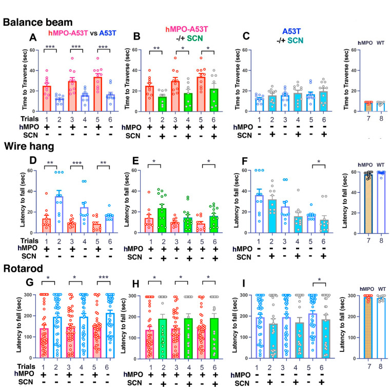
Parkinson's disease (PD) is due to the oxidation of alpha synuclein (αSyn) contributing to motor impairment. We developed a transgenic mouse model of PD that overexpresses the mutated human αSyn gene (A53T) crossed to a mouse expressing the human MPO gene. This model exhibits increased oxidation and chlorination of αSyn leading to greater motor impairment. In the current study, the hMPO-A53T mice were treated with thiocyanate (SCN-) which is a favored substrate of MPO as compared to chlorine. We show that hMPO-A53T mice treated with SCN- have less chlorination in the brain and show an improvement in motor skills compared to the nontreated hMPO-A53T mice. Interestingly, in the hMPO-A53T mice we found a possible link between MPO-related disease and the glymphatic system which clears waste including αSyn from the brain. The untreated hMPO-A53T mice exhibited an increase in the size of periventricular glymphatic vessels expressing the glymphatic marker LYVE1 and aquaporin 4 (AQP4). These vessels also exhibited an increase in MPO and HOCl-modified epitopes in the glymphatic vessels correlating with loss of ependymal cells lining the ventricles. These findings suggest that MPO may significantly promote the impairment of the glymphatic waste removal system thus contributing to neurodegeneration in PD. Moreover, the inhibition of MPO chlorination/oxidation by SCN- may provide a potential therapeutic approach to this disease.

Keywords: Parkinson’s disease; alpha synuclein; aquaporin 4; carbamylation; endothelial hyaluronan receptor 1; glial fibrillary acidic protein; glymphatics; hypochlorous acid; myeloperoxidase; nitration; reactive oxygen species.


Conflict of interest statement

The authors declare no conflict of interest.

Figures

Figure 1
Figure 1
Impaired motor abilities in hMPO-A53T mice compared to A53T. (A) Balance beam analysis was performed with genotypes hMPO-A53T (n = 10) (lanes 1,3,5) versus A53T (n = 10) (lanes 2,4,6). Only male mice were used in these experiments. Three consecutive trials were performed for each group with rest intervals of 5 min. (B) hMPO-A53T (n = 10) (lanes 1,3,5) vs. hMPO-A53T SCN treated (n = 8) (lanes 2,4,6). (C) A53T (n = 10) (lanes 1,3,5) vs. A53T SCN treated (n = 10) (lanes 2,4,6). hMPO mice (n = 10) (lane 7) vs. WT (C57Bl/6) (n = 9) (lane 8). Only the third trial is shown for WT and hMPO mice. Trials A lanes 1,3,5 are the same as B lanes 1,3,5. Trials A lanes 2,4,6 are the same as C lanes 1,3,5. (DF) The wire hang was performed with the indicated genotypes as in panels A,B,C (n = 10 A53T, n = 10 hMPO-A53T, n = 10 hMPO, n = 10 WT). Three trials with rest intervals were performed for each group. Only the third trial is shown for WT and hMPO mice. (GI) Rotarod analysis was performed with the genotypes indicated in panels A,B,C (n = 39–44 for each group, n = 10 hMPO, n = 10 WT). Three consecutive trials were performed for each group with rest intervals of 5 min. Only the third trial is shown for WT and hMPO mice. Behavior data were analyzed using a one-way analysis of variance (ANOVA) followed by Dunnets post- hoc test using GraphPad Prism v9. Data are represented as mean +/− S.E.M. (* p < 0.01, ** p < 0.005, *** p < 0.001).
Figure 2
Figure 2
Antibodies to hMPO, 2D10G9 (detects HOCl modified epitopes), and LYVE1 (glymphatic vessel marker) colocalize in dystrophic vessels bordering the lateral ventricles in the hMPO-A53T mouse brain. (AC) Immunofluorescence staining of a whole mount sagittal section of hMPO-A53T mouse brain shows colocalization of 2D10G9 ((A), Alexafluor 594, red) and LYVE1 ((B), Alexafluor 488, green) and the merged image in Panel (C). Immunofluorescence is visible in vessels around the lateral ventricles (LV), and 4th ventricle under the cerebellum. The box (C) shows an example of location of vessels as seen in (DF). Scale bar: (AC) 1.5 mm. (DF) Higher magnification (20×) of these regions shows the vessels around the ventricle stained for 2D10G9 ((D), Alexafluor 594, red) and LYVE1 glymphatic/lymphatic vessel marker ((E), Alexafluor 488, green) and the merged image (F). Scale bar: (DF) 0.1 mm. (GI) Higher magnification of panels of (DF) (100×, oil immersion) shows the atypical structure of the vessels which have lumens surrounded by walls of filamentous and granular components. Some vessels appear to open to the lateral ventricle (LV). Scale bar: (GI) 0.03 mm.
Figure 3
Figure 3
Thiocyanate treatment reduces size and number of 2D10G9 MPO modified HOCl epitopes in the MPO A53T mice but not in A53T lacking hMPO. (AD) Coronal sections of hMPO-A53T brain show colocalization of MPO-generated HOCl epitopes (using 2D10G9) (A) and LYVE1 (B) with merged image in (C) and another merged image from a more anterior position (D). Panel (B) shows location of the lateral ventricles (LV), dorsal third ventricle (D3V) and third ventricle (3V). (M) 2D10G9 staining of numerous vessels surrounding the dorsal third ventricle. Scale bar (AD) 1 mm. (E-H) 2D10G9 immunostaining of periventricular vessels in hMPO-A53T brain from mice that were not treated with SCN. Images were made at 10× (E), 20× (F), 40×, (G), and 100× (H) objectives. Scale bar for E–L: E 0.1 mm. (IL) Immunoreactivity of 2D10G9 in periventricular vessels in hMPO-A53T brain from mice that were treated with SCN. Images were made at 10× (E), 20×, (F) 40× (G), and 100× (H) objectives. Choroid Plexus (CP, Panel (J)). (NQ) 2D10G9 immunofluorescence staining of hMPO-A53T lateral ventricle from mice not treated with SCN (N), compared to hMPO-A53T mice treated with SCN (O). D10 staining of A53T mice not treated with SCN (P), compared to A53T mice treated with SCN (Q). Controls show D10 staining of the lateral ventricle from MPO transgenic mice lacking the A53T transgene (R) and WT mouse brain (S). Objective was 20×. Scale bar: (NS) 0.1 mm. (T) 2D10G9 immunofluorescence staining was quantitated for the periventricular regions from hMPO-A53T and A53T brain with or without SCN treatment. Lane 1 shows the relative 2D10G9 fluorescence signal in hMPO-A53T mice not treated with SCN (n = 7). Lane 2 shows the signal for hMPO-A53T mice treated with SCN (n = 7). Lane 3 shows the signal in A53T mice not treated with SCN (n = 8). Lane 4 shows the signal in A53T mice treated with SCN (n = 7). Little to no signals were obtained for sections of hMPO transgenic mouse brain lacking A53T (lane 5) or WT mouse brain (lane 6). Statistical analysis was conducted in Panel T using GraphPad Prism software (v9) and Student’s t-test for comparing the means of two samples (p < 0.01 **).
Figure 4
Figure 4
Thiocyanate treatment of hMPO-A53T mice reduces levels of nitrated aSyn in glymphatic vessels surrounding the ventricles. Immunostaining with antibodies to nitrated αSyn (nSyn14) was performed on brain sections from hMPO-A53T and A53T mice that had been treated (B,D) or not treated (A,C) with SCN. Higher levels of nSyn14 staining were detected in vessels in hMPO-A53T brain lacking SCN treatment (A) than hMPO-A53T mice treated with SCN (B). A53T brain lacking hMPO (C) had less nSyn14 reactivity than hMPO-A53T brain (A). There was no difference in levels of nSyn14 reactivity in A53T brain with SCN treatment (D) or without SCN treatment (C). As controls, there were no significant 2D10G9 staining in brains of hMPO KO (C57Bl/6) (E) or WT mice (F). Scale bar 0.1 mm. (G). Coronal section of hMPOA53T brain immunostained for nitrated aSyn in vessels surrounding the lateral ventricles (LV), dorsal third ventricle (D3V) and third ventricle (3V). (H) Levels of nitrated αSyn detected by nSyn14 antibody and Alexafluor594, red in areas around the third ventricle. Numbers of mouse brains examined were 5 to 7 for each genotype and treatment. Lane 1 shows hMPO A53T mice not treated with SCN. Lane 2 shows hMPO-A53T mice that had SCN treatment. Lane 3 shows A53T mice that lacked SCN treatment. Lane 4 shows A53T mice that had SCN treatment. Control lane 5 shows MPO KO (C57Bl/6) mice that lacked SCN treatment. Lane 6 shows hMPO mice that lacked SCN treatment. Statistical analysis was conducted in Panel H using GraphPad Prism software (v9) and Student’s t-test for comparing the means of two samples (p < 0.01 **). (I) hMPO-A53T brain was immunostained for GFAP astrocyte marker (green) and 2D10G9 HOCl oxidized epitopes(red) alongside the 3rd ventricle. One vessel is enlarged in (J) showing astrocytic processes indicative of astrocytes.
Figure 5
Figure 5
Colocalization for LYVE1, AQP4, MPO, GFAP, ntSyn14, HOCl oxidized epitopes, and carbsyn in periventricular vessels can be toxic to ependymal cells in hMPO-A53T mice. (AD) Panels (AD) show colocalization of MPO (B) and GFAP (C), marker for activated astrocytes, with merged image in (A) in glymphatic vessels alongside the ependymal cell nuclei of the 3rd ventricle. Scale bar 0.01 mm. The boxed area in (A) is enlarged in (D) to show loss of ependymal DAPI stained nuclei adjacent to the glymphatic vessels. (EH) Panels E through H show colocalization in the glymphatic vessels of LYVE1 (F), marker of glymphatic vessels and 2D10G9 HOCl modified epitopes (G) with merge in (E). The boxed areas in (F) are enlarged in (H) to show loss of ependymal DAPI stained nuclei next to vessels. The scale is the same as in Figure 5A. Panels (I) through (M) show colocalization of nitrated aSyn (NTSYN14) and AQP4, marker of astrocytic end feet that encase glymphatic vessels, with merged image in (I). The boxed area in (J) and in (K) are enlarged in (M) and (L), respectively, to show loss of ependymal nuclei close to the vessels. The scale is the same as in Figure 5A. Panels (NQ) show colocalization of 2D10G9 HOCl modified epitopes and carbamylated αSyn (carbsyn), a marker of hMPO oxidation. The boxed area in (O) is enlarged in (Q) to show loss of DAPI stained ependymal nuclei near the vessels. (R) shows lack of staining for 2D10G9 and carb-syn around control wildtype (WT) brain which exhibits healthy DAPI stained nuclei in the ependymal nuclei. Objective 20×. Scale bar under A, 0.01 mm.
Figure 6
Figure 6
Colocalization of AQP4 and HOCl-modifed epitopes in vessels near the lateral ventricles suggest a role for MPO in glymphatic impairment (AC). Immunostaining of hMPO-A53T brain shows 2D10G9 and AQP4 costained in a subset of ependymal cells lining the lateral ventricle (LV) (A). Scale bar under C, 70 um. (B) AQP4 staining is seen in the ependymal cells and a few vessels in the boxed area. (C) 2D10G9 staining is seen in adjacent vessels in the boxed area and in some ependymal cells. (D) The small box in Panel (A) is enlarged in (D) to show two 2D10G9 positive vessels (green) with AQP4 (red) in filaments or astrocytic sheath encircling the vessel. The lower Panel shows a region from the big box in A with 2D10G9 stained vessels (green) colocalizing with AQP4 (red). (EI) Panel (E) shows AQP4 (red) and 2D10G9 (green) staining along a vessel near the ependymal cells (EC) of the lateral ventricle (LV). The boxed area is enlarged in panel (F) showing what appears to be distinct layers of AQP4 and 2D10G9 staining. Panel (G) shows another example of AQP4 and 2D10G9 in layered staining along a vessel. Panel (H) show AQP4 (red) in vessels whose ends interact with 2D10G9 (green) stained vessels. Panel (I) shows AQP4 again in vessels that contact 2D10G9 vessels without colocalizing. Panel (J) show colocalization of GFAP (red) astrocyte marker, and LYVE1 (green) glymphatic marker in the vessels along the 3rd ventricle (3V) (not DAPI stained). Panel (K) shows LYVE1 staining only, which allows visualization of lumens in some vessels. Panel (L) shows the boxed area in Panel (J) enlarged to reveal lumens in LYVE1+ vessels around GFAP astrocyte marker (red).20× and 40× objectives was used in (AE), (HK), and 100× objective in (F,G,L).

Similar articles

Cited by

References

    1. Goetz C.G., McGhiey A. The movement disorder society and movement disorders: A modern history. Mov. Disord. 2011;26:939–946. doi: 10.1002/mds.23689. - DOI - PubMed
    1. Poewe W., Seppi K., Tanner C.M., Halliday G.M., Brundin P., Volkmann J., Schrag A.E., Lang A.E. Parkinson disease. Nat. Rev. Dis. Prim. 2017;3:17013. doi: 10.1038/nrdp.2017.13. - DOI - PubMed
    1. Chavarria C., Souza J.M. Oxidation and nitration of alpha-synuclein and their implications in neurodegenerative diseases. Arch. Biochem. Biophys. 2013;533:25–32. doi: 10.1016/j.abb.2013.02.009. - DOI - PubMed
    1. He Y.X., Yu Z.W., Chen S.D. Alpha-synuclein nitration and its implications in Parkinson’s disease. ACS Chem. Neurosci. 2018;10:777–782. doi: 10.1021/acschemneuro.8b00288. - DOI - PubMed
    1. Perry T.L., Yong V.W. Idiopathic Parkinson’s disease, progressive supranuclear palsy and glutathione metabolism in the substantia nigra of patients. [(accessed on 21 October 2022)];Neurosci. Lett. 1986 67:269–274. doi: 10.1016/0304-3940(86)90320-4. Available online: https://www.ncbi.nlm.nih.gov/pubmed/3737015. - DOI - PubMed