. 2022 Nov 28;11(12):2359.
doi: 10.3390/antiox11122359.

The Sulfide-Responsive SqrR/BigR Homologous Regulator YgaV of Escherichia coli Controls Expression of Anaerobic Respiratory Genes and Antibiotic Tolerance

Affiliations

The Sulfide-Responsive SqrR/BigR Homologous Regulator YgaV of Escherichia coli Controls Expression of Anaerobic Respiratory Genes and Antibiotic Tolerance

Rajalakshmi Balasubramanian et al. Antioxidants (Basel). .

Abstract

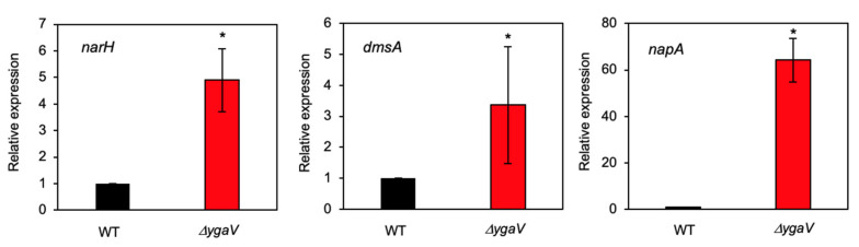
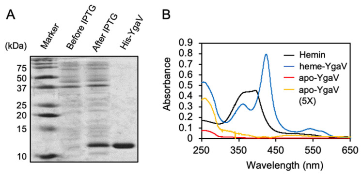
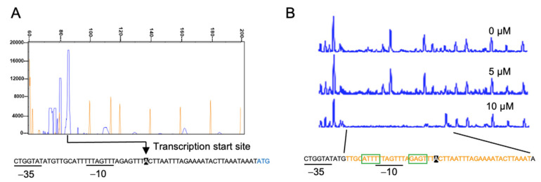
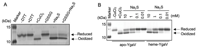
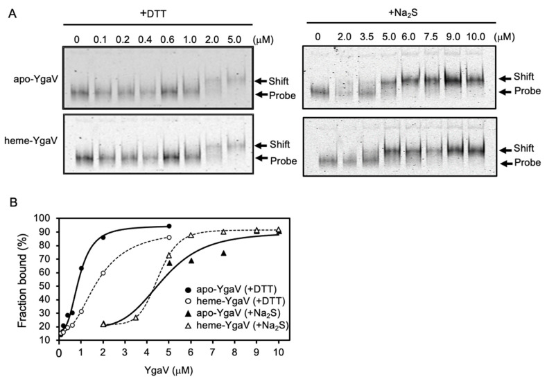
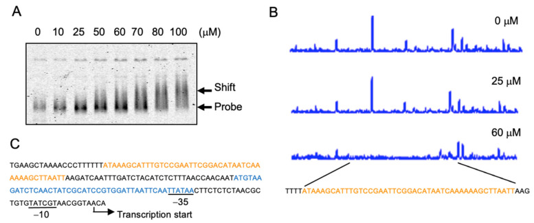
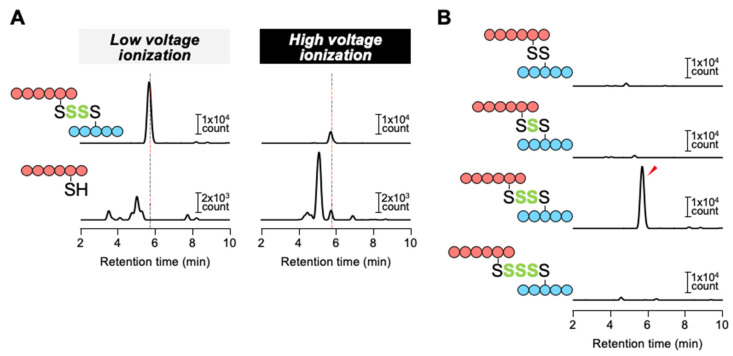
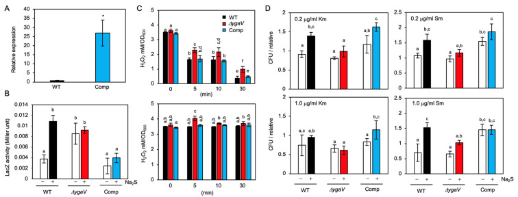
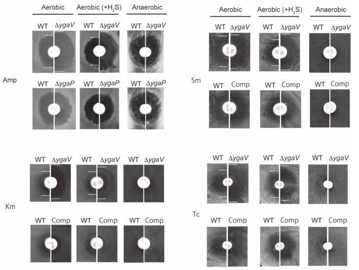
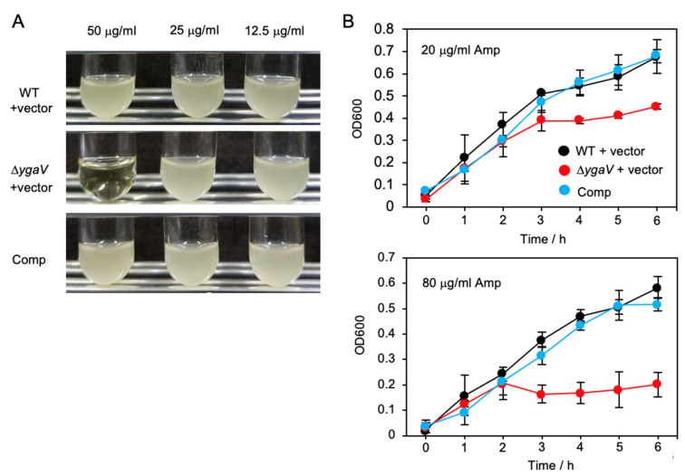
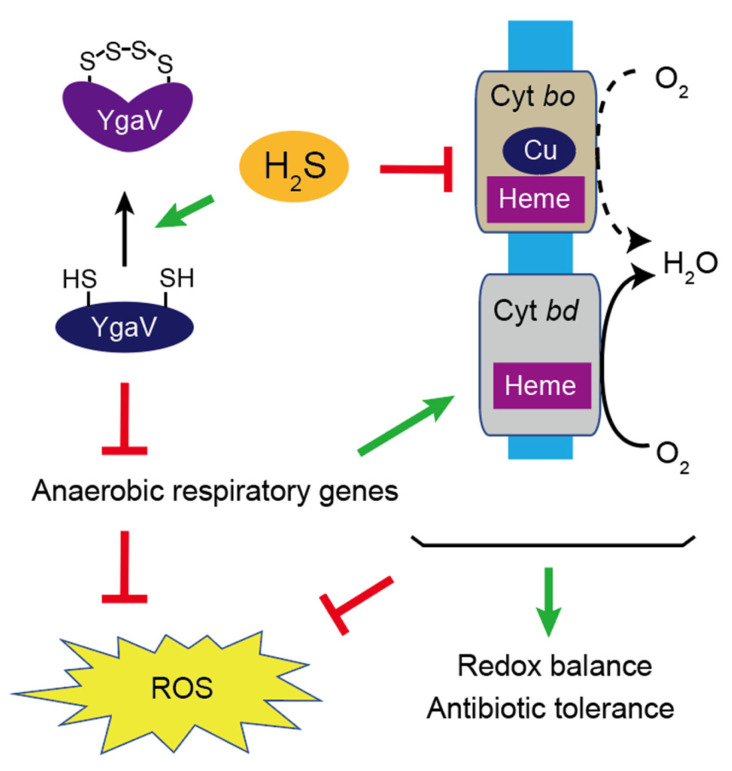
Compositions and activities of bacterial flora in the gastrointestinal tract significantly influence the metabolism, health, and disease of host humans and animals. These enteric bacteria can switch between aerobic and anaerobic growth if oxygen tension becomes limited. Interestingly, the switching mechanism is important for preventing reactive oxygen species (ROS) production and antibiotic tolerance. Studies have also shown that intracellular and extracellular sulfide molecules are involved in this switching control, although the mechanism is not fully clarified. Here, we found that YgaV, a sulfide-responsive transcription factor SqrR/BigR homolog, responded to sulfide compounds in vivo and in vitro to control anaerobic respiratory gene expression. YgaV also responded to H2O2 scavenging in the enteric bacterium Escherichia coli. Although the wild-type (WT) showed increased antibiotic tolerance under H2S-atmospheric conditions, the ygaV mutant did not show such a phenotype. Additionally, antibiotic sensitivity was higher in the mutant than in the WT of both types in the presence and absence of exogenous H2S. These results, therefore, indicated that YgaV-dependent transcriptional regulation was responsible for maintaining redox homeostasis, ROS scavenging, and antibiotic tolerance.

Keywords: Escherichia coli; SqrR; YgaV; antibiotics; hydrogen sulfide; reactive sulfur species.


Conflict of interest statement

The authors declare no conflict of interest.

Figures

Figure 1
Figure 1
Relative mRNA levels of narH, dmsA and napA genes in WT and ygaV mutant (ΔygaV) as determined by quantitative real-time PCR analysis. The WT levels were set to 1.0. The gyrA gene was used as an internal standard. The values are means ± SD of three biological replicates. * p < 0.05, t-test.
Figure 2
Figure 2
Purification and reconstitution of YgaV: (A) SDS-PAGE analysis of purified His-tagged YgaV. Cell extracts of E. coli expressing N-terminally His-tagged YgaV, before and after IPTG induction, are also analyzed. Proteins were stained with Coomassie brilliant blue; (B) Absorption spectra of hemin, heme-reconstituted YgaV (heme-YgaV), purified YgaV before reconstitution (apo-YgaV), and 5-times concentrated purified YgaV before reconstitution (apo-YgaV, 5×).
Figure 3
Figure 3
Characterization of the ygaVP promoter: (A) Primer extension analysis of the ygaVP promoter. Signal of the reverse transcripts and size markers are blue and red, respectively. ((A), bottom) The RNA polymerase σ-subunit recognition site (−10 and −35) and the translational start site of YgaV (blue) are shown. Reverse transcription was conducted with purified RNA and the fluorescent–labeled primer. The synthesized cDNA was mixed with the 600 LIZ Size Standard (Applied biosystem), and then analyzed using a 3730xl DNA analyzer (Applied biosystems). Finally, fragment analysis was conducted with Peak scanner software v.1.0 (Applied biosystem); (B) DNase I footprint analysis of YgaV. Binding to the ygaVP promoter DNA with different concentrations of reduced YgaV. Regions corresponding to the DNase I protection regions are shown in red. Motifs similar to the putative RcSqrR-recognition sequences are indicated by green boxes (for details, see text).
Figure 4
Figure 4
Mobility shifts of YgaV caused by thiol modification on SDS-PAGE gels: (A) The purified apo-YgaV was treated with 1 mM DTT, 0.5 mM CuCl2, 1 mM GSSG, and/or 10 mM Na2S. The proteins were precipitated by trichloroacetic acid (TCA) treatment, labeled with AMS, and then applied for SDS-PAGE; (B) The AMS modification was performed with apo- and heme-reconstituted YgaV with different concentrations of Na2S. The apo-YgaV was also treated with 0.5 mM CuCl2 as a control. Proteins were stained with Coomassie brilliant blue.
Figure 5
Figure 5
DNA-binding characteristics of YgaV: (A) Gel mobility shift analysis of DTT- and Na2S-treated apo- and heme-reconstituted YgaV to the ygaVP promoter DNA. The fluorescent-labeled DNA probe (50 nM) was incubated with various concentrations of purified apo-YgaV and Heme-YgaV for 30 min at room temperature. Subsequently, proteins were pretreated with 1 mM DTT or 10-mM Na2S to be reduced and oxidized, respectively. Then, 5% PAGE was used to separate samples. After electrophoresis, gels were analyzed using an image analyzer; (B) Binding isotherms of DTT-treated apo- and heme-reconstituted YgaV, and Na2S-treated apo- and heme-reconstituted YgaV.
Figure 6
Figure 6
YgaV binding to the katG promoter: (A) Gel mobility shift analysis of DTT-treated apo-YgaV to the fluorescent-labeled katG promoter DNA. The protein-DNA mixtures were separated by 5% PAGE. After electrophoresis, gels were analyzed using an image analyzer; (B) DNase I footprint analysis of DTT-reduced apo-YgaV with the katG promoter DNA. Regions corresponding to the DNase I protection regions are shown in red. (C) The DNA sequence of the katG promoter region. OxyR and YgaV-binding sites are indicated by blue and red, respectively. The transcription start site and the RNA polymerase recognition sites (−10 and −35) are also shown.
Figure 7
Figure 7
Detection of oxidized polysulfur structure of Na2S-treated YgaV by iCOPS method: (A) The complex of ILC31ML (blue) and KNVYC98P (red) with an intra-tetrasulfide bridge was detected under low voltage ionization conditions (30 V), while the oxidized polysulfur bond was disrupted by high voltage ionization (60 V), resulting in the formation of free KNVYC98P peptide which was detected at the same retention time as the intact peptide complex (dashed line); (B) Various lengths of oxidized polysulfur bridges between ILC31ML and KNVYC98P peptides were investigated under low-voltage ionization conditions (30 V). A significant signal of the intra-tetrasulfide bridge-containing complex was only detected (red arrow).
Figure 8
Figure 8
Phenotypes of the complementing strain of the ygaV mutant (Comp), which harbors the multicopy plasmid encoding ygaV gene: (A) Relative mRNA levels of ygaV in WT and the ygaV complementing strain as determined by quantitative real-time PCR analysis. The WT levels were set to 1.0. The gyrA gene was used as an internal standard. The values are means ± SD of three biological replicates. * p < 0.05, t test; (B) Promoter activities of the ygaVP operon. LacZ (β-galactosidase) activity measurement with the ygaV-lacZ fusion in WT, ygaV mutant and the complementing strain. Cells grown at mid-log phase (OD600 = ~0.3) were treated or untreated with 0.2 mM Na2S (final concentration) and harvested at late-log phase (OD600 = 0.8~0.9). The values are means ± SD of three biological replicates; (C) H2O2 levels in WT, ygaV mutant and the complementing strain. Cells grown at mid-log phase (OD600 = 0.5–0.6) were treated with 5.0 μg mL−1 kanamycin (Km) (upper) or streptomycin (Sm) (bottom) (final concentration) for 5, 10 and 30 min, and then harvested. The values are means ± SD of three biological replicates; (D) Mean colony-forming-unit (CFU) values of WT, ygaV mutant and the complementing strain exposed to 0.2 μg mL−1 or 1.0 μg mL−1 Km or Sm (final concentration) for 30 min in the presence or absence of 0.2 mM Na2S (final concentration). One of the CFU values of WT at 0.2 μg mL−1 Km or Sm was set as 1. Different letters (a, b, c, d, e and f) indicate significant differences between groups (p < 0.05; Tukey’s test).
Figure 9
Figure 9
Phenotypic characterization of ygaV and ygaP mutants. A paper disk saturated with Amp, Km, Sm and Tc was placed on E. coli WT, ygaV complementing strain (Comp), ygaV and/or ygaP mutants’ lawns that were grown under aerobic, anaerobic, and aerobic H2S-atmosphere conditions for 18 h. Zone borders are marked with dotted lines to easily see the differences.
Figure 10
Figure 10
Inhibited growth of the ygaV mutant by Amp: (A) The Amp-resistance empty pPAB404 vector was introduced into WT and ygaV mutants. The complementing strain of ygaV mutant (Comp) harbors the pPAB404 encoding ygaV. Colonies of each strain on agar-solidified plates containing 50 μg mL−1 Amp were picked, placed in liquid medium containing 50, 25 and 12.5 μg mL−1 Amp, and then incubated for 16 h; (B) Overnight cultures of the three strains shown in (A) in liquid medium containing 20 μg mL−1 Amp, were diluted 20-times with fresh LB medium containing 80 and 20 μg mL−1 Amp, and then growth of the strains was monitored by measuring optical density at 600 nm (OD600).
Figure 11
Figure 11
A schematic model for YgaV-dependent control of gene expression and its physiological function. H2S inhibits activity of the energy-efficient cytochrome bo oxidase, which causes ROS accumulation. In the presence of H2S, two cysteine residues of YgaV form an intramolecular tetrasulfide bond to derepress anaerobic-respiratory and ROS-scavenging genes, which contributes to redox homeostasis, ROS scavenging and antibiotic tolerance.

Similar articles

Cited by

References

    1. Sikalidis A.K. Amino acids and immune response: A role for cysteine, glutamine, phenylalanine, tryptophan and arginine in t-cell function and cancer? Pathol. Oncol. Res. 2015;21:9–17. doi: 10.1007/s12253-014-9860-0. - DOI - PubMed
    1. Sikalidis A.K., Maykish A. The gut microbiome and type 2 diabetes mellitus: Discussing a complex relationship. Biomedicines. 2020;8:8. doi: 10.3390/biomedicines8010008. - DOI - PMC - PubMed
    1. Unden G., Bongaerts J. Alternative respiratory pathways of Escherichia coli: Energetics and transcriptional regulation in response to electron acceptors. Biochim. Biophys. Acta. 1997;1320:217–234. doi: 10.1016/S0005-2728(97)00034-0. - DOI - PubMed
    1. Gunsalus R., Park S.-J. Aerobic-anaerobic gene regulation in Escherichia coli: Control by the ArcAB and Fnr regulons. Res. Microbiol. 1994;145:437–450. doi: 10.1016/0923-2508(94)90092-2. - DOI - PubMed
    1. Imlay J.A. The molecular mechanisms and physiological consequences of oxidative stress: Lessons from a model bacterium. Nat. Rev. Microbiol. 2013;11:443–454. doi: 10.1038/nrmicro3032. - DOI - PMC - PubMed

LinkOut - more resources