. 2022 Nov 30;11(12):2376.
doi: 10.3390/antiox11122376.

Colon-Targeted eNAMPT-Specific Peptide Systems for Treatment of DSS-Induced Acute and Chronic Colitis in Mouse

Affiliations

Colon-Targeted eNAMPT-Specific Peptide Systems for Treatment of DSS-Induced Acute and Chronic Colitis in Mouse

Jae-Sung Kim et al. Antioxidants (Basel). .

Abstract

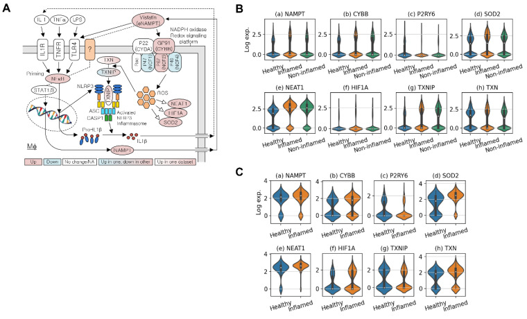
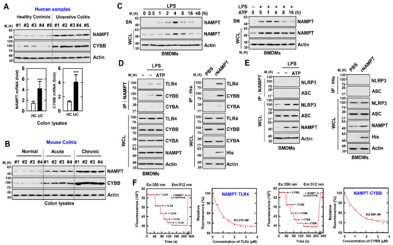
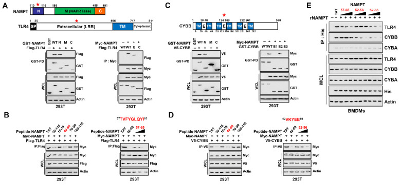
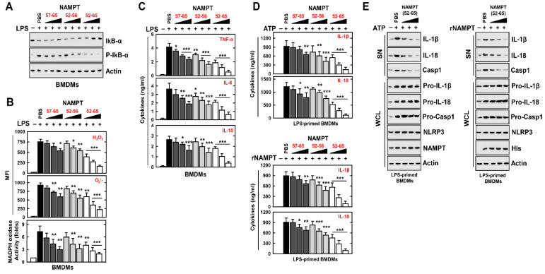
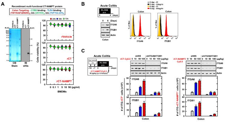
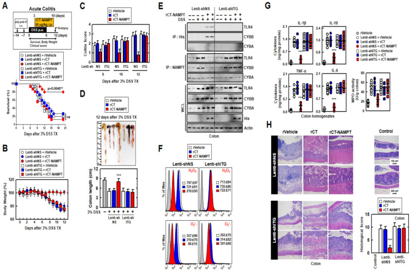
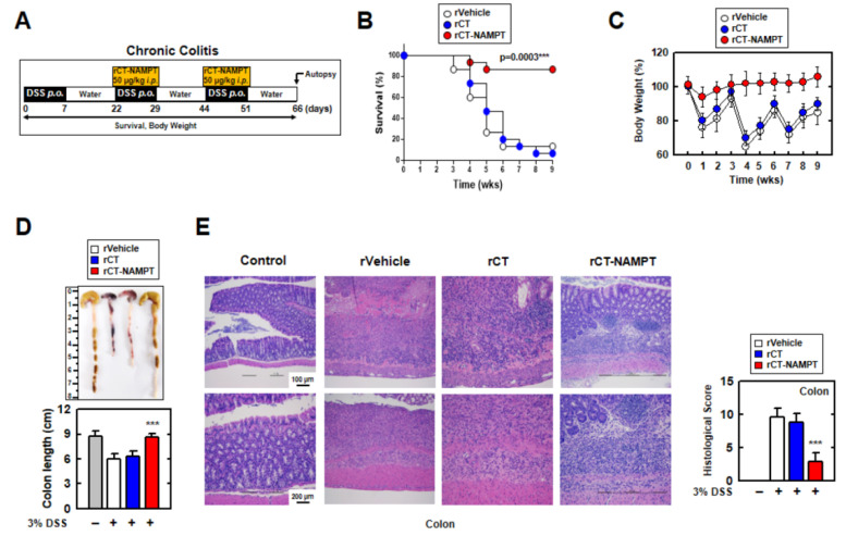
Nicotinamide phosphoribosyl transferase (NAMPT) is required to maintain the NAD+ pool, among which extracellular (e) NAMPT is associated with inflammation, mainly mediated by macrophages. However, the role of (e) NAMPT in inflammatory macrophages in ulcerative colitis is insufficiently understood. Here our analyses of single-cell RNA-seq data revealed that the levels of NAMPT and CYBB/NOX2 in macrophages were elevated in patients with colitis and in mouse models of acute and chronic colitis. These findings indicate the clinical significance of NAMPT and CYBB in colitis. Further, we found that eNAMPT directly binds the extracellular domains of CYBB and TLR4 in activated NLRP3 inflammasomes. Moreover, we developed a recombinant 12-residue TK peptide designated colon-targeted (CT)-conjugated multifunctional NAMPT (rCT-NAMPT), comprising CT as the colon-targeting moiety, which harbors the minimal essential residues required for CYBB/TLR4 binding. rCT-NAMPT effectively suppressed the severity of disease in DSS-induced acute and chronic colitis models through targeting the colon and inhibiting the interaction of NAMPT with CYBB or TLR4. Together, our data show that rCT-NAMPT may serve as an effective novel candidate therapeutic for colitis by modulating the NLRP3 inflammasome-mediated immune signaling system.

Keywords: CYBB; NAMPT; NLRP3 inflammasome; TLR4; colitis.


Conflict of interest statement

The authors declare no conflict of interest.

Figures

Figure 1
Figure 1
eNAMPT plays a key role in ulcerative colitis (A) NAMPT’s association with TLR4 or NADPH oxidase-driven NLRP3 inflammasome activation. (B) Violin plots for the expression of key genes in macrophages around NLRP3 inflammasome activation in SCP259. (C) Violin plots for the expression of key genes around NLRP3 inflammasome activation in GSE182270.
Figure 2
Figure 2
eNAMPT directly binds with CYBB or TLR4 (A) Normal human cases alongside UC patients were employed for IB with αNAMPT and αCYBB. WCLs (whole cell lysates) were utilized for IB with αActin (upper). Representative gel images derived from five independent healthy controls and patients are shown. NAMPT mRNA and CYBB mRNA expression measured by quantitative real-time PCR (lower). Five of ten normal human and UC patients’ data are shown. Statistical significance was evaluated by the Student’s t-test coupled with the Bonferroni adjustment (*** p < 0.001) versus human normal. (B) Colon of normal, acute and chronic colitis mouse were analyzed for IB with αNAMPT and αCYBB. WCLs were used for IB with αActin. Biological replicates (n = 10) for each condition were performed. (C) BMDMs were activated with LPS (100 ng/mL) for the durations indicated (left), then primed with LPS (100 ng/mL) for 4 h before being stimulated with ATP (1 mM) for the times indicated (right). IB in supernatant (SN) with αNAMPT and WCL with αNAMPT or αActin. (D,E) BMDMs were primed with LPS and stimulated with ATP (left) or incubated with rNAMPT (1 μg/mL) for 2 h (right). BMDMs were treated with IP with αNAMPT or αHis, trailed by IB with αTLR4, αCYBB or αCYBA (D) and IB with αASC or αNLRP3 (E). WCLs were used for IB with αTLR4, αCYBB, αCYBA, αASC, αNLRP3 or αActin. (F) Titration of fluorescently labeled TLR4 or CYBB with unlabeled NAMPT, using curve fit analysis to determine Kd (219 and 896 nM). The data come from five independent experiments that yielded comparable results.
Figure 3
Figure 3
NAMPT (aa 57-65 and aa 52-65) is an essential region for binding to TLR4 and CYBB. (A) Schematic diagram of the structures of NAMPT and TLR4 (upper). The 293T cells were transfected with GST-NAMPT or its mutants and Flag-TLR4. 293T cells were subjected to GST pull-down, followed by IB with αFlag. WCLs were used for IB with αGST, αFlag or αActin (lower, left). The 293T cells were transfected with Flag-TLR4 or its mutants and Myc-NAMPT. The 293T cells were used for IP with αMyc, followed by IB with αFlag. WCLs were used for IB with αMyc, αFlag or αActin (lower, right). (B) 293T cells were transfected with Myc-NAMPT and Flag-TLR4 and treated NAMPT peptide or its mutants for 6 h (5 µM, left; 1, 5, 10 µM, right). The 293T cells were used for IP with αFlag, followed by IB with αMyc. WCLs were used for IB with αMyc, αFlag or αActin. (C) Schematic diagram of the structures of CYBB (upper). The 293T cells were transfected with GST-NAMPT or its mutants and V5-CYBB (lower, left). The 293T cells were transfected with GST-CYBB or its mutants and Myc-NAMPT (lower, right). The 293T cells were subjected to GST pull-down, followed by IB with αV5 or αMyc. WCLs were used for IB with αGST, αMyc, αV5 or αActin. (D) 293T cells were transfected with Myc-NAMPT and V5-CYBB and treated NAMPT peptide or its mutants for 6 h (5 µM, left; 1, 5, 10 µM, right). The 293T cells were used for IP with αV5, followed by IB with αMyc. WCLs were used for IB with αMyc, αFlag or αActin. (E) BMDMs were incubated with rNAMPT (1, 5, 10 μg/mL) for 2 hr. BMDMs were subjected to IP with αHis, followed by IB with αTLR4, αCYBB or αCYBA. WCLs were used for IB with αTLR4, αCYBB or αCYBA, αHis or αActin. The data come from seven independent experiments that yielded comparable results (AE). Ref star: Binding domain with binding partners.
Figure 4
Figure 4
NAMPT peptides inhibit the signal 1 and 2 activation of the NLRP3 inflammasome. (AC) BMDMs were stimulated with LPS for 4 h, pretreated with rNAMPT for 1 h (A,B) or 18 h (C). (A) WCLs were used for IB with αIkB-α, αP-IkB-α or αActin. (B) FACS analysis for H2O2 (probe for 2′, 7′-dichlorofluorescin diacetate) or O2- (probe for Dihydroethidium). Quantitative analysis of mean fluorescence intensities of H2O2 and O2- (upper). NADPH oxidase activity (lower). (C) Culture supernatants were harvested and analyzed for cytokine ELISA. (D,E) LPS-primed BMDMs were treated with rNAMPT for 1 h, and then activated with ATP for 30 min. (D) ELISA for IL-1β and IL-18. (E) IB in SN with αIL-1β p17, αIL-18 p18 or αCasp1 p10 and WCL with αPro-IL-1β, αPro-IL-18, αPro-Casp1, or αActin. The data come from seven independent experiments that yielded comparable results (A,E). The data come from three independent experiments that yielded comparable results. (BD). Data are given as means ± SD of three experiments. Significant differences (* p < 0.05; ** p < 0.01; *** p < 0.001) versus LPS + PBS.
Figure 5
Figure 5
rCT-NAMPT targets the inflamed colon. (A) Schematic illustration of rCT-NAMPT (upper). Bacterially purified 6xHis-rCT-NAMPT, rVehicle or rCT were analyzed by Coomassie Blue staining or IB with αHis (lower, left). BMDMs were treated with rVehicle, rCT, or rCT-NAMPT for the periods and concentrations stated, and cell viability was determined using the MTT test (lower, right). (B) DSS-induced model for colitis. Mice were subjected to 3% DSS for 6 days and were evaluated at day 6 (left). Colon harvests were used for IB with αITGA6, αITGB1 or αActin and analysis of the number of ITGA6+ or ITGB1+ cells by FACS. (C) The scheme of the acute colitis model transduced with Lenti-shNS or Lenti-shITG virus and treated with 3% DSS. Intraperitoneal administration of rCT-NAMPT conjugated with Cy5.5 for 1 h and colon harvest (left). Colon harvests were used for IB with αITGA6, αITGB1 or αActin and analysis of the number of ITGA6+ or ITGB1+ cells was done by FACS. The data come from three independent experiments that yielded comparable results (AC).
Figure 6
Figure 6
rCT-NAMPT has a medicinal effect against acute DSS-induced colitis in mice. (A) Scheme of the acute model of colitis transduced with Lenti-shNS or Lenti-shITG virus and subjected to 3% DSS with rCT-NAMPT (50 μg/kg) (upper). The survival of mice was monitored for 12 days; mortality was measured for n = 15 mice per group (lower). The statistical differences between the rVehicle-treated animals are noted (log-rank test). The data come from two different experiments that yielded comparable results. (B) Weight loss (n = 8). (C) Colitis scores were obtained from clinical parameters (weight loss, stool consistency, bleeding) (n = 8). (D) Image (upper) and length (lower) of colon in 3% DSS-treated mice with rVehicle, rCT or rCT-NAMPT (n = 8). (E) Colon was used for IP with αHis or αNAMPT, followed by IB with αTLR4, αCYBB or αCYBA. WCLs were used for IB with αTLR4, αCYBB, αCYBA, αHis or αActin. (F) FACS analysis for cellular ROS from colon (n = 8). (G) Levels of cytokines and MPO activity in colon homogenates (n = 10). (H) Hematoxylin and eosin (H&E) staining of the colon (left) (n = 10): representative imaging Histopathology scores were assessed in 3% DSS-treated mice with rVehicle, rCT, or rCT-NAMPT using H&E staining, as indicated in techniques (Materials and Methods). Statistical significance was assessed using the Student’s t-test with the Bonferroni correction (*** p < 0.001) in comparison to rVehicle.
Figure 7
Figure 7
rCT-NAMPT relieves chronic DSS-induced colitis in a mouse model. (A) Scheme of the chronic colitis model subjected to 3% DSS with rCT-NAMPT (50 μg/kg). (B) Mice survival was tracked for 9 weeks, and mortality was calculated for n = 15 mice per group. The statistical differences between the rVehicle-treated animals are noted (log-rank test). The data come from two different experiments that yielded comparable results. (C) Weight loss of rVehicle, rCT or rCT-NAMPT in mice (n = 15). (D) Image (upper) and length (lower) of colon in 3% DSS-induced chronic colitis mice with rVehicle, rCT or rCT-NAMPT. (E) H&E staining of the colon (left) (n = 8): a representative image. H&E staining was used to evaluate histopathology scores in 3% DSS-induced chronic colitis mice with rVehicle, rCT, or rCT-NAMPT. The Student’s t-test with the Bonferroni adjustment was used to establish statistical significance when compared to rVehicle (*** p < 0.001).

Similar articles

Cited by

References

    1. Nielsen O.H. New strategies for treatment of inflammatory bowel disease. Front. Med. (Lausanne) 2014;1:3. doi: 10.3389/fmed.2014.00003. - DOI - PMC - PubMed
    1. Kaplan G.G., Ng S.C. Understanding and Preventing the Global Increase of Inflammatory Bowel Disease. Gastroenterology. 2017;152:313–321.e2. doi: 10.1053/j.gastro.2016.10.020. - DOI - PubMed
    1. Nielsen O.H., Ainsworth M.A. Tumor necrosis factor inhibitors for inflammatory bowel disease. N. Engl. J. Med. 2013;369:754–762. doi: 10.1056/NEJMct1209614. - DOI - PubMed
    1. Vulliemoz M., Brand S., Juillerat P., Mottet C., Ben-Horin S., Michetti P., on behalf of Swiss IBDnet, an official working group of the Swiss Society of Gastroenterology TNF-Alpha Blockers in Inflammatory Bowel Diseases: Practical Recommendations and a User’s Guide: An Update. Digestion. 2020;101((Suppl. S1)):16–26. doi: 10.1159/000506898. - DOI - PubMed
    1. Chiarugi A., Dolle C., Felici R., Ziegler M. The NAD metabolome—A key determinant of cancer cell biology. Nat. Rev. Cancer. 2012;12:741–752. doi: 10.1038/nrc3340. - DOI - PubMed

LinkOut - more resources