. 2022 Dec 2;11(12):2396.
doi: 10.3390/antiox11122396.

Mangosteen Pericarp Extract Supplementation Boosts Antioxidant Status via Rebuilding Gut Microbiota to Attenuate Motor Deficit in 6-OHDA-Induced Parkinson's Disease

Affiliations

Mangosteen Pericarp Extract Supplementation Boosts Antioxidant Status via Rebuilding Gut Microbiota to Attenuate Motor Deficit in 6-OHDA-Induced Parkinson's Disease

Bira Arumndari Nurrahma et al. Antioxidants (Basel). .

Abstract

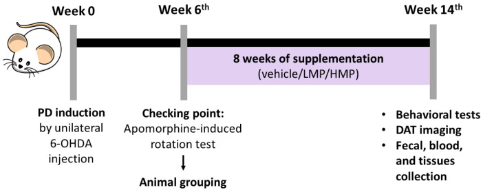
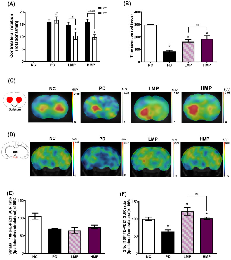
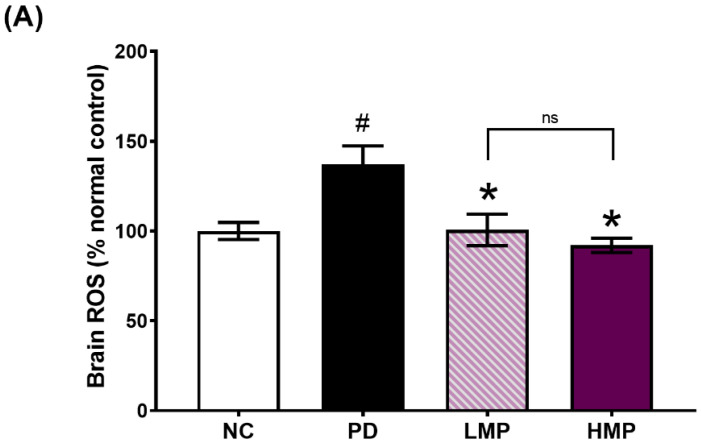
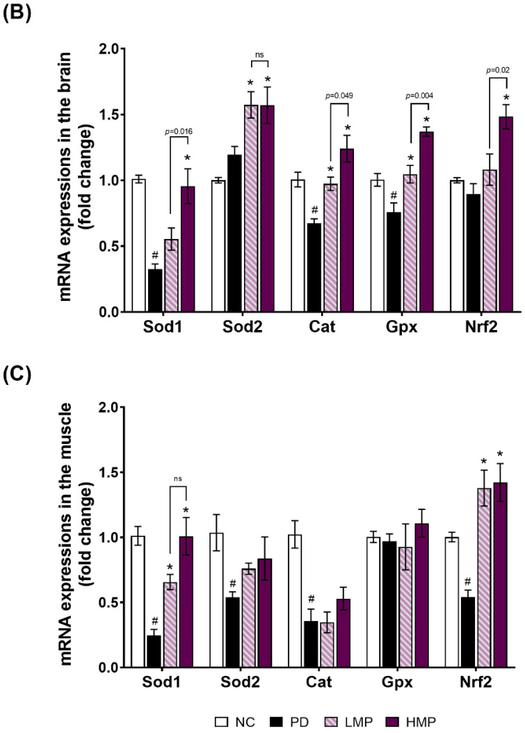
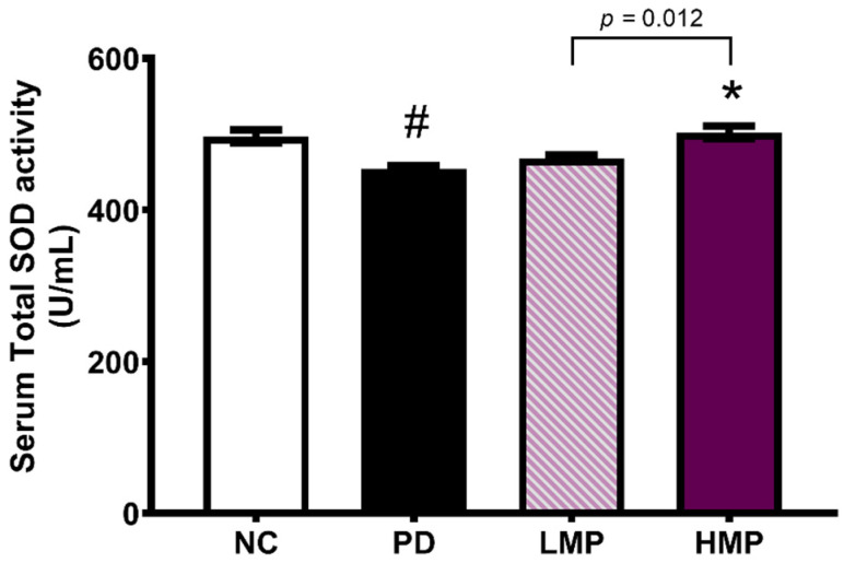
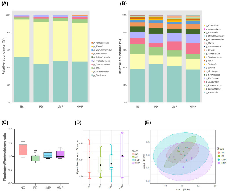
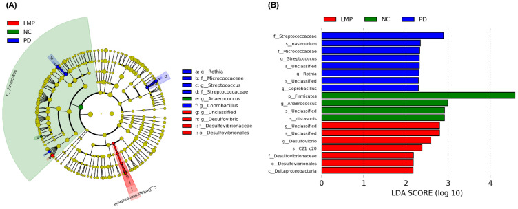
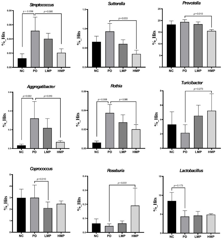
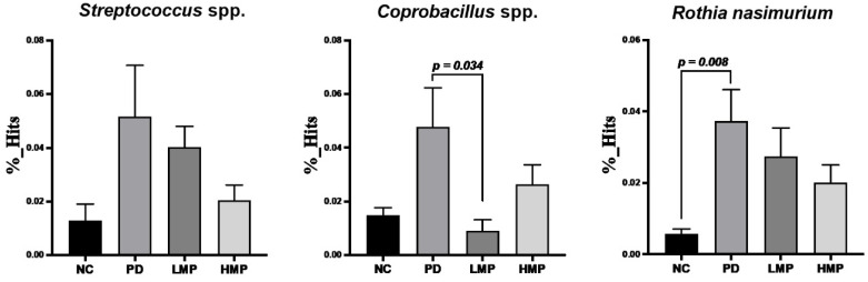
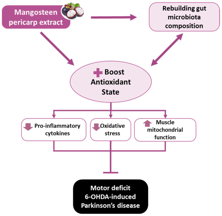
Oxidative stress and gut dysbiosis have been known to precede Parkinson's disease (PD). An antioxidant-rich product, mangosteen pericarp (MP), has the ability to counterbalance excessive free radicals and the imbalanced gut microbiota composition, suggesting the MP's capacity to delay PD progression. In this study, we explored the effects of two doses of MP extract in a unilateral 6-hydroxydopamine (6-OHDA)-induced PD rat model. We revealed that the 8-week supplementation of a low dose (LMP) and a high dose of the MP extract (HMP) improved motor function, as observed in decreased contralateral rotation, improved time spent on rod, and higher dopamine binding transporter (DAT) in the substantia nigra pars compacta (SNc). The MP extract, especially the HMP, also increased antioxidant-related gene expressions, restored muscle mitochondrial function, and remodeled fecal microbiota composition, which were followed by reduced reactive oxygen species levels in brain and inflammation in plasma. Importantly, bacterial genera Sutterella, Rothia, and Aggregatibacter, which were negatively correlated with antioxidant gene expressions, decreased in the HMP group. It is imperative to note that in addition to directly acting as an antioxidant to reduce excessive free radicals, MP extract might also increase antioxidant state by rebuilding gut microbiota, thereby enhanced anti-inflammatory capacity and restored mitochondrial function to attenuate motor deficit in 6-OHDA-induced PD-like condition. All in all, MP extract is a potential candidate for auxiliary therapy for PD.

Keywords: 6-hydroxydopamine; Parkinson’s disease; antioxidant; fecal microbiota composition; mangosteen pericarp.


Conflict of interest statement

The authors declare no conflict of interest.

Figures

Figure 1
Figure 1
The timeline of the animal experiment.
Figure 2
Figure 2
Effects of mangosteen pericarp (MP) extract supplementation on motor function. (A) Number of contralateral rotations recorded in the apomorphine-induced rotation test at week 0 and week 8 of supplementation. (B) Evaluation of motor coordination and balance skills in the rotarod test after 8 weeks of supplementation. (C) Representative images of the standard uptake ratio (SUR) of [18F]FE-PE21 in the striatum, illustrating the dopamine transporter (DAT) binding activity. (D) Representative images of the SUR of [18F]FE-PE21 in the substantia nigra pars compacta (SNc). (E) Quantitation of the SUR ratio in the striatum. (F) Quantitation of the SUR ratio in the SNc. Data are average ± SEM for n = 5 rats/group and were compared using one-way ANOVA with Tukey’s post hoc test; # p < 0.05 for Parkinson’s disease (PD) group vs. normal control (NC) group. * p < 0.05 for low-dose mangosteen pericarp (LMP) and high-dose mangosteen pericarp (HMP) groups vs. PD group. ns, not significant.
Figure 3
Figure 3
Levels of ROS in the brain (A) and antioxidant-related gene expression in (B) the brain and (C) muscle after 8 weeks of supplementation in the rat model of PD. All data are average ± SEM for n = 5 rats/group and were analyzed using one-way ANOVA with Tukey’s post hoc test. # p < 0.05 for the Parkinson’s disease (PD) group vs. normal control (NC) group, * p < 0.05 for the low-dose mangosteen pericarp (LMP) and high-dose mangosteen pericarp (HMP) groups vs. the PD group. Sod, superoxide dismutase; Cat, catalase; Gpx, glutathione peroxidase; Nrf2, nuclear factor-erythroid factor 2-related factor 2. ns, not significant.
Figure 3
Figure 3
Levels of ROS in the brain (A) and antioxidant-related gene expression in (B) the brain and (C) muscle after 8 weeks of supplementation in the rat model of PD. All data are average ± SEM for n = 5 rats/group and were analyzed using one-way ANOVA with Tukey’s post hoc test. # p < 0.05 for the Parkinson’s disease (PD) group vs. normal control (NC) group, * p < 0.05 for the low-dose mangosteen pericarp (LMP) and high-dose mangosteen pericarp (HMP) groups vs. the PD group. Sod, superoxide dismutase; Cat, catalase; Gpx, glutathione peroxidase; Nrf2, nuclear factor-erythroid factor 2-related factor 2. ns, not significant.
Figure 4
Figure 4
Serum levels of total superoxide dismutase (SOD) after 8 weeks of supplementation in the rat model of PD. All data are average ± SEM for n = 5 rats/group and were analyzed using one-way ANOVA with Tukey’s post hoc test. # p < 0.05 for the Parkinson’s disease (PD) group vs. normal control (NC) group; * p < 0.05 for low-dose mangosteen pericarp (LMP) and high-dose mangosteen pericarp (HMP) groups vs. the PD group.
Figure 5
Figure 5
Comparison of mitochondrial function and mitochondria-related mRNA expression in muscles after 8 weeks of supplementation in the rat model of PD. (A) The basal oxygen consumption rate (OCR) was quantified to evaluate mitochondrial respiration. (B) Basal extracellular acidification flux (ECAR) was used to evaluate the rate of glycolysis. (C) mtDNA copy numbers (Nd1 and Atp6). (D) Expression of mitochondrial biogenesis-related mRNAs: PPARG coactivator 1 alpha (Pgc1a), nuclear respiratory factor 1 (Nrf1), mitochondrial transcription factor A (Tfam). Results are average ± SEM for n = 5 rats/group and were analyzed using one-way ANOVA with Tukey’s post hoc test. # p < 0.05 for the Parkinson’s disease (PD) group vs. the normal control (NC) group. * p < 0.05 for the low-dose mangosteen pericarp (LMP) and high-dose mangosteen pericarp (HMP) groups vs. the PD group. ns, not significant.
Figure 6
Figure 6
Comparison of the relative bacterial abundances and analysis of bacterial diversity based on 16S sequencing analysis of fecal samples after 8 weeks of supplementation in the rat model of PD. (A) Distribution of the top ten most abundant bacteria at the phylum level across all groups. (B) Distribution of the top twenty most abundant bacteria at the genus level across all groups. (C) Firmicutes to Bacteroidetes ratios of each group. # p < 0.05 for the Parkinson’s disease (PD) group vs. the normal control (NC) group. (D) Analysis of alpha diversity based on the Shannon index. (E) Analysis of beta-diversity based on principal coordinates analysis (PCoA) of the Bray–Curtis index. The ellipses illustrate 95% confidence intervals for the normal control (NC) versus Parkinson’s disease (PD) versus low-dose mangosteen pericarp (LMP) versus high-dose mangosteen pericarp (HMP).
Figure 7
Figure 7
Linear discriminant analysis (LDA) effect size (LEfSe) of fecal microbiota enrichment in the normal control (NC), Parkinson’s disease (PD), low-dose mangosteen pericarp (LMP), and high-dose mangosteen pericarp (HMP) groups after 8 weeks of supplementation. (A) The circles of the phylogenetic tree cladogram demonstrate the classification of the fecal microbiota from the phylum to genus level. The diameter of the circles illustrates relative abundance. The typescripts A to J in A represent 10 different taxa. (B) LDA scores for differentially abundant taxa between the NC, PD, LMP, and HMP groups. Taxa with an LDA score >2 (log 10) were included in the LEfSe analysis. There was no distinct taxonomic enrichment between the HMP group and other groups; thus, the HMP group is not shown in the phylogenetic tree cladogram. C, class; F, family; G, genus; O, order; P, phylum.
Figure 8
Figure 8
Analysis of the abundance of specific bacteria at the genus level after 8 weeks of supplementation in the low-dose mangosteen pericarp (LMP) and high-dose mangosteen pericarp (HMP). Results are average ± SEM for n = 5 rats/group and were analyzed using the unpaired t-test or Mann–Whitney test.
Figure 9
Figure 9
Comparison of the abundance of specific bacterial species identified in rats with 6-OHDA-induced Parkinson’s disease after 8 weeks of supplementation with the low-dose mangosteen pericarp (LMP) and high-dose mangosteen pericarp (HMP). Results are average ± SEM for n = 5 rats/group and were analyzed using unpaired t-tests.
Figure 10
Figure 10
The proposed mechanism of action by which mangosteen pericarp extract delays motor deficit in 6-OHDA-induced PD.

Similar articles

Cited by

References

    1. Ou Z., Pan J., Tang S., Duan D., Yu D., Nong H., Wang Z. Global trends in the incidence, prevalence, and years lived with disability of Parkinson’s disease in 204 countries/territories from 1990 to 2019. Front. Public Health. 2021;9:776847. doi: 10.3389/fpubh.2021.776847. - DOI - PMC - PubMed
    1. Maiti P., Manna J., Dunbar G.L. Current understanding of the molecular mechanisms in Parkinson’s disease: Targets for potential treatments. Transl. Neurodegener. 2017;6:28. doi: 10.1186/s40035-017-0099-z. - DOI - PMC - PubMed
    1. Sun M.F., Shen Y.Q. Dysbiosis of gut microbiota and microbial metabolites in Parkinson’s Disease. Ageing Res. Rev. 2018;45:53–61. doi: 10.1016/j.arr.2018.04.004. - DOI - PubMed
    1. Henchcliffe C., Beal M.F. Mitochondrial biology and oxidative stress in Parkinson disease pathogenesis. Nat. Clin. Pract. Neurol. 2008;4:600–609. doi: 10.1038/ncpneuro0924. - DOI - PubMed
    1. Zuo L., Motherwell M.S. The impact of reactive oxygen species and genetic mitochondrial mutations in Parkinson’s disease. Gene. 2013;532:18–23. doi: 10.1016/j.gene.2013.07.085. - DOI - PubMed

LinkOut - more resources