

Complex Interplay between Autophagy and Oxidative Stress in the Development of Endometriosis
- PMID: 36552692
- PMCID: PMC9774576
- DOI: 10.3390/antiox11122484
Complex Interplay between Autophagy and Oxidative Stress in the Development of Endometriosis
Abstract
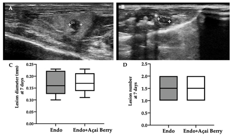
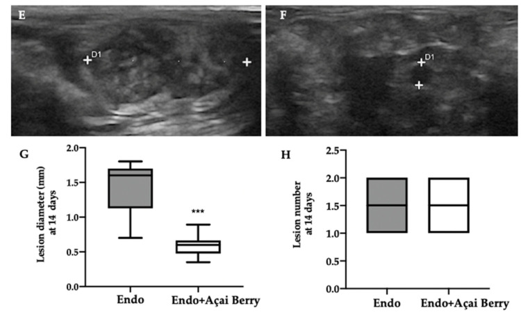
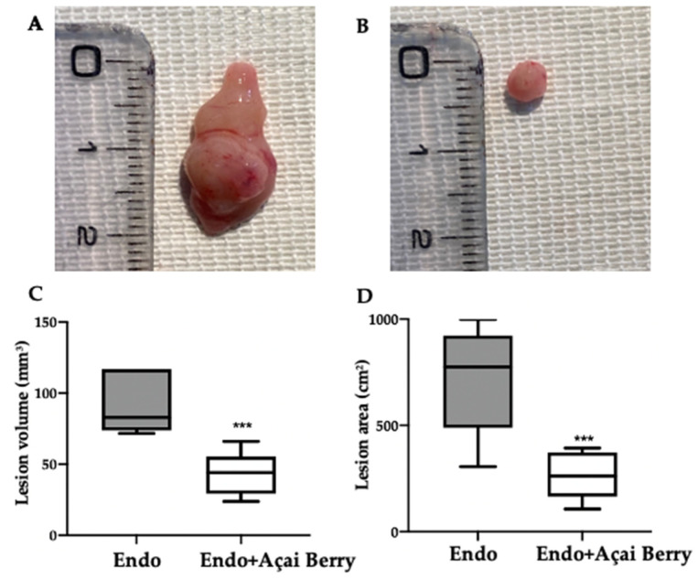
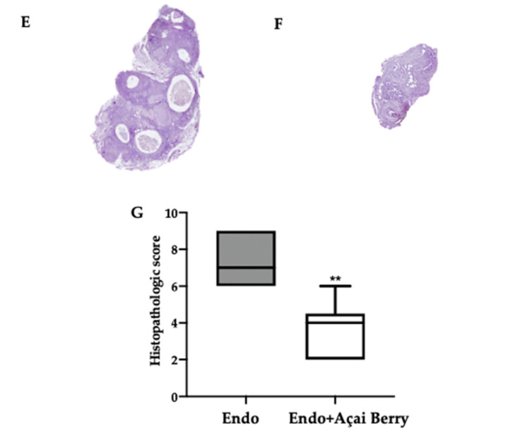
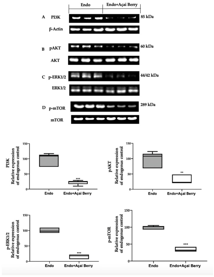
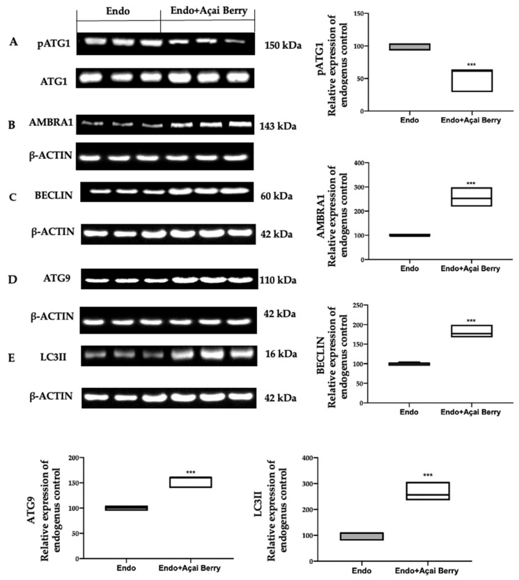
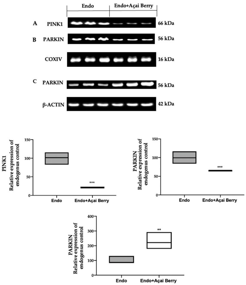
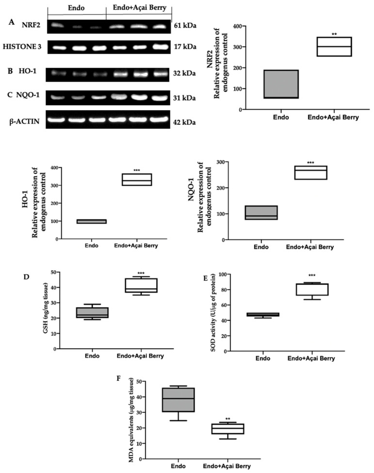
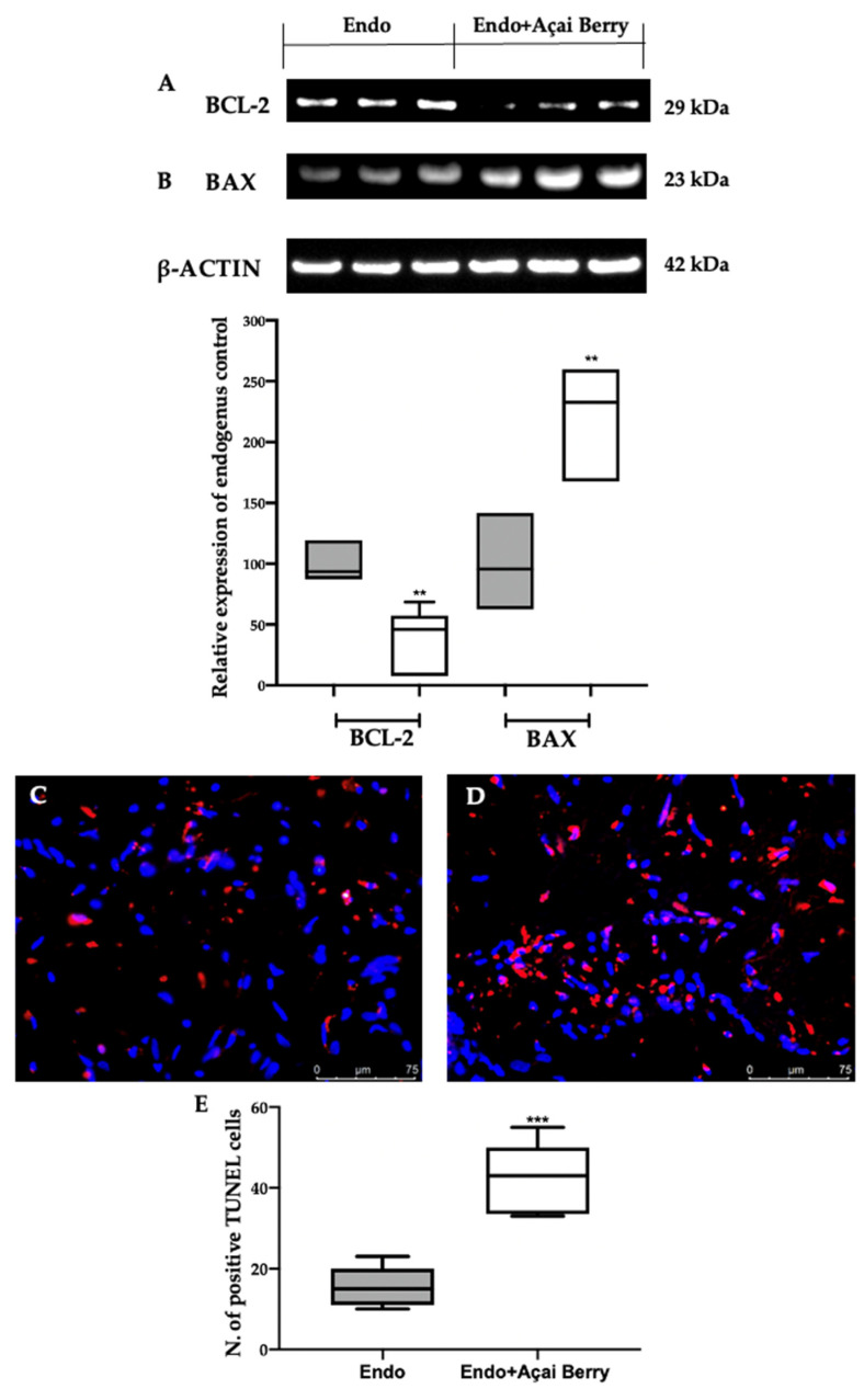
Endometriosis (Endo) is a chronic gynecological disease. This paper aimed to evaluate the modulation of autophagy, oxidative stress and apoptosis with Açai Berries in a rat model of endometriosis. Endometriosis was induced with an intraperitoneal injection of minced uterus tissue from a donor rat into a recipient one. The abdominal high-frequency ultrasound (hfUS) analysis was performed at 7 and 14 days from the endometriosis induction to evaluate the growth of the lesion during the experiment. Seven days from the induction, once the lesions were implanted, an Açai Berry was administered daily by gavage for the next seven days. At the end of the experiment, the hfUS analysis showed a reduced lesion diameter in animals given the Açai Berry. A macroscopical and histological analysis confirmed this result. From the molecular point of view, Western blot analyses were conducted to evaluate the autophagy induction. Samples collected from the Endo group showed impaired autophagy, while the Açai Berry administration inhibited PI3K and AKT and ERK1/2 phosphorylation and promoted autophagy by inactivating mTOR. Additionally, Açai Berry administration dephosphorylated ATG1, promoting the activity of the ATG1/ULK1 complex that recruited Ambra1/Beclin1 and Atg9 to promote autophagosome nucleation and LC3II expression. Açai Berry administration also restored mitophagy, which increased Parkin cytosolic expression. The Açai Berry increased the expression of NRF2 in the nucleus and the expression of its downstream antioxidant proteins as NQO-1 and HO-1, thereby restoring the oxidative imbalance. It also restored the impaired apoptotic pathway by reducing BCL-2 and increasing BAX expression. This result was also confirmed by the TUNEL assay. Overall, our results displayed that Açai Berry administration was able to modulate autophagy, oxidative stress and apoptosis during endometriosis.
Keywords: apoptosis; autophagy; endometriosis; mitophagy; oxidative stress.
Conflict of interest statement
The authors declare no conflict of interest.
Figures
Similar articles
-
Molecular mechanism of autophagy and apoptosis in endometriosis: Current understanding and future research directions.Reprod Med Biol. 2024 Apr 20;23(1):e12577. doi: 10.1002/rmb2.12577. eCollection 2024 Jan-Dec. Reprod Med Biol. 2024. PMID: 38645639 Free PMC article. Review.
-
Regulation of Apoptosis and Oxidative Stress by Oral Boswellia Serrata Gum Resin Extract in a Rat Model of Endometriosis.Int J Mol Sci. 2022 Dec 5;23(23):15348. doi: 10.3390/ijms232315348. Int J Mol Sci. 2022. PMID: 36499679 Free PMC article.
-
Autophagy and Mitophagy Promotion in a Rat Model of Endometriosis.Int J Mol Sci. 2021 May 11;22(10):5074. doi: 10.3390/ijms22105074. Int J Mol Sci. 2021. PMID: 34064854 Free PMC article.
-
Açai Berry Attenuates Cyclophosphamide-Induced Damage in Genitourinary Axis-Modulating Nrf-2/HO-1 Pathways.Antioxidants (Basel). 2022 Nov 28;11(12):2355. doi: 10.3390/antiox11122355. Antioxidants (Basel). 2022. PMID: 36552563 Free PMC article.
-
Impaired autophagy and APP processing in Alzheimer's disease: The potential role of Beclin 1 interactome.Prog Neurobiol. 2013 Jul-Aug;106-107:33-54. doi: 10.1016/j.pneurobio.2013.06.002. Epub 2013 Jul 1. Prog Neurobiol. 2013. PMID: 23827971 Review.
Cited by
-
Modulation of the Proliferative Pathway, Neuroinflammation and Pain in Endometriosis.Int J Mol Sci. 2023 Jul 21;24(14):11741. doi: 10.3390/ijms241411741. Int J Mol Sci. 2023. PMID: 37511500 Free PMC article.
-
Açaí (Euterpe oleracea Mart.) in Health and Disease: A Critical Review.Nutrients. 2023 Feb 16;15(4):989. doi: 10.3390/nu15040989. Nutrients. 2023. PMID: 36839349 Free PMC article. Review.
-
Prohibitin2/PHB2, Transcriptionally Regulated by GABPA, Inhibits Cell Growth via PRKN/Parkin-dependent Mitophagy in Endometriosis.Reprod Sci. 2023 Dec;30(12):3629-3640. doi: 10.1007/s43032-023-01316-7. Epub 2023 Aug 16. Reprod Sci. 2023. PMID: 37587393
-
Molecular mechanism of autophagy and apoptosis in endometriosis: Current understanding and future research directions.Reprod Med Biol. 2024 Apr 20;23(1):e12577. doi: 10.1002/rmb2.12577. eCollection 2024 Jan-Dec. Reprod Med Biol. 2024. PMID: 38645639 Free PMC article. Review.
-
Bioactive Compounds of the Mediterranean Diet as Nutritional Support to Fight Neurodegenerative Disease.Int J Mol Sci. 2023 Apr 15;24(8):7318. doi: 10.3390/ijms24087318. Int J Mol Sci. 2023. PMID: 37108480 Free PMC article. Review.
References
LinkOut - more resources
Full Text Sources
Research Materials
Miscellaneous
